ORIGINAL RESEARCH
PEDIATRICS
Morphologic Evolution and Coordinated Development of the
Fetal Lateral Ventricles in the Second and Third Trimesters
X Z. Li,XF. Xu,XZ. Zhang,XX. Lin,XG. Teng,XF. Zang, andXS. Liu
ABSTRACT
BACKGROUND AND PURPOSE: Few investigators have studied the lateral ventricle formation related to the development of the calcarine sulcus. Our purpose was to establish the relationship between the lateral ventricles and the calcarine sulcus in the second and third trimesters.
MATERIALS AND METHODS: Fetal brain MR imaging (3T and 7T) was performed in 84 fetuses at 14 –35 gestational weeks. The lateral ventricles and calcarine sulcus were 3D-reconstructed, and quantitative measurements were obtained.
RESULTS:The lateral ventricle volume decreases slowly at 14 –23 gestational weeks and then increases rapidly at 24 –35 gestational weeks. The depth and length of the calcarine sulcus develop with the increase in gestational weeks, leading to be squeezed in the lateral ventricle posterior horn. A linear correlation occurs between the calcarine sulcus length and posterior horn length: Right-length⫽2.4204 (LPH)⫺ 27.5706, Left-length⫽2.0939 (LPH)⫺23.4099.
CONCLUSIONS: The variation of lateral ventricle volume evolved from a slow to rapid increase at 14 –35 gestational weeks. The shrinkage in the lateral ventricle posterior horn is accompanied by the development of the calcarine sulcus, resulting in a better linear correlation between the calcarine sulcus length and the posterior horn length. The present results are valuable in elucidating the evolution of lateral ventricle development and provide clues for the diagnosis of lateral ventricle abnormalities in the prenatal examination.
ABBREVIATIONS:LAPP⫽width between the anterior and posterior parts; LBIH⫽width between the bilateral inferior horns; LCI⫽length between the central part and inferior horn; LHCP⫽intraventricular height at the central part; LPH⫽length of the posterior horn; LTAP⫽total anteroposterior length
F
etal brain development is a highly complex and delicate pro-cess, during which the size and the shape of the brain change rapidly. The lateral ventricles occupy most of the fetal brain, and their changes are closely related to the changes in brain structures. At present, studies on the development of the lateral ventricles are focused on the second trimester, and the data on the thirdtrimes-ter are still lacking. It would be valuable to study the changes in the lateral ventricles within a wide range of gestational ages.
Many ultrasonographic and in utero MR imaging studies on the size of the lateral ventricles are limited to subjective descrip-tions and 2D measurements, which cannot reflect the size and the shape of lateral ventricles correctly.1A number of recent studies
have started using postmortem specimens to study the develop-ment in the lateral ventricles and have found that the changes in the lateral ventricles are related to the development of the sur-rounding structures during fetal development.2The volumes of
the basal ganglia and the ganglionic eminences increase with ges-tational weeks, whereas the volume of the lateral ventricles de-crease later in the second trimester.3Fetal total lateral ventricle
and thalamus volumes were correlated at 22–38 gestational weeks.4A recent study found that the inward folding and
devel-opment of the neonatal cerebral surface were considered the main cause of the decrease in the volume of the lateral ventricles.5It was
observed that the changes in the lateral ventricles are consistent with not only the growth of internal surrounding structures but also the external cortical folding. The calcarine sulcus is an impor-tant and consistent sulcus in the occipital lobe, which is
consid-Received October 27, 2018; accepted after revision February 13, 2019.
From the Research Center for Sectional and Imaging Anatomy (Z.L., F.X., Z.Z., X.L., S.L.), Institute of Brain and Brain-Inspired Science, Shandong University Cheeloo Medical College, Shandong, China; Department of Medical Imaging (Z.Z., X.L.), Pro-vincial Hospital Affiliated with Shandong University, Shandong, China; Department of MR Imaging (X.L.), Shandong Medical Imaging Research Institute, Shandong, Chi-na; and Department of Radiology (G.T., F.Z.), Zhong Da Hospital, Southeast Univer-sity School of Clinical Medicine, Jiangsu, China.
This work was supported by the National Natural Science Foundation of China (No. 31771328, 31571237).
Please address correspondence to Shuwei Liu, MD, Research Center for Sectional and Imaging Anatomy, Institute of Brain and Brain-Inspired Science, Shandong Uni-versity Cheeloo Medical College, 44 Wen-hua Xi Rd, 250012 Jinan, Shandong, China; e-mail: [email protected]
Indicates open access to non-subscribers at www.ajnr.org
ered the most valuable landmark for the recognition of the medial surface of the occipital lobe.6On the basis of the location
corre-lation of the calcarine sulcus and the lateral ventricles, some schol-ars have pointed out that there is a certain relationship between them during their development.
Animal-based research confirmed that the decrease in ventric-ular volume was accompanied by an increase in the depth of the calcarine sulcus in cynomolgus monkey fetuses.7The study
con-cluded that the degree of infolding of the calcarine sulcus can be used as the anatomic landmark for evaluating the cerebral matu-ration. Then, the correlation investigation was conducted be-tween the morphologic maturation of the calcarine sulcus and the width of lateral ventricles in human fetuses with isolated mild ventriculomegaly at 20 –36 gestational weeks.8 The study
indi-cated that there was a negative correlation between the fetal cal-carine sulcus depth and the width of the lateral ventricles in fe-tuses with isolated mild ventriculomegaly. It was also found that the lateral ventricle volume began to decrease after the appearance of the calcarine sulcus in the gyral development of the human brain.9Studies have depicted the relationship between the lateral
ventricles and calcarine sulcus in animals and fetuses with isolated mild ventriculomegaly.7,8This relationship is also fascinating and
interesting in healthy fetuses and needs to be studied to further elucidate the evolution of the fetal brain. Thus, the present study planned to first investigate the development of the lateral ventri-cles and then explore the developmental correlation during the normal human fetal development across 14 –35 gestational weeks using high-field-strength MR imaging.
MATERIALS AND METHODS
SubjectsA total of 161 fetuses at 14 –35 gestational weeks were selected from medically indicated or spontaneous abortions within 24 hours, fetal deaths attributed to maternal diseases, stillbirths caused by abnormal delivery, and premature deaths caused by diseases outside the brain such as respiratory diseases, from hos-pitals in the Shandong Province of China. According to the strict inclusion criteria that were established on the basis of the size of the cerebrum and the developmental status of the sulci, lateral ventricles, and corpus callosum in previous studies,10,1184
spec-imens were included in this study to minimize the influence of brain deformation on experimental results. Previous studies have confirmed the clinical application of the volume and the sulcal width in fetuses with formalin fixation.12,13Therefore, all the
specimens were immersed in 10% formalin and scanned by MR imaging within 2 months after the fetal death.
The gestational age of fetuses at 14 –35 gestational weeks was estimated according to their crown–rump length and/or preg-nancy records and was expressed as weeks from the last menstrual period.14TheTablepresents the number of subjects in each
ges-tational week, including the numbers of males and females. The specific protocol of this study was approved by the Human Re-search Ethics Committees of the School of Medicine of Shandong University. The parents’ consent to donate the fetal cadaver was obtained.
Image Acquisition
The specimens were scanned by keeping the brain in situ without destroying the ventricular system and subarachnoid space to en-sure that the extrauterine environment was consistent with the intrauterine environment of the lateral ventricles. We performed 3T and 7T MR imaging at 2 stages for clear images and accurate 3D reconstruction. We used 7T MR imaging to scan the fetus at 14 –22 gestational weeks because the structure of the fetus was immature during this period. The 3T MR images of the fetal brain at 23–35 gestational weeks were sufficient to show the structural boundaries. Postmortem MR imaging ensures the consistency of measurement and the accuracy of results, avoiding the effects of motion artifacts, field strength limitations, and so forth.15Finally,
41 specimens at 14 –22 gestational weeks were scanned by 7T mi-cro-MR imaging (70/16 PharmaScan; Bruker Biospin, Ettlingen, Germany). The inner diameter of the rat body coil used was 60 mm. The acquisition parameters of T1-weighted images were the following: TR/TE, 384.4/15.8 ms; matrix size, 512⫻ 512; slice thickness, 0.8 mm; number of excitations, 1; FOV, 6⫻6 cm; voxel size, 0.8⫻0.12⫻0.12 mm3. The acquisition parameters of T2-weighted slice images were the following: TR/TE, 17,000/50 ms; matrix size, 256⫻256; slice thickness, 0.5 mm; number of exci-tations, 4; FOV, 6⫻6 cm; voxel size, 0.5⫻0.23⫻0.23 mm3.14
Due to the coil limitations, 43 specimens at 23–35 gestational weeks were scanned with a Signa 3T MR imaging scanner (GE Healthcare, Milwaukee, Wisconsin). The acquisition parameters of T1-weighted slice images were the following: TR/TE, 2580.0/ 23.4 ms; matrix size, 512⫻512; slice thickness, 2 mm; number of excitations, 1; voxel size, 2⫻1.9⫻1.9 mm3. The acquisition parameters of T2-weighted slice images were the following: TR/ TE, 4600.0/111.6 ms; matrix size, 512⫻512; slice thickness, 2 mm; number of excitations, 1; voxel size, 2⫻1.9⫻1.9 mm3.
Image Processing
This work was performed using manual segmentation. The se-lected structures were segmented on horizontal, sagittal, and cor-onal planes using the inner interface in Amira 5.2.2 software (www.amira.com) (Fig 1). Segmenting these structures was per-formed manually by 2 experienced anatomists based on a histol-ogy atlas of second-trimester fetal brains.16Then, the 3D
recon-struction models were obtained as shown inFig 2.
Distribution of gestational age and number of specimens in each age selected (nⴝ84)
GA Total No. Sex (Male/Female) GA Total No. Sex (Male/Female)
14 2 1:1 25 4 4:0
15 4 2:2 26 5 5:0
16 6 3:3 27 1 1:0
17 5 1:4 28 3 1:2
18 3 2:1 29 4 1:3
19 3 2:1 30 3 0:3
20 8 2:6 31 3 0:3
21 5 1:4 32 1 0:1
22 7 2:5 33 3 0:3
23 6 4:2 34 1 0:1
24 6 4:2 35 1 0:1
Note:—GA indicates gestational age in weeks.
Data Measurement
The lateral ventricle volume was obtained by Amira software, and we measured the following lengths of the 3D ventricle model: total anteroposterior length (LTAP), width between the anterior and posterior parts (LAPP), length of the posterior horn (LPH), intraven-tricular height at the central part (LHCP), length between the central part and the inferior horn (LCI),and width between the bilateral inferior horns (LBIH) (Fig 2). These measures reflect the elongation and narrowing of the ventricles and also reflect the changes of sur-rounding structures around the lateral ventricles.2
Sulcal depth is defined as the distance between the superficial and deep ridges (Fig 3A,-B). When the sulcus is curved, the sum of the paths is the sulcal depth (Fig 3E). The length of the calcarine sulcus was measured on the sagittal plane (Fig 3C). It is the sum of the anterior and the posterior parts of the calcarine sulcus (Fig 3D). For the sulcal depth and length, the maximum value on the multiple layers was obtained by 2 anatomists, and the mean value was used as the last measurement.
Statistical Analysis
We used 3T and 7T MR imaging data to obtain high-precision segmentation and measurement data. Thus, all data were ana-lyzed with SPSS software, Version 17.0 (IBM, Armonk, New York) in 1 group. Simple correlation analysis was used to analyze the correlation between the lateral ventricle posterior horn length and the measurement of the calcarine sulcus. A 2-samplettest for
dependent samples was used to test sex differences. The differ-ences in the hemispheres were compared using the paired-sample
ttest for dependent samples.P⬍.05 was considered statistically significant.
RESULTS
[image:3.594.301.534.223.458.2]Morphologic Evolution of the Lateral Ventricles
Figure 4delineates the morphologic evolution of the lateral ven-tricles at 14 –31 gestational weeks. The lateral venven-tricles were im-mature at 14 gestational weeks, and they gradually developed into 4 parts at 21 gestational weeks: the anterior horn, inferior horn, posterior horn, and central part. The anterior horn, inferior horn, and central part became thin at 27 gestational weeks. The poste-rior horn became flat at 31 gestational weeks.
FIG 1. Segmentation of the brain structures with Amira 5.2.2.A, seg-mentation on the axial plane.B, Segmentation on the coronal plane.
FIG 2. Length measurements of the lateral ventricle.Arrowsindicate the length.A, LTAP.B, LAPP.C, LPH.D, LCI.E, LHCP.F, LBIH.
[image:3.594.53.286.376.494.2] [image:3.594.85.503.531.714.2]Lateral Ventricular Volume
The left, right, and total ventricular volume were obtained after 3D reconstruction (Fig 5). The variation of ventricular volume
can be divided into 2 stages: a stable stage at 14 –23 gestational weeks and a fluctuant stage at 24 –35 gestational weeks. The total volume of the lateral ventricles declined slowly at the stable stage and then increased to the first peak of 2945.68 mm3at 26 gestational weeks. Then the lateral ventricle volume fluctu-ated from 26 to 35 gestational weeks, reaching the maximum value of 4321 mm3at 33 gestational weeks and a low value of 2023.288 mm3at 35 gestational weeks. After a slow descent, the left vol-ume increased to the first peak of 1215.7 mm3at 26 gestational weeks and then fluctuated twice until 33 gestational weeks, reaching the maximum value of 2820.015 mm3. The right volume was smaller than the volume of the left lat-eral ventricles at the fluctuant stage. The right volume decreased slowly at the stable stage and rose to 1729.98 mm3 at 26 gestational weeks. After a relatively stable plateau, the right volume reached 1527.73 mm3at 33 gestational weeks and then dropped to 912.968 mm3 at 35 gestational weeks.
[image:4.594.56.374.46.278.2]Length Parameters
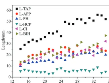
Figure 6represents the variation of 6 length parameters with the gestational weeks, where the LTAPobviously increased from 25.46 to 52.28 mm, reflecting the development of the whole brain. There was no obvious change in the LHCP, which may corre-spond to the relatively stable development of the body of the corpus callosum.17,18For the other 4 curves, the L
BIHincreased from 13.74 to 37.53 mm. A smaller magnitude of increase oc-curred in the LCIand LAPP, 10.785–32.45 and 11.61–23.81 mm, respectively.
[image:4.594.64.272.315.467.2]Depth and Length of the Calcarine Sulcus
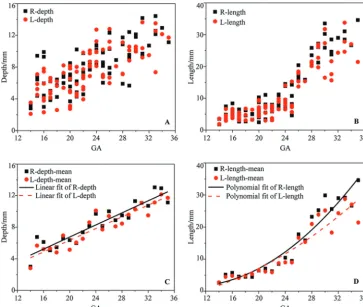
Figure 7reflects the changes in the depth and length of the calca-rine sulcus at 14 –35 gestational weeks with an increasing trend. The distribution of the sulcus length was more concentrated than that of the depth, showing a smaller individual difference. The depth had a linear increase compared with the smooth variation in the length at 14 –23 gestational weeks (Fig 7A). At 14 gestational weeks, the average depth reached 2.82 mm and then increased to 5.67 mm at 15 gestational weeks and 8.61 mm at 23 gestational weeks. At 24 –35 gestational weeks, the average depth increased from 10.11 to 12.71 mm with a linear growth law (Fig 7C). The length development was nonlinear growth with the increase in gestational weeks, which was not consistent with the depth of the calcarine sul-cus. After a platform period from 14 –23 gestational weeks, the length of the calcarine sulcus increased rapidly (Fig 7D).
Location Correlation
The calcarine sulcus and lateral ventricles were reconstructed in 1 space to show the spatial position relationship (Fig 8). The calca-FIG 4.The morphologic evolution of fetal lateral ventricles at 14 –31 weeks. ah indicates anterior
horn; ph, posterior horn; ih, inferior horn; cp, central part; GW, gestational weeks.
FIG 5. Volume measurements of the lateral ventricles. L-LV indicates the left lateral ventricle volume; R-LV, right lateral ventricle volume; T-LV, total lateral ventricle volume; GA, gestational age.
FIG 6. Quantitative measurements of the lateral ventricles.
[image:4.594.72.264.516.663.2]rine sulcus was close to the posterior part of the lateral ventricles (Fig 8A1,B1,andC1). The inner side of the calcarine sulcus was parallel to the cerebral median fissure. The lateral ventricles be-came thin with the increase of gestational weeks, and the calcarine sulcus became wider and longer. The posterior view showed that part of the 2 structures overlapped and the morphologic charac-teristics were not obvious. On the lateral view, the calcarine sulcus and lateral ventricles overlapped before 21 gestational weeks. With the development of the calcarine sulcus, the posterior part gradually extended longer backward. The occipital part of the lateral ventricles narrowed with the lengthening of the calcarine sulcus.
Measurements Correlation
According to the results of location correlation, the length and depth of the left and right calcarine sulcus and the posterior horn length in the lateral ventricles were selected for analysis in this study. The depth and length of the calcarine sulcus were linearly related to the posterior horn length (Fig 9). The length and depth of the calcarine sulcus can be expressed by the following equation: Right-length⫽2.4204 (LPH) – 27.5706, Right-depth⫽0.5145 (LPH) – 2.3072, Left-length ⫽ 2.0939 (LPH) – 23.4099, Left-depth ⫽ 0.5902(LPH)–2.3403.
Sex and Side Difference
There was almost no difference between the left and right lateral ventricle volume before 24 gestational weeks and a large differ-ence after 24 gestational weeks (Fig 5). However, statistics showed that there was no hemispheric difference in the lateral ventricle
volume during the entire period (P⬎.05). The depth and length of the calcarine sulcus in the right hemisphere were greater than those in the left, which was statistically significant (allP⬍.05). There were no sexual dimorphisms in those measurements (all,
P⬎.05).
DISCUSSION
Formation of the Fetal Lateral Ventricle
The morphologic evolution of the lateral ventricles is affected by many aspects such as the cortical folding and the development of subcortical structures.3 At 10 –13 gestational weeks, the lateral
ventricles account for most of the brain because of the vesicular period of the ventricular system.19In this study, it was found that
the lateral ventricle morphology still had the characteristics of the vesicular period at 14 gestational weeks (Fig 4A). The shape of the lateral ventricles evolves from an embryonic crescent into the im-mature lateral ventricles with 4 horns.20With the development of
the fetal brain, the lateral ventricles begin to lose the features of the vesicular period and gradually separates into 4 distinct parts at 16 gestational weeks.2The increase in L
APP, LCI, and LBIHindicates the development of the basal nuclei and thalamus at this stage, resulting in the narrowing of the anterior horn of the lateral ven-tricles (Fig 4B, -C). It is evident that the slope of the LPHis less than that of the LTAP, findings in agreement with the shrinkage of the lateral ventricular occipital region (Fig 6,Fig 4D). Eventually, the lateral ventricle morphology development approaches that of adults.
[image:5.594.113.476.46.352.2]tional weeks (Fig 5), which is in agree-ment with the study by Clouchoux et al.21However, Huang et al3found that
the volume of the lateral ventricles first increased and then decreased at 13–21 gestational weeks. Kinoshita et al19
ob-served that the volume of the lateral ven-tricles increased at 7–23 gestational weeks. The results of the lateral ventricle volume are different in these studies. One reason is the differences in the sam-ple sizes and individual fetuses. Another reason could be the differences in the structural anatomic definition and in-clusion of the third ventricle or the ca-vum septum pellucidum. It is conceiv-able that the decrease in volume is associated with the change of the ven-tricular morphology. At 14 gestational weeks, the vesicular effect of the ventri-cles gradually disappears and the vol-ume of the lateral ventricles is reduced correspondingly. Meanwhile, the vol-ume of the germinal matrix and subcor-tical structures such as the basal nuclei also increases with the gestational week.22It is inferred that the shrinkage
in the lateral ventricles is accompanied by the expansion of the germinal matrix and basal nuclei. The lateral ventricles and these subcortical structures develop together and affect each other in the morphologic evolution of the lateral ventricles.
At 23–35 gestational weeks, the vol-ume of the lateral ventricles increases with the fluctuating trend. The LTAP grows faster than the LAPP, LHCP, and LCI, reflecting the growth in the brain volume being faster than that of the basal ganglia, thalamus, and other struc-tures around the ventricles (Fig 6). Thus, the lateral ventricle volume increases rapidly at 23–26 gestational weeks. This finding is in agreement with a previous study that found that fetal brain volume increases linearly and subcortical struc-tures such as germinal matrix volume decrease at this stage.22After 26
gesta-tional weeks, the fetal brain volume in-creases quickly and the germinal matrix decreases. The germinal matrix gradu-ally develops into the caudate, putamen, globus pallidus, and basal ganglia.3The
increase in LAPP, LBIH, and LCI repre-sents these subcortical structures begin-ning to expand. Then the increased ve-locity of the lateral ventricle volume FIG 8. The location of the calcarine sulcus in 3D space: superior view (A1,B1,C1), posterior view
(A2,B2,C2), lateral view (A3,B3,C3).A, Twenty-one GW (A1,A2,A3).B, Thirty-one GW (B1,B2,B3). C, Thirty-five GW (C1,C2,C3). The left lateral ventricle is green, the right lateral ventricle is red, and the calcarine sulcus is pink. GW indicates gestational weeks.
FIG 9. The correlation between the LPHand the depth and length of the calcarine sulcus.
[image:6.594.57.372.48.361.2] [image:6.594.55.382.442.715.2]becomes slower. It is inferred that the individual differences in development are more obvious at this stage, resulting in the in-crease in the volume of the lateral ventricles with the fluctuating trend.
A difference occurred in the development of left and right lateral ventricle volume after 24 gestational weeks, which may be related to the lateralization of cerebral hemisphere development. Lateral differences in the cerebral hemispheres already exist at birth. The growth of the cerebral hemisphere structures may af-fect the lateral ventricles, resulting in this large difference in lateral ventricle volume. Meanwhile, this difference may also be related to other features such as the number of specimens and the method of measurement and so forth. However, the cerebral lateralization is a very complex problem that requires more data to reach a conclusion. The change in the lateral ventricle volume is stable at 14 –23 gestational weeks and fluctuates after 23 gestational weeks. The lateral ventricle volume can reflect the development of the lateral ventricles comprehensively compared with the lateral ven-tricle width on a 2D level.4It is suggested that the lateral ventricle
volume measured by 3D sonography techniques needs to be used to improve prenatal diagnostic accuracy after 23 gestational weeks.
Correlation Analysis Between the Lateral Ventricles and the Calcarine Sulcus
The growth of the calcarine sulcus length accelerated noticeably after a slow developmental period. It is consistent with the varia-tion of the lateral ventricle volume (Fig 7D). This finding indicates that there is a certain relationship between the calcarine sulcus and lateral ventricles. Thus, we elucidated the relationship be-tween the lateral ventricles and the calcarine sulcus, including the location and measurement correlations.
The calcarine sulcus is located at the inner side of the lateral ventricle occipital part. The inner sides of the calcarine sulcus on both sides are parallel (Fig 8A1). The calcarine sulcus gradually increases backward and exceeds the occipital part of the lateral ventricles (Fig 8B1,-C1). The curvature of the posterior calcarine sulcus is related to the acceleration of cortical folding in the later developmental period (Fig 8A3–C3).5The calcarine sulcus depth
increases and extends outward. The lateral ventricle occipital part becomes flattened, and the size relatively decreases (Fig 8A3–C3). Combined with the development of the fetal brain, the varia-tion of calcarine sulcus length and the volume of lateral ventricles is associated with the whole-brain development.23At 14 –23
ges-tational weeks, the whole calcarine sulcus does not exceed the boundary of the lateral ventricle occipital part. The lateral ventri-cles and the calcarine sulcus gradually develop with fetal brain development. At 24 –35 gestational weeks, the calcarine sulcus posterior part size exceeds the lateral ventricle occipital part and becomes a wave type. Both of them are in a relatively slow growth period; then, the growth becomes faster. The variation of the cal-carine sulcus length and lateral ventricle volume is roughly con-sistent with the trend of fetal brain volume.
On the basis of the above analysis, the lateral ventricle occipital part is affected by the calcarine sulcus in its development. As the increase in the calcarine sulcus depth, the posterior horn length becomes smaller. There is a better linear correlation between the
left calcarine sulcus length and the posterior horn length repre-sented by the value of the LPH(Fig 9). The fitting effect between the LPHand calcarine sulcus length is better than that between the LPHand the calcarine sulcus depth, which is consistent with the phenomenon that the point dispersion degree of the calcarine sulcus depth is greater than that of calcarine sulcus length (Fig 9
andFig 7A, -B). This feature indicates that the stability of the calcarine sulcus length is better than that of the calcarine sulcus depth. The linear correlation between the lateral ventricle and the calcarine sulcus is helpful to elucidate the development of the lateral ventricles and improve the prenatal diagnosis of fetal ventriculomegaly.
CONCLUSIONS
The lateral ventricle volume increases, with a slow-to-rapid growth period at 14 –35 gestational weeks. The shrinkage in the lateral ventricle posterior horn is accompanied by the develop-ment of the calcarine sulcus, resulting in a better linear correlation between the calcarine sulcus length and the posterior horn length. The relevant results are helpful in understanding the evolution of the lateral ventricles and providing a basis for the diagnosis of lateral ventricle abnormality. Our future work is to study the relationship between the fetal lateral ventricles and the calcar-ine sulcus in patients with ventriculomegaly, to provide more valuable data for a differential diagnosis and prenatal exami-nation of ventriculomegaly.
ACKNOWLEDGMENTS
We thank Derong Duan, Qing Xia, Wenjia Liang, and Wenjun Wang for language help during the revision.
REFERENCES
1. Perlman S, Shashar D, Hoffmann C, et al.Prenatal diagnosis of fetal ventriculomegaly: agreement between fetal brain ultrasonography and MR imaging.AJNR Am J Neuroradiol2014;35:1214 –18CrossRef Medline
2. Taketani K, Yamada S, Uwabe C, et al.Morphological features and length measurements of fetal lateral ventricles at 16 –25 weeks of gestation by magnetic resonance imaging.Congenit Anom (Kyoto) 2015;55:99 –102CrossRef Medline
3. Huang H, Xue R, Zhang J, et al.Anatomical characterization of hu-man fetal brain development with diffusion tensor magnetic reso-nance imaging.J Neurosci2009;29:4263–73CrossRef Medline
4. Kyriakopoulou V, Vatansever D, Elkommos S, et al.Cortical over-growth in fetuses with isolated ventriculomegaly.Cereb Cortex 2014;24:2141–50CrossRef Medline
5. Dubois J, Benders M, Cachia A, et al.Mapping the early cortical folding process in the preterm newborn brain.Cereb Cortex2008; 18:1444 –54Medline
6. Baffero GM, Crovetto F, Fabietti I, et al.Prenatal ultrasound predic-tors of postnatal major cerebral abnormalities in fetuses with ap-parently isolated mild ventriculomegaly.Prenat Diagn 2015;35: 783– 88CrossRef Medline
7. Fukunishi K, Sawada K, Kashima M, et al.Correlation between for-mation of the calcarine sulcus and morphological maturation of the lateral ventricle in cynomolgus monkey fetuses.Acta Neurobiol Exp (Wars) 2011;71:381– 86Medline
8. Li H, Liang H, Wu H.Magnetic resonance imaging-based correla-tion analysis between calcarine sulcus development and isolated fetal ventriculomegaly. Congenit Anom (Kyoto) 2017;57:52–56
9. Chi JG, Dooling EC, Gilles FH.Gyral development of the human brain.Ann Neurol1977;1:86 –93CrossRef Medline
10. Zhang Z, Hou Z, Lin X, et al.Development of the fetal cerebral cortex in the second trimester: assessment with 7T postmortem MR imaging.AJNR Am J Neuroradiol2013;34:1462– 67CrossRef Medline
11. Zhang Z, Liu S, Lin X, et al.Development of fetal cerebral cortex: assessment of the folding conditions with post-mortem magnetic resonance imaging. Int J Dev Neurosci2010;28:537– 43CrossRef Medline
12. Tubbs RS, Shoja MM, Aggarwal A, et al.Choroid plexus of the fourth ventricle: review and anatomic study highlighting anatomical vari-ations.J Clin Neurosci2016;26:79 – 83CrossRef Medline
13. Ballesteros MC, Hansen PE, Soila K, et al.MR imaging of the devel-oping human brain, Part 1: prenatal development.Radiographics 1993;13:611–22CrossRef Medline
14. Zhang H, Zhang Z, Yin X, et al.Early development of the fetal central sulcus on 7.0T magnetic resonance imaging.Int J Dev Neurosci2016; 48:18 –23CrossRef Medline
15. Habas PA, Scott JA, Roosta A, et al.Early folding patterns and asym-metries of the normal human brain detected from in utero MRI. Cereb Cortex2012;22:13–25CrossRef Medline
16. Bayer SA, Altman J. The human brain during the second trimester. In: Atlas of Human Central Nervous System Development, Indianapolis: CRC Press; 2005
17. Rakic P, Yakovlev PI.Development of the corpus callosum and cavum septi in man. J Comp Neurol 1968;132:45–72CrossRef Medline
18. Achiron R, Achiron A.Development of the human fetal corpus callosum: a high-resolution, cross-sectional sonographic study. Ul-trasound Obstet Gynecol2001;18:343– 47CrossRef Medline
19. Kinoshita Y, Okudera T, Tsuru E, et al.Volumetric analysis of the germinal matrix and lateral ventricles performed using MR images of postmortem fetuses.AJNR Am J Neuroradiol 2001;22:382– 88
Medline
20. Shiraishi N, Katayama A, Nakashima T, et al.Morphology and mor-phometry of the human embryonic brain: a three-dimensional analysis.Neuroimage2015;115:96 –103CrossRef Medline
21. Clouchoux C, Guizard N, Evans AC, et al.Normative fetal brain growth by quantitative in vivo magnetic resonance imaging.Am J Obstet Gynecol2012;206:173.e1– 08CrossRef Medline
22. Meng H, Zhang Z, Geng H, et al.Development of the subcortical brain structures in the second trimester: assessment with 7.0-T MRI.Neuroradiology2012;54:1153–59CrossRef Medline
23. Scott JA, Habas PA, Rajagopalan V, et al.Volumetric and surface-based 3D MRI analyses of fetal isolated mild ventriculomegaly: brain morphometry in ventriculomegaly.Brain Struct Funct2013; 218:645–55CrossRef Medline