Timothy J. Foster
J Clin Invest.
2004;
114(12)
:1693-1696.
https://doi.org/10.1172/JCI23825
.
There has been some debate about the disease-invoking potential of
Staphylococcus
aureus
strains and whether invasive disease is associated with particularly virulent
genotypes, or “superbugs.” A study in this issue of the
JCI
describes the genotyping of a
large collection of nonclinical, commensal
S. aureus
strains from healthy individuals in a
Dutch population. Extensive study of their genetic relatedness by amplified restriction
fragment typing and comparison with strains that are associated with different types of
infections revealed that the
S. aureus
population is clonal and that some strains have
enhanced virulence. This is discussed in the context of growing interest in the mechanisms
of bacterial colonization, antibiotic resistance, and novel vaccines.
Commentary
Find the latest version:
platelet P-selectin and tumor cell sialylated mucins that has been shown to be essential for metastatic spread in mouse models (17). Finally, the findings of Boucharaba et al. lay the groundwork to suggest that inhibition of platelet-derived LPA action on its cog-nate receptors expressed by tumor cells may be another promising therapeutic target, especially for bone metastasis. The devel-opment and clinical testing of this class of specific modulators of platelet function will be necessary before a verdict can be reached regarding the importance of platelets in the progression of disease in cancer patients.
Address correspondence to: Joan Mas-sagué, Memorial Sloan-Kettering Cancer Center, 1275 York Avenue, Box 116, New York, New York 10021, USA. Phone: (212) 639-8975; Fax: (212) 717-3298; E-mail: [email protected].
1. Rickles, F.R., and Falanga, A. 2001. Molecular basis for the relationship between thrombosis and
can-cer. Thromb. Res. 102:V215–V224.
2. Nash, G.F., Turner, L.F., Scully, M.F., and Kakkar, A.K. 2002. Platelets and cancer. Lancet Oncol. 3:425–430. 3. Karpatkin, S., and Pearlstein, E. 1981. Role of
platelets in tumor cell metastases. Ann. Intern. Med.
95:636–641.
4. Nieswandt, B., Hafner, M., Echtenacher, B., and Mannel, D.N. 1999. Lysis of tumor cells by natural killer cells in mice is impeded by platelets. Cancer Res. 59:1295–1300.
5. Trikha, M., and Nakada, M.T. 2002. Platelets and cancer: implications for antiangiogenic therapy.
Semin. Thromb. Hemost. 28:39–44.
6. Boucharaba, A., et al. 2004. Platelet-derived lysophosphatidic acid supports the progres-sion of osteolytic bone metastases in breast can-cer. J. Clin. Invest. 114:1714–1725. doi:10.1172/ JCI200422123.
7. Jurasz, P., Alonso-Escolano, D., and Radomski, M.W. 2004. Platelet-cancer interactions: mechanisms and pharmacology of tumour cell-induced platelet aggre-gation. Br. J. Pharmacol. doi:10.1038/sj.bjp.0706013. 8. FitzGerald, G.A. 1991. Mechanisms of platelet acti-vation: thromboxane A2 as an amplifying signal for other agonists. Am. J. Cardiol. 68:11B–15B. 9. Brass, L.F. 2003. Thrombin and platelet activation.
Chest. 124:18S–25S.
10. Mills, G.B., and Moolenaar, W.H. 2003. The emerg-ing role of lysophosphatidic acid in cancer. Nat. Rev. Cancer. 3:582–591.
11. Ishii, I., Fukushima, N., Ye, X., and Chun, J. 2004.
Lysophospholipid receptors: signaling and biology.
Annu. Rev. Biochem. 73:321–354.
12. van Corven, E.J., Groenink, A., Jalink, K., Eichholtz, T., and Moolenaar, W.H. 1989. Lysophosphatidate-induced cell proliferation: identification and dis-section of signaling pathways mediated by G pro-teins. Cell. 59:45–54.
13. Mundy, G.R. 2002. Metastasis to bone: causes, con-sequences and therapeutic opportunities. Nat. Rev. Cancer. 2:584–593.
14. Yin, J.J., et al. 1999. TGF-β signaling blockade inhibits PTHrP secretion by breast cancer cells and bone metastases development. J. Clin. Invest.
103:197–206.
15. Kang, Y., et al. 2003. A multigenic program medi-ating breast cancer metastasis to bone. Cancer Cell.
3:537–549.
16. Falanga, A. 2004. The effect of anticoagulant drugs on cancer. J. Thromb. Haemost. 2:1263–1265. 17. Varki, N.M., and Varki, A. 2002. Heparin inhibition
of selectin-mediated interactions during the hema-togenous phase of carcinoma metastasis: rationale for clinical studies in humans. Semin. Thromb. Hemost. 28:53–66.
18. Amirkhosravi, A., et al. 2003. Inhibition of tumor cell-induced platelet aggregation and lung metas-tasis by the oral GpIIb/IIIa antagonist XV454.
Thromb. Haemost. 90:549–554.
19. Bakewell, S.J., et al. 2003. Platelet and osteoclast beta3 integrins are critical for bone metastasis.
Proc. Natl. Acad. Sci. U. S. A. 100:14205–14210.
The
Staphylococcus aureus
“superbug”
Timothy J. FosterMicrobiology Department, Moyne Institute of Preventive Medicine, Trinity College, Dublin, Ireland.
There has been some debate about the disease-invoking potential of
Staphy-lococcus aureus
strains and whether invasive disease is associated with
par-ticularly virulent genotypes, or “superbugs.” A study in this issue of the
JCI
describes the genotyping of a large collection of nonclinical, commensal
S. aureus
strains from healthy individuals in a Dutch population. Extensive
study of their genetic relatedness by amplified restriction fragment typing
and comparison with strains that are associated with different types of
infec-tions revealed that the
S. aureus
population is clonal and that some strains
have enhanced virulence (see the related article beginning on page 1732).
This is discussed in the context of growing interest in the mechanisms of
bacterial colonization, antibiotic resistance, and novel vaccines.
Nasal colonization
Staphylococcus aureus is a common commen-sal of humans and its primary habitat is the moist squamous epithelium of the anterior nares (1). About 20% of the population are always colonized with S. aureus, 60% are
intermittent carriers, and 20% never carry the organism. As there is considerable evi-dence that carriage is an important risk factor for invasive infection (1, 2), it is sur-prising that so little is known about the bacterial factors that promote coloniza-tion of squamous epithelial surfaces and the host factors that determine whether an individual can be colonized or not.
Methicillin-resistant S. aureus
Healthy individuals have a small but finite risk of contracting an invasive infection caused by S. aureus, and this risk is increased among carriers. Hospital patients who are
catheterized or who have been treated surgi-cally have a significantly higher rate of infec-tion. In some, but not all, developed coun-tries, many nosocomial infections are caused by S. aureus strains that are multiply resis-tant to antibiotics — known as methicillin-resistant Staphylococcus aureus (MRSA) (3, 4) — although the acronym MRSA is somewhat misleading because the semisynthetic β -lac-tam methicillin is no longer used to treat
S. aureus infections. In MRSA, the horizon-tally acquired mecA gene encodes a penicil-lin-binding protein, PBP2a, which is intrin-sically insensitive to methicillin and all
β-lactams that have been developed, includ-ing the isoxazoyl penicillins (e.g., oxacillin) that superceded methicillin, in addition to the broad spectrum β-lactams (third-gener-ation cephalosporins, cefamycins, and car-bapenems) that were introduced primarily to treat infections caused by Gram-negative bacteria (4) (Figure 1). In contrast to noso-comial MRSA strains, which are usually multidrug resistant, the recently emerged community-acquired MRSA (CA-MRSA) strains are susceptible to drugs other than
β-lactams (5). Nonstandard abbreviations used: AFLP, amplified
fragment length polymorphism; CA-MRSA, communi-ty-acquired MRSA; MLST, multilocus sequence typing; MRSA, methicillin-resistant Staphylococcus aureus; PVL; Panton-Valentine leukocidin; WTA, wall teichoic acid. Conflict of interest: The author has declared that no conflict of interest exists.
The term MRSA is synonymous with mul-tidrug-resistant S. aureus because many nos-ocomial MRSA strains are resistant to most commonly used antibiotics. The glycopep-tide vancomycin was the last available drug to which this organism had remained uniform-ly sensitive until recent reports of low-level glycopeptide resistance (6) and, very recent-ly, the transfer of high-level vancomycin resistance from Enterococcus to S. aureus (7). Although new drugs, including linezolid and synercid, have recently been introduced to treat MRSA infections (8), there is a worry-ing lack of novel drugs in the pipeline.
Virulence factors
S. aureus strains can express a wide array of potential virulence factors (Figure 1), includ-ing surface proteins that promote
adher-ence to damaged tissue (9), bind proteins in blood to help evade antibody-mediated immune responses (9), and promote iron uptake (10). The organism also expresses a number of membrane-damaging toxins and superantigen toxins that can cause tissue damage and the symptoms of septic shock, respectively (11). There is a growing realiza-tion that S. aureus has multiple mechanisms for evading both innate immunity mediated by polymorphonuclear leukocytes (12, 13) and induced immunity mediated by both B and T cells (11, 14). Some virulence fac-tors are expressed by genes that are located on mobile genetic elements called pathoge-nicity islands (e.g., toxic shock syndrome toxin–1 and some enterotoxins; ref. 15) or lysogenic bacteriophages (e.g., Panton-Val-entine leucocidin [PVL]; refs. 15, 16) and
factors associated with suppressing innate immunity such as the chemotaxis inhibitory protein and staphylokinase (ref. 13), which are integrated in the bacterial chromosome.
The S. aureus population is clonal
The study by Melles et al. reported in this issue of the JCI (17) examines the major questions about natural populations of S. aureus con-cerning clonality and virulence. The authors examined 829 S. aureus strains from healthy donors from the city of Rotterdam in The Netherlands. Selective amplified fragment length polymorphism (AFLP) amplification analysis was used to compare genetic relat-edness of strains, and this analysis revealed the existence of 3 major and 2 minor genetic clusters, subsequently confirmed by mul-tilocus sequence typing (MLST) (Figure 2). The authors therefore concluded that the S. aureus population is clonal. These clusters corresponded to the predominant groups identified in a recent study of carriage iso-lates from the county of Oxfordshire in the United Kingdom using MLST (18). Thus the same clonal lineages appear to be dominant in 2 distinct geographic locations.
Evidence for clones with enhanced virulence
Melles et al. (17) also examined a smaller number of isolates from individuals with invasive disease (bacteremia and deep-seat-ed abscesses) as well as those from individ-uals with severe impetigo. There was clear evidence that some clonal types are more virulent than others in that they appeared more frequently among disease isolates than among carriage isolates (Figure 2). This sharply contrasts with the earlier Oxfordshire study, in which no evidence for hypervirulent clones was found (18). In the Melles et al. study, bacteremia in elderly patients was significantly more frequently caused by 1 strain in cluster IVa (17). In addition, strains causing severe skin infec-tions were significantly more frequently found to be members of cluster IVb. This could be due to lysogenization of a progen-itor IVb strain with the bacteriophage that encodes PVL (16), a toxin that is strongly associated with severe skin infections (19).
The question remains: Why did this study find evidence for virulent clones, whereas the Oxfordshire study did not? Perhaps this discrepancy can be attributed to the fact that Melles et al. tested a larger number of strains (17). Furthermore, the Oxford-shire study (18) was confined to isolates obtained from patients with invasive
infec-Figure 1
tions requiring admission to hospital and thus excluded impetigo and skin infections. Another factor might have been be the prev-alence of MRSA strains present in the noso-comial invasive isolates from individuals in the UK; in contrast, of the invasive strains isolated from Dutch individuals, none were identified as MRSA. This is consistent with the conclusion that all strains of S. aureus
have the potential to cause infection and that some are more virulent than others.
Evolution of MRSA
MRSA strains have emerged by acquisition of mobile genetic elements called SCCmec
cassettes, which carry the mecA gene that encodes PBP2a. There are 5 different cas-settes (SCCmec types I–V; refs. 3, 20, and 21). It is now clear that major MRSA clones were created on multiple occasions by acquisition of SCCmec by prevalent strains that have continued to flourish (22). None of the Dutch carriage or invasive disease isolates were found to harbor the mecA gene (17). Nevertheless, the authors examined a variety of MRSA strains from other sources and found SCCmec-containing strains in each of their major genetic clusters. This is consistent with previous studies (22) that
established that MRSA strains have arisen many times by transfer of SCCmec cassettes into susceptible host strains.
PVL is a 2-component cytolytic toxin with high affinity for human leukocytes (11). It has been associated with S. aureus
strains causing severe skin infections (19) and with necrotizing pneumonia in previ-ously healthy youths (23). In the Melles et al. study (17), PVL was rarely found in the carriage isolates (0.6%) but was present in a significantly high number of strains that caused abscesses and arthritis. PVL is also expressed by the newly emerged CA-MRSA strains, which appear to have enhanced vir-ulence (5, 23). However, Melles et al. did not examine CA-MRSA strains in this study.
Future prospects for combating
S. aureus
[image:4.585.53.364.82.321.2]Given the problems caused by the develop-ment of antibiotic-resistant S. aureus, vac-cination may well have a significant role to play in controlling this organism in the future. A number of companies are develop-ing products intended for active or passive immunization against S. aureus infections, including a capsular polysaccharide vaccine that has been subjected to a clinical trial
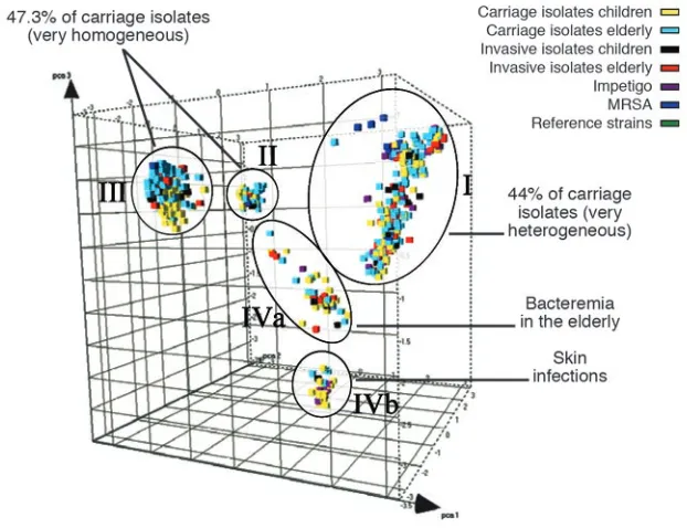
Figure 2
Principal component analysis of 1,056 S. aureus strains reveals genetic clusters of hyperviru-lent clones (17). The different cubes, plotted here in a 3D space and colored according to their source, represent each S. aureus strain analyzed in the Melles et al. study. The 5 circles indicate the 3 major (I, II, and III) and 2 minor (IVa and IVb) different phylogenetic clusters identified by AFLP. While strains from each of the genetic clusters are essentially able to cause invasive disease, some clusters contain proportionally more invasive isolates.
with hemodialysis patients (24), a mono-clonal antibody (25), and human immuno-globulin that is enriched for antibodies that recognize clumping factor A (26).
An increased understanding of how
S. aureus colonizes the nares could allow improved methods for controlling nasal and skin carriage. Recent studies of mutant strains defective in wall teichoic acid (WTA) in a rat model of nasal colo-nization implicated WTA in colocolo-nization (27). Also, several different surface pro-teins can promote adherence of S. aureus
to squamous epithelial cells isolated from the nares (28, 29) and could act as adhesins involved in nasal colonization. Another mystery that deserves greater attention is the question of why some members of the population never carry S. aureus, while others are persistent carriers.
The study by Melles et al. (17), in combi-nation with MLST analysis (18), provides a solid foundation for analysis of novel hyper-virulent or epidemic drug-resistant S. aureus
clones that might arise in the future. One thing seems certain: S. aureus will continue to respond to challenges imposed by humans’ continued attempts to combat its carriage and development of related disease.
Address correspondence to: Timothy J. Foster, Microbiology Department, Moyne Institute of Preventive Medicine, Trinity College, Dublin 2, Ireland. Phone: 353-1-6082014; Fax: 353-1-6799294; E-mail: [email protected].
1. Peacock, S.J., de Silva, I., and Lowy, F.D. 2001. What determines nasal carriage of Staphylococcus aureus?
Trends Microbiol. 9:605–610.
2. von Eiff, C., Becker, K., Machka, K., Stammer, H., and Peters, G. 2001. Nasal carriage as a source of
Staphylococcus aureus bacteremia. Study Group.
N. Engl. J. Med. 344:11–16.
3. Hiramatsu, K., Cui, L., Kuroda, M., and Ito, T. 2001. The emergence and evolution of methicillin-resistant
Staphylococcus aureus. Trends Microbiol. 9:486–493. 4. Chambers, H.F. 1997. Methicillin resistance in
staphylococci: molecular and biochemical basis and clinical implications. Clin. Microbiol. Rev.
10:781–791.
5. Vandenesch, F., et al. 2003. Community-acquired methicillin-resistant Staphylococcus aureus carrying Panton-Valentine leukocidin genes: worldwide emergence. Emerging Infect. Dis. 9:978–984. 6. Hiramatsu, K. 2001. Vancomycin-resistant
Staphylo-coccus aureus: a new model of antibiotic resistance.
Lancet Infect. Dis. 1:147–155.
7. Weigel, L.M., et al. 2003. Genetic analysis of a high-level vancomycin-resistant isolate of Staphylococcus aureus. Science. 28:1569–1571.
8. Eliopoulos, G.M. 2003. Quinupristin-dalfopristin and linezolid: evidence and opinion. Clin. Infect. Dis.
36:473–481.
9. Foster, T.J., and Höök, M. 1998. Surface protein adhesins of Staphylococcus aureus. Trends Microbiol.
10. Mazmanian, S.K., et al. 2003. Passage of heme-iron across the envelope of Staphylococcus aureus. Science.
299:906–909.
11. Bohach, G.A., and Foster, T.J. 1999. Staphylococcus aureus exotoxins. In Gram positive bacterial pathogens.
V.A. Fischetti, R.P. Novick, J.J. Ferretti, and J.I. Rood, editors. American Society for Microbiology. Washington, D.C., USA. 367–378.
12. Fedtke, I., Gotz, F., and Peschel, A. 2004. Bacterial evasion of innate host defenses--the Staphylococcus aureus lesson. Int. J. Med. Microbiol. 294:189–194. 13. de Haas, C.J., et al. 2004. Chemotaxis inhibitory
protein of Staphylococcus aureus, a bacterial antiin-flammatory agent. J. Exp. Med. 199:687–695. 14. Goodyear, C.S., and Silverman, G.J. 2003. Death by
a B cell superantigen: in vivo VH-targeted apoptotic supraclonal B cell deletion by a staphylococcal Toxin. J. Exp. Med. 197:1125–1139.
15. Novick, R.P. 2003. Mobile genetic elements and bacterial toxinoses: the superantigen-encoding pathogenicity islands of Staphylococcus aureus. Plasmid. 49:93–105.
16. Narita, S., et al. 2001. Phage conversion of Pan-ton-Valentine leukocidin in Staphylococcus aureus: molecular analysis of a PVL-converting phage, phiSLT. Gene. 268:195–206.
17. Melles, D.C., et al. 2004. Natural population
dynamics and expansion of pathogenic clones of
Staphylococcus aureus. J. Clin. Invest. 114:1732–1740. doi:10.1172/JCI200423083.
18. Feil, E.J., et al. 2003. How clonal is Staphylococcus aureus? J. Bacteriol. 185:3307–3316.
19. Lina, G., et al. 1999. Involvement of Panton-Val-entine leukocidin-producing Staphylococcus aureus
in primary skin infections and pneumonia. Clin. Infect. Dis. 29:1128–1132.
20. Ito, T., et al. 2004. Novel type V staphylococcal cas-sette chromosome mec driven by a novel cascas-sette chromosome recombinase, ccrC. Antimicrob. Agents Chemother. 48:2637–2651.
21. Ma, X.X., et al. 2002. Novel type of staphylococ-cal cassette chromosome mec identified in com-munity-acquired methicillin-resistant Staphylo-coccus aureus strains. Antimicrob. Agents Chemother.
46:1147–1152.
22. Enright, M.C., et al. 2002. The evolutionary his-tory of methicillin-resistant Staphylococcus aureus
(MRSA). Proc. Natl. Acad. Sci. U. S. A. 99:7687–7692. 23. Gillet, Y., et al. 2002. Association between Staphy-lococcus aureus strains carrying gene for Panton-Valentine leukocidin and highly lethal necrotising pneumonia in young immunocompetent patients.
Lancet. 359:753–759.
24. Fattom, A.I., Horwith, G., Fuller, S., Propst, M.,
and Naso, R. 2004. Development of StaphVAX, a polysaccharide conjugate vaccine against S. aureus
infection: from the lab bench to phase III clinical trials. Vaccine. 17:880–887.
25. Hall, A.E., et al. 2003. Characterization of a protec-tive monoclonal antibody recognizing Staphylococ-cus aureus MSCRAMM protein clumping factor A.
Infect. Immun. 71:6864–6870.
26. Vernachio, J., et al. 2003. Anti-clumping fac-tor A immunoglobulin reduces the duration of methicillin-resistant Staphylococcus aureus bacteremia in an experimental model of infective endocarditis.
Antimicrob. Agents Chemother. 47:3400–3406. 27. Weidenmaier, C., et al. 2004. Role of teichoic acids
in Staphylococcus aureus nasal colonization, a major risk factor in nosocomial infections. Nat. Med.
10:243–245.
28. Roche, F.M., Meehan, M., and Foster, T.J. 2003. The Staphylococcus aureus surface protein SasG and its homologues promote bacterial adherence to human squamous nasal epithelial cells. Microbiol-ogy. 149:2759–2767.
29. O’Brien, L.M., Walsh, E.J., Massey, R.C., Peacock, S.J., and Foster, T.J. 2002. Staphylococcus aureus
clumping factor B (ClfB) promotes adherence to human type I cytokeratin 10: implications for nasal colonization. Cell. Microbiol. 4:759–770.
Acid sensing in renal epithelial cells
Stephen L. GluckDivision of Nephrology, UCSF, San Francisco, California, USA.
The kidney adjusts net acid excretion to match production with exquisite
precision, despite little or no change in the plasma bicarbonate
concen-tration. The acid-sensing pathway that signals the kidney to increase acid
secretion involves activation of the proto-oncogene c-Src. A new study in
this issue shows that proline-rich tyrosine kinase 2 (Pyk2) is responsible for
acid-induced activation of c-Src and is essential for acid sensing in renal
epi-thelial cells (see the related article beginning on page 1782). The findings
implicate a broader role for Pyk2 in acid-base homeostasis in bone and other
tissues beyond the kidney.
Although the principal product of metabo-lism in mammalian cells is the volatile acid carbon dioxide, humans on a typical West-ern diet produce about 70 millimoles of nonvolatile acid per day. Remarkably, vary-ing metabolic acid production over a range of 0–150 millimoles is accompanied by a matching increase in net acid excretion by the kidney with a change of only 1 mM in plasma bicarbonate concentration (1). The adaptive responses that enable the kidney to increase net acid excretion in response to
increased acid generation have been stud-ied extensively in animal models of meta-bolic acidosis. In the proximal tubule, aci-dosis increases the activity of luminal and basolateral proteins involved in bicarbon-ate transport (2, 3), ammonia generation (4), and the reabsorption and metabolism of citrate (5). In the collecting duct, acido-sis suppresses bicarbonate secretion (6) and stimulates recruitment of proton pumps to the luminal membrane of intercalated cells (7). Of the acid-base transporters in the proximal tubule, the luminal sodium/ hydrogen exchanger 3 (NHE3) has a promi-nent role, and the mechanism by which its activity increases during metabolic acidosis has been examined in some detail. Meta-bolic acidosis acutely increases the kinetic activity of NHE3 through direct pH effects and by phosphorylation (8), while chronic
acidosis increases the number of NHE3 transporters (9).
Acid-base transporter kinetics cannot account for precise pH sensing
How does the kidney “know” to adjust net acid excretion with such precision with only minimal changes in plasma bicarbonate con-centration? Available data in the physiology literature suggests that transporter kinetics alone cannot account for this degree of sen-sitivity. In the proximal tubule, a reduction in extracellular bicarbonate induces a fall in intracellular pH, which directly activates the sodium/hydrogen exchanger through an intracellular pH regulatory site (10). This requires a change in intracellular pH of about 0.1 to achieve a 50% increase in the rate of transport or an approximately 5% change in the rate of transport in response to a change in extracellular bicarbonate concentration of 1 mM. Both the luminal vacuolar H+-ATPase and the basolateral
sodium bicarbonate cotransporter in the proximal tubule are even less responsive to changes in intracellular pH (11–13). This suggests that a bicarbonate (or pH) sensor that can amplify luminal proton secretion must be present.
Nonstandard abbreviations used: FAK, focal adhe-sion kinase; NHE3, sodium/hydrogen exchanger 3; OKP, opossum kidney clone P; Pyk2, proline-rich tyro-sine kinase 2.
Conflict of interest: The author has declared that no conflict of interest exists.