A novel therapy for colitis utilizing PPAR-
gg
ligands to inhibit the epithelial inflammatory
response
Chinyu G. Su, … , Mitchell A. Lazar, Gary D. Wu
J Clin Invest.
1999;
104(4)
:383-389.
https://doi.org/10.1172/JCI7145
.
Peroxisome proliferator-activated receptor
g
(PPAR-
g
), a member of the nuclear hormone
receptor superfamily originally shown to play a critical role in adipocyte differentiation and
glucose homeostasis, has recently been implicated as a regulator of cellular proliferation
and inflammatory responses. Colonic epithelial cells, which express high levels of PPAR-
g
protein, have the ability to produce inflammatory cytokines that may play a role in
inflammatory bowel disease (IBD). We report here that PPAR-
g
ligands dramatically
attenuate cytokine gene expression in colon cancer cell lines by inhibiting the activation of
nuclear factor-
k
B via an I
k
B-
a
–dependent mechanism. Moreover, thiazolidinedione ligands
for PPAR-
g
markedly reduce colonic inflammation in a mouse model of IBD. These results
suggest that colonic PPAR-
g
may be a therapeutic target in humans suffering from IBD.
J. Clin. Invest.
104
:383-389(1999).
Article
Introduction
Delicately balanced mechanisms maintain the intestin-al mucosa in a quiescent state of inflammation. In humans, loss of this balance leads to inflammatory bowel disease (IBD), a chronic debilitating disease that affects millions of people worldwide. The intestinal inflammatory phenotype observed in many immune receptor and cytokine knockout mice models lends sup-port to the notion that cytokines help to regulate and maintain this quiescent state (1). It is now apparent that the intestinal epithelium, which is the interface between the highly antigenic luminal environment and the mucosal immune system, plays an active role in the immune responsiveness of the intestinal mucosa. Intesti-nal epithelial cells not only express various immune receptors traditionally believed to be expressed primari-ly by myeloid cell lineages (2), but they can also produce a wide array of immunomodulatory substances such as cytokines and complement factors (3–5). Indeed, specif-ic perturbation of the intestinal epithelium can lead to intestinal inflammation (6, 7).
Many immune response genes are regulated by homo-and heterodimeric complexes of the nuclear factor-κB/Rel (NF-κB/Rel) family of transcription factors. We have pre-viously shown that binding of proteins to the NF-κB ele-ment in the IL-8 promoter is critical for IL-1β–mediated activation of this promoter in intestinal cell lines (8). In most cells, NF-κB/Rel factors are maintained in an inac-tive state in the cytoplasm bound to members of the IκB family of inhibitory proteins (9). Upon cell stimulation by a variety of immunomodulatory substances such as IL-1,
TNF-α, PMA, or oxygen radicals, these IκB proteins are rapidly phosphorylated and degraded via the ubiquitin-proteosome pathway (10). This enables NF-κB/Rel pro-teins to translocate from the cytoplasm into the nucleus and activate gene transcription. Inhibitors of NF-κB acti-vation, such as glucocorticoids, have been shown to be very potent anti-inflammatory agents (11).
Recently, ligands for the peroxisome proliferator-acti-vated receptor γ(PPAR-γ) have been shown to inhibit the expression of various cytokines in monocytes and macrophages, principally by preventing the activation of NF-κB/Rel by an unknown mechanism (12, 13). PPAR-γ is a member of the nuclear hormone receptor superfami-ly whose ligands include several prostanoids including 15-deoxy-∆12,14prostaglandin J
2(15d-PGJ2), polyunsaturated
fatty acids, a variety of nonsteroidal anti-inflammatory drugs (NSAIDs), and a new class of oral antidiabetic agents, the thiazolidinediones (TZDs) (14–16). PPAR-γ lig-ands are best characterized as regulators of adipocyte dif-ferentiation and glucose homeostasis (17, 18).
PPAR-γis also expressed at high levels in both the colonic epithelium and in colon cancer cell lines (19–23). The function of PPAR-γin the colonic epithelium is cur-rently unknown. This is reinforced by recent studies that report that PPAR-γligands can either increase or decrease colonocyte proliferation depending on the model system studied (21–24). Here we show that the role of PPAR-γin the colon may not be solely, or even primarily, to regulate growth and differentiation, but rather to regulate immune responsiveness. PPAR-γligands dramatically interfere with the activation of NF-κB and the ability of
A novel therapy for colitis utilizing PPAR-
γ
ligands
to inhibit the epithelial inflammatory response
Chinyu G. Su,
1Xiaoming Wen,
1Shannon T. Bailey,
2Wen Jiang,
1Shamina M. Rangwala,
2Sue A. Keilbaugh,
1Anne Flanigan,
3Sreekant Murthy,
3Mitchell A. Lazar,
2and Gary D. Wu
11Division of Gastroenterology, and
2Division of Endocrinology, Diabetes, and Metabolism, University of Pennsylvania School of Medicine, Philadelphia,
Pennsylvania 19104, USA
3Division of Gastroenterology and Hepatology, Hahnemann University Hospital, Philadelphia, Pennsylvania 19102, USA
Address correspondence and reprint requests to: Gary D. Wu, 600 Clinical Research Building, 415 Curie Boulevard, Philadelphia, Pennsylvania 19104-6144, USA. Phone: (215) 898-0158; Fax: (215) 573-2024; E-mail: [email protected].
Chinyu G. Su and Xiaoming Wen contributed equally to this work.
Received for publication April 21, 1999, and accepted in revised form June 30, 1999.
Peroxisome proliferator-activated receptor γ(PPAR-γ), a member of the nuclear hormone receptor super-family originally shown to play a critical role in adipocyte differentiation and glucose homeostasis, has recently been implicated as a regulator of cellular proliferation and inflammatory responses. Colonic epithelial cells, which express high levels of PPAR-γprotein, have the ability to produce inflammatory cytokines that may play a role in inflammatory bowel disease (IBD). We report here that PPAR-γligands dramatically attenuate cytokine gene expression in colon cancer cell lines by inhibiting the activation of nuclear factor-κB via an IκB-α–dependent mechanism. Moreover, thiazolidinedione ligands for PPAR-γmarkedly reduce colonic inflammation in a mouse model of IBD. These results suggest that colonic PPAR-γmay be a therapeutic target in humans suffering from IBD.
colonocytes to express immunomodulatory cytokines. Moreover, PPAR-γ ligands interfere with the colonic inflammatory process in vivo and, thus, have promise as preventive and therapeutic agents against IBD.
Methods
Cell culture.Caco-2 and HT-29 cells (American Type Culture Collection, Rockville, Maryland, USA) were maintained in DMEM containing 10% FBS and penicillin/streptomycin. RNA analysis. Caco-2 and HT-29 cells were treated with various concentrations of 15d-PGJ2, BRL 49653,
ethyl acetate, or DMSO for 24 hours. After RNA isola-tion and blotting, the Northern blots were hybridized with cDNA probes for IL-8, monocyte chemoattractant protein-1 (MCP-1), and ribosomal 7S (25).
Protein analysis.Proteins were isolated from cells in cul-ture by suspension in RIPA buffer (1×PBS, 1% Nonidet P-40, 0.5% sodium deoxycholate, 0.1% SDS) containing 100
µg/mL PMSF, 60 µg/mL aprotinin, and 1 mM sodium orthovanadate. Cell lysates were centrifuged, and the supernatants were collected and subjected to Western analysis using a 1:200 dilution of an affinity-purified Iκ
B-αantibody (sc-203; Santa Cruz Biotechnology Inc., Santa Cruz, California, USA). After a wash in 1×TTBS contain-ing 0.1% Tween and a 1-hour incubation at room temper-ature with a horseradish peroxidase–conjugated anti-rab-bit antibody (1:5,000 dilution; Boehringer Mannhein Biochemicals, Indianapolis, Indiana, USA), the proteins were viewed by enhanced chemiluminescence (Amersham Pharmacia Biotech, Piscataway, New Jersey, USA).
Electrophoretic mobility shift assays (EMSAs) were performed using Caco-2 cells treated with either methyl acetate (solvent) or 15d-PGJ2(30 µM) for 24 hours. The
cells were stimulated with complete medium containing IL-1β (5 ng/mL) for various periods as indicated. Nuclear
proteins were isolated from both unstimulated and IL-1β–stimulated Caco-2 cells. Double-stranded oligonu-cleotides spanning the NF-κB element of the IL-8 pro-moter (8) were labeled with 32P. Binding reactions, each
containing 10 µg of nuclear proteins, were performed as described previously (26). Nuclear proteins were subse-quently separated on a 4% polyacrylamide gel.
Transient cell transfections.Caco-2 cells were transiently cotransfected with either the IL-8 promoter luciferase reporter plasmid (wt)LUC (8) or the PPAR-γ–regulated reporter plasmid acyl-CoA X 3-TK-LUC (27) and
pCMV-β-gal (transfection control), using the calcium phos-phate precipitation method (28). Forty-eight hours after transfection, the cells were treated with various concen-trations of 15d-PGJ2for 24 hours. Cells were stimulated
with IL-1β(5 ng/mL) for 6 hours, followed by assays for luciferase and β-gal activity as described previously (29). Animal treatments.Female 8-week-old Swiss-Webster mice, weighing 25–30 g, were fed standard mice chow pellets and had access to tap water. Acute colitis was induced by feeding the mice 4% dextran sodium sulfate (DSS; molecular mass = 30–40 kDa dissolved in drink-ing water) for 7 days. Two treatment studies were per-formed, one with troglitazone (Rezulin; Parke-Davis, Morris Plains, New Jersey,USA) and the other with BRL 49653. The 2 studies contained 8 animals in each treat-ment group. In the first protocol, troglitazone was sus-pended in 0.75% methylcellulose solution. Zero, 10, 30, and 100 mg/kg/d of troglitazone (0.1 mL/mouse) was administered by gavage on the day DSS feeding was ini-tiated. After induction of colitis, DSS was discontinued on day 7. Therapy with troglitazone was continued until the mice were sacrificed on day 15. In the second proto-col, colitis was initiated with DSS for 7 days, followed by therapy with BRL 49653 (suspended in 0.75%
methyl-Figure 1
PPAR-γ ligands inhibit cytokine gene expression in colon cancer cell lines. (a) The Caco-2 colon cancer cell line was treated with various concentrations of 15d-PGJ2 for 24 hours before immune
stimulation with IL-1βfor 90 minutes. (b) Quantitation of IL-8 mRNA expression in Caco-2 cells treated with various concen-trations of 15d-PGJ2. Mean ± SD of 3
inde-pendent determinations. (cand d) HT-29 cells were treated for 24 hours with various concentrations of either 15d-PGJ2(c) or
[image:3.612.166.536.466.740.2]cellulose at 0 or 20 mg/kg/d gavaged orally) for an addi-tional 8 days, at which time the animals were sacrificed for histological analysis. A disease activity index (DAI) was determined on a daily basis for each animal, and consists of a calculated score based on change in body weight, stool consistency, and intestinal bleeding as described previously (30). Parameters for the DAI were measured by an investigator (A. Flanigan) blinded to the protocol. A significant decrease in the DAI is considered an end point of successful therapy.
Histology and immunohistochemistry.Mouse colonic tis-sues embedded in OCT were cut into 5-µm sections and dried and fixed in 4% paraformaldehyde. After a rinse in 1×PBS, they were treated with 1.5% H2O2in methanol
for 20 minutes at room temperature. The sections were then microwaved for 12 minutes in 10 mM citrate acetate (pH 6.0). Blocking was performed for 10 minutes with 1×PBS containing 5% normal goat serum, and the sections were incubated with an affinity-purified poly-clonal antibody for PPAR-γ(31) at 1:500 dilution for 30 minutes. After washing with 1×PBS, the slides were incubated with a biotinylated goat anti-rabbit secondary antibody (Vector Laboratories, Burlingame, California, USA) at a dilution of 1:200 for 30 minutes. Staining was detected by using the horseradish peroxidase VECTAS-TAIN Elite ABC kit (Vector Laboratories) and 3,3′ -diaminobenzidine (DAB; Sigma Chemical Co., St. Louis, Missouri, USA). The slides were counterstained in hema-toxylin, dehydrated in ethanol and xylene, and mounted with mounting media. Blocking experiments used 10 µg of immunogen peptide preincubated with the PPAR-γ antibody at 4°C overnight.
Results
Treatment of the Caco-2 intestinal cell line, previously shown to express PPAR-γ,with 15d-PGJ2strongly
inhibit-ed IL-1β–induced expression of both the neutrophil chemoattractant IL-8 and MCP-1 (Figure 1a). The average of 3 independent experiments showed this inhibitory effect to be dose dependent, with an EC50of 12 µM
(Fig-ure 1b), which is near the Kdof 15d-PGJ2binding to
PPAR-γ(15, 32). 15d-PGJ2also inhibited the induction of IL-8
expression in a second intestinal cell line, HT-29, in response to stimulation with either IL-1β(Figure 1c) or TNF-α(data not shown). A different ligand for PPAR-γ, the TZD BRL 49653, also inhibited IL-8 gene expression in a dose-dependent fashion, with an EC50of 10–50 µM
(Figure 1d). Interestingly, although the PPAR-αisoform has been shown to be expressed in colon cancer cell lines (33), treatment of either Caco-2 or HT-29 cells with the PPAR-α–specific ligand WY 14643 did not inhibit the expression of IL-8 (data not shown). This finding is direct contrast to that observed in vascular smooth muscle cells, where ligands for PPAR-α,but not PPAR-γ,inhibit the inflammatory response (34). Therefore, although both PPAR-αand PPAR-γheterodimerize with retinoid X recep-tor and bind to similar cis-acting elements, their effects on immune responsiveness appear to be tissue specific.
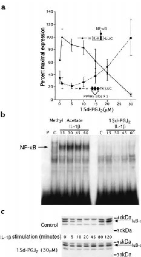
We next investigated the mechanism by which PPAR-γ ligands inhibit IL-8 gene expression in colon cancer cells. Consistent with its effect on IL-8 gene expression, 15d-PGJ2inhibited the ability of IL-1βto activate a reporter
gene regulated by the IL-8 promoter in a dose-depend-ent fashion (Figure 2a). Moreover, the EC50of 25 µM for
[image:4.612.305.538.43.467.2]inhibition of the IL-8 promoter showed excellent agree-ment with the activation of transcription from a well-characterized positive PPAR-γresponse element from the acyl-CoA oxidase gene (27), strongly suggesting that the effects of the ligand are mediated by PPAR-γ. Differences Figure 2
15d-PGJ2inhibits the transcriptional activation of the IL-8 promoter by
preventing the activation of NF-κB via an IκB-α–dependent pathway. (a) Caco-2 cells cotransfected with either an IL-8 promoter luciferase reporter gene or the PPAR-γ–regulated reporter plasmid, acyl-CoA X 3-TK-LUC, were treated with various concentrations of 15d-PGJ2for 24
hours, followed by stimulation with IL-1β(5 ng/mL). (b) EMSAs of nuclear extracts isolated from Caco-2 cells treated with either 15d-PGJ2
or methyl acetate for 24 hours and followed by IL-1βstimulation (5 ng/mL) for the times indicated. A double-stranded oligonucleotide span-ning the NF-κB element in the IL-8 promoter was used as a probe. (c) Iκ
B-αWestern blot of proteins isolated from Caco-2 cells treated with 15d-PGJ2or methyl acetate for 24 hours and followed by IL-1βstimulation (5
in mRNA stability, translation, and enzymatic activity, alone or in combination, may account for the difference between the EC50in this reporter gene study and that
observed for the inhibition of IL-8 mRNA expression (Figure 1, a and b).
We have previously shown that site-directed mutagen-esis of the NF-κB element within the context of the IL-8 promoter completely eliminates activation in response to stimulation with IL-1β(8). Transfection experiments confirm that this mutated promoter remains completely inactive when Caco-2 cells are pretreated with 10–30 µM 15d-PGJ2(data not shown). EMSAs show that the
treat-ment of Caco-2 cells with 15d-PGJ2dramatically reduced
nuclear protein binding to the NF-κB element of the IL-8 promoter (Figure 2b). In response to immune stimula-tion, degradation of the IκB family of proteins plays a critical role in the activation of NF-κB (9). The rapid and transient expression of IL-8 mRNA in Caco-2 cells in response to IL-1βstimulation is consistent with regula-tion by IκB-αexpression. An immunoblot of IκB-αin Caco-2 cells showed that IκB-α protein was rapidly degraded within 10 minutes after stimulation with IL-1β, followed by reappearance of the protein at 80 minutes due to autoregulatory induction of this gene by NF-κB (35) (Figure 2c). In contrast, IκB-αwas resistant to degra-dation in IL-1β–stimulated Caco-2 cells treated with 15d-PGJ2(Figure 2c), consistent with the absence of NF-κB
binding noted on EMSA (Figure 2b).
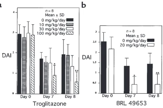
To determine whether ligands for PPAR-γcan attenu-ate intestinal inflammation in vivo, we studied the effect of 2 different TZD PPAR-γligands on a well-established murine model of colonic inflammation that is com-monly used to screen pharmacologic agents (1). Animals fed 4% DSS develop colonic inflammation within 7 days; after termination of DSS administration, colonic inflammation slowly resolves (30). The DAI provides a well-characterized method of quantitating disease sever-ity in this model that correlates well with histological healing (30). Two treatment studies were performed, one with troglitazone and the other with BRL 49653. In the first study, mice were treated throughout the entire pro-tocol with various concentrations of troglitazone. The DAI for each study group demonstrated that 7 and 8 days after DSS was discontinued, troglitazone (100
mg/kg/d) led to a 47% and 70% decrease in the DAI, respectively, compared with the placebo-treated group (Figure 3a). Also noted was a trend toward a decrease in the DAI observed in mice treated with an intermediate dose of troglitazone (30 mg/kg/d), suggestive of a dose-dependent response. Therapeutic efficacy was also deter-mined for BRL 49653 in the same model. BRL 49653 was administered once colitis was established after 7 days of DSS. This eliminated the possibility that the compound altered water consumption while the disease was being initiated with DSS during the first 7 days. Figure 3b shows that BRL dramatically decreased the DAI by 67% and 70% on days 7 and 8, respectively.
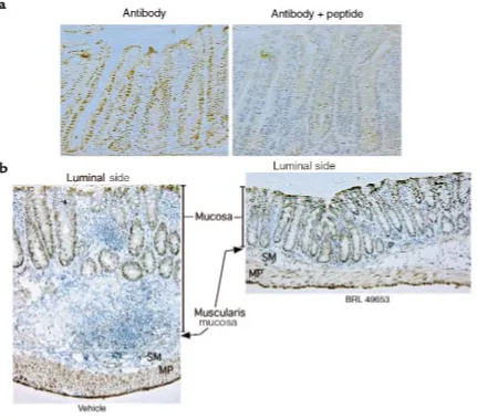
Immunohistochemistry of the normal colon showed that PPAR-γis expressed predominantly by epithelial cells located in the proliferative crypt compartment (Fig-ure 4a). The specificity of this nuclear staining was con-firmed by effective competition using the immunogen peptide. Immunohistochemistry was also performed on tissues obtained from animals treated with DSS (Figure 4b). Colonic tissue from animals that received vehicle alone showed intense mucosal inflammation with a chronic inflammatory infiltrate, edema, and marked thickening of the bowel wall. In contrast, histological sections from the colon of animals treated with 8 days of BRL 49653 showed a remarkable decrease in inflamma-tion with only a mild inflammatory infiltrate, no edema, and normal bowel wall thickness. Immunostaining of these tissue sections demonstrated that PPAR-γ was expressed primarily in the colonic epithelium, even in the presence of intense inflammation (Figure 4b). The inflammatory cells expressed comparatively little
PPAR-γ, consistent with the colonic epithelium being the tar-get of PPAR-γligands.
Discussion
[image:5.612.271.534.49.224.2]There is significant interest in the biologic conse-quences of PPAR-γactivation in the colon. Because of the differentiating and antiproliferative effects of PPAR-γin adipose and breast tissue (17, 18, 36, 37), it has been proposed that ligands for this receptor may have therapeutic potential in chemoprevention or the treatment of colorectal neoplasia (21, 24). Recent stud-ies, however, suggest that these same ligands actually Figure 3
enhance colon polyp and tumor formation in the min mouse model of familial adenomatous polyposis coli (22, 23). Here we show that PPAR-γligands have anoth-er major effect on colon epithelial cells, namely, to reg-ulate the inflammatory response. Several historical observations are consistent with the possible utility of PPAR-γactivation in the treatment of colonic inflam-mation. First, polyunsaturated fatty acids, such as pro-vided in fish oil, have been shown to be potent activa-tors of PPAR-γand to have efficacy in the treatment of IBD (38, 39). Second, there is evidence that TZDs may decrease insulin resistance by decreasing the produc-tion of TNF-αby adipose tissue and/or by inhibiting TNF-α–mediated signal transduction (40). Inhibition of TNF-αactivity by the use of anti-TNF antibodies has been shown to be an effective treatment modality for refractory Crohn’s disease (41).
We show that 2 different classes of ligands for PPAR-γ potently inhibit cytokine expression in 2 different
PPAR-γ–expressing colon cell lines (21, 24). The increased
potency of 15d-PGJ2relative to BRL 49653 contrasts
with the direct binding affinities of these compounds for PPAR-γ,but is consistent with the relative potencies of these compounds for negative regulation of genes involved in the inflammatory response (12, 13). At pres-ent, it is not known why the EC50for BRL 49653 is
greater than that for 15d-PGJ2. It is possible that
cofac-tors necessary for transcriptional repression interact with ligand-bound PPAR-γ differently than those involved in transcriptional activation (42). We also do not exclude the possibility that these compounds inhib-it cytokine gene expression through addinhib-itional mecha-nisms independent of PPAR-γ. Indeed, a PPAR-γ –inde-pendent pathway may be operative in neuronal cells, where investigators have recently shown that 15d-PGJ2
inhibits inducible nitric oxide synthase (iNOS) expres-sion and promoter activity by inhibiting transcriptional activation via NF-κB (43). iNOS expression in microglial cells was not inhibited by TZDs, and the nuclear translo-cation and DNA-binding activities of NF-κB were
unaf-Figure 4
[image:6.612.90.521.299.678.2]fected by 15d-PGJ2. These results, however, are
distinct-ly different from our findings in intestinal cell lines, where BRL 49653 does inhibit IL-8 expression. Further-more, we show that in Caco-2 cells, 15d-PGJ2inhibits the
nuclear translocation and subsequent DNA binding of NF-κB via an IκB-α–dependent pathway by inhibiting the immune response–induced degradation of IκB-α. This is also distinct from the putative non-PPAR path-way of iNOS regulation in neuronal cells. Although fur-ther investigation will be required to elucidate the upstream mechanisms by which PPAR-γinhibits the degradation of IκB-α, inhibition of both IL-1β– and TNF-α–induced IL-8 gene expression suggests that acti-vation of PPAR-γalters functional elements common to both these pathways. Targeting of NF-κB as a therapeu-tic approach to IBD is already established (44).
Although previous studies have shown that activation of PPAR-γinhibits the inflammatory response in mono-cytes and macrophages, immunolocalization of PPAR-γ primarily to colonocytes, even in the presence of inflam-mation, strongly suggests that these epithelial cells are the target of PPAR-γligands in this model. This provides evidence that immunomodulation of the intestinal epithelium alone may be efficacious in the treatment of intestinal inflammatory states. We do not exclude the possibility that a component of the anti-inflammatory effect is mediated by the action of TZDs on the systemic immune system. However, the fact that DSS adminis-tered rectally can also induce colitis suggests that the inflammatory response is initiated by an effect localized to the colon rather than involving an alteration in the systemic immune system.
The role that PPAR-γligands may play in neoplasia need not be necessarily viewed as a disparate effect. Indeed, it is well known that chronic intestinal inflammation not only alters patterns of cellular proliferation and differentiation in the intestinal epithelium, but is also a significant factor in the development of colorectal neoplasia (45, 46). At the present time, the effect of PPAR-γagonists on colonic epithelial cell growth is controversial. Two studies show that these ligands have potent antineoplastic and antipro-liferative activities both in vivo and in vitro (21, 24). In con-trast, the 2 studies showing that TZD ligands increase the number of colonic polyps utilize the minmouse model of intestinal polyposis (22, 23). The proneoplastic effect observed, therefore, occurs in a single murine model relat-ed to an adenomatous polyposis coli gene (APC) muta-tion. The relevance of the minmouse model to IBD is uncertain, particularly because APC mutations are rare in colitis-induced neoplasia (47). Furthermore, highly unsat-urated fatty acids, which are known natural ligands for PPAR-γ, are well known not only to reduce the risk of colonic tumors, but also as an effective treatment for intestinal inflammation (38, 48, 49), supporting the anti-neoplastic role of PPAR-γ. At this point, therefore, it is pre-mature to conclude that PPAR-γ ligands will induce colonic neoplasia in any background, but particularly in the setting of intestinal inflammation. In fact, an equally compelling case could be made that these compounds may protect against the development of neoplasia associ-ated with colitis. Further investigation into the effect of PPAR-γligands on neoplastic transformation in the
set-ting of IBD may provide important insights into the biol-ogy of the colonic epithelium.
PPAR-γligands represent a novel approach to the treat-ment of IBD. The TZD ligands used in the animal stud-ies were well within the doses needed for insulin sensiti-zation and have been shown to be well tolerated by mice (21). The DAIs used to quantitate disease activity in our animal studies are comprehensive functional measures that are analogues to the subjective clinical symptoms observed in humans with ulcerative colitis. Many anti-inflammatory pharmaceuticals have been studied in the DSS model of colitis that show efficacy paralleling the clinical response observed in humans (30, 50–53). In fact, the results with either troglitazone or BRL 49653 show a greater level of therapeutic efficacy than that observed with Olsalazine (Dipentum; Pharmacia and Upjohn, Kalamazoo, Michigan, USA); (150 mg/kg/d) (51). There-fore, the highly significant anti-inflammatory effect observed with the DSS model provides the single best indication that PPAR-γligands may have clinical effica-cy in patients with IBD.
Acknowledgments
This work was supported by National Institutes of Health grants DK-47709 and AI-39368 (to G.D. Wu); Center Grant P30 DK-50306; DK-49780 and DK-49210 (to M.A. Lazar).
1. Elson, C.O., Sartor, R.B., Tennyson, G.S., and Riddell, R.H. 1995. Exper-imental models of inflammatory bowel disease. Gastroenterology.
109:1344–1367.
2. Reinecker, H.C., and Podolsky, D.K. 1995. Human intestinal epithelial cells express functional cytokine receptors sharing the commonγc chain of the interleukin 2 receptor. Proc. Natl. Acad. Sci. USA.92:8353–8357. 3. McCormick, B.A., et al. 1995. Surface attachment of Salmonella typhimuri-umto intestinal epithelia imprints the subepithelial matrix with gradi-ents chemotactic for neutrophils. J. Cell Biol.131:1599–1608. 4. Jung, H.C., et al. 1995. A distinct array of proinflammatory cytokines is
expressed in human colon epithelial cells in response to bacterial inva-sion. J. Clin. Invest.95:55–65.
5. Andoh, A., Fujiyama, Y., Bamba, T., and Hosoda, S. 1993. Differential cytokine regulation of complement C3, C4, and factor B synthesis in human intestinal epithelial cell line, Caco-2. J. Immunol.151:4239–4247. 6. Hermiston, M.L., and Gordon, J.I. 1995. Inflammatory bowel disease and adenomas in mice expressing a dominant negative N-cadherin. Science.
270:1203–1207.
7. Baribault, H., Penner, J., Iozzo, R.V., and Wilson-Heiner, M. 1994. Col-orectal hyperplasia and inflammation in keratin 8-deficient FVB/N mice. Genes Dev.8:2964–2973.
8. Wu, G.D., Lai, E.J., Huang, N., and Wen, X. 1997. Oct-1 and C/EBP bind to overlapping elements within the IL-8 promoter: the role of Oct-1 as a transcriptional repressor. J. Biol. Chem.272:2396–2403.
9. Verma, I.M., Stevenson, J.K., Schwarz, E.M., Antwerp, D.V., and Miyamo-to, S. 1995. Rel/NF-κB/IκB family: intimate tales of association and dis-sociation. Genes Dev.9:2723–2735.
10. Brown, K., Gerstberger, S., Carlson, L., Franzoso, G., and Siebenlist, U. 1995. Control of IκB-αproteolysis by site-specific, signal-induced phos-phorylation. Science.267:1485–1488.
11. Auphan, N., DiDonate, J.A., Rosette, C., Helmberg, A., and Karin, M. 1995. Immunosuppression by glucocorticoids: inhibition of NF-κB activity through induction of IκB synthesis. Science.270:286–290. 12. Ricote, M., Li, A.C., Willson, T.M., Kelly, C.J., and Glass, C.K. 1998. The
peroxisome proliferator-activated receptor-γis a negative regulator of macrophage activation. Nature.391:79–82.
13. Jiang, C., Ting, A.T., and Seed, B. 1998. PPAR-γagonists inhibit produc-tion of monocyte inflammatory cytokines. Nature.391:82–86. 14. Yu, K., et al. 1995. Differential activation of peroxisome
proliferator-acti-vated receptors by eicosanoids. J. Biol. Chem.270:23975–23983. 15. Kliewer, S.A., et al. 1995. A prostaglandin J2 metabolite binds
peroxi-some proliferator-activated receptor γand promotes adipocyte differen-tiation. Cell.83:813–819.
17. Tontonoz, P., Hu, E., and Spiegelman, B.M. 1994. Stimulation of adipo-genesis in fibroblasts by PPARγ2, a lipid-activated transcription factor.
Cell.79:1147–1156.
18. Chawla, A., Schwarz, E.J., Dimaculangan, D.D., and Lazar, M.A. 1994. Peroxisome proliferator-activated receptor γ(PPARγ): adipose predomi-nant expression and induction early in adipocyte differentiation.
Endocrinology.135:798–800.
19. Fajas, L., et al. 1997. The organization, promoter analysis, and expression of the human PPARγgene. J. Biol. Chem.272:18779–18789.
20. Dubois, R.N., et al. 1998. The nuclear eicosanoid receptor, PPARγ, is aberrantly expressed in colonic cancers. Carcinogenesis.19:49–53. 21. Sarraf, P., et al. 1998. Differentiation and reversal of malignant changes
in colon cancer through PPARγ. Nat. Med.4:1046–1052.
22. Saez, E., et al. 1998. Activators of the nuclear receptor PPARγenhance colon polyp formation. Nat. Med.4:1058–1061.
23. Lefebvre, A.M., et al. 1998. Activation of the peroxisome proliferator-acti-vated receptor γ promotes the development of colon tumors in C57BL/6J-APCMin/+ mice. Nat. Med.4:1053–1057.
24. Brockman, J.A., Gupta, R.A., and Dubois, R.N. 1998. Activation of PPARγ leads to inhibition of anchorage independent growth of human col-orectal cancer cells. Gastroenterology.115:1049–1055.
25. Huang, N., Katz, J.P., Martin, D.R., and Wu, G.D. 1997. Inhibition of IL-8 gene expression in Caco-2 cells by compounds which induce histone hyperacetylation. Cytokine.9:27–36.
26. Traber, P.G., Wu, G.D., and Wang, W. 1992. Novel DNA-binding proteins regulate intestine-specific transcription of the sucrase-isomaltase gene.
Mol. Cell. Biol.12:3614–3627.
27. Adams, M., Reginato, M.J., Shao, D., Lazar, M.A., and Chatterjee, V.K. 1997. Transcriptional activation by peroxisome proliferator-activated receptor γis inhibited by phosphorylation at a consensus mitogen-acti-vated protein kinase site. J. Biol. Chem.272:5128–5132.
28. Chen, C., and Okayama, H. 1987. High-efficiency transformation of mammalian cells by plasmid DNA. Mol. Cell. Biol.7:2031–2034. 29. Wu, G.D., Wang, W., and Traber, P.G. 1992. Isolation and
characteriza-tion of the human sucrase-isomaltase gene and demonstracharacteriza-tion of intes-tine-specific transcriptional elements. J. Biol. Chem.267:7863–7870. 30. Cooper, H.S., Murthy, S.N.S., Shah, R.S., and Sedergran, D. 1993.
Clini-copathologic study of dextran sulfate sodium experimental murine coli-tis. Lab. Invest.69:238–249.
31. Xue, J.C., Schwartz, E.J., Chawla, A., and Lazar, M.A. 1996. Distinct stages in adipogenesis revealed by retinoid inhibition of differentiation after induction of PPARγ. Mol. Cell. Biol.16:1567–1575.
32. Shao, D., et al. 1998. Interdomain communication regulating ligand binding by PPAR-γ. Nature.396:377–380.
33. Suzuki, R., Suruga, K., Goda, T., and Takase, S. 1998. Peroxisome prolif-erator enhances gene expression of cellular retinol-binding protein, type II in Caco-2 cells. Life Sci.62:861–871.
34. Staels, B., et al. 1998. Activation of human aortic smooth-muscle cells is inhibited by PPARαbut not by PPARγactivators. Nature.393:790–793. 35. Sun, S.C., Ganchi, P.A., Ballard, D.W., and Greene, W.C. 1993. NF-κB controls expression of inhibitor IκBα: evidence for an inducible autoreg-ulatory pathway. Science.259:1912–1915.
36. Tontonoz, P., Hu, E., Graves, R.A., Budavari, A.I., and Spiegelman, B.M. 1994. mPPARγ2: tissue-specific regulator of an adipocyte enhancer.
Genes Dev.8:1224–1234.
37. Mueller, E., et al. 1998. Terminal differentiation of human breast cancer through PPARγ. Mol. Cell.1:465–470.
38. Kliewer, S.A., et al. 1997. Fatty acids and eicosanoids regulate gene expression through direct interactions with peroxisome proliferator-acti-vated receptors αand γ. Proc. Natl. Acad. Sci. USA.94:4318–4323. 39. Belluzzi, A., et al. 1996. Effect of enteric-coated fish-oil preparation on
relapses in Crohn’s disease. N. Engl. J. Med.334:1557–1560.
40. Peraldi, P., Xu, M., and Spiegelman, B.M. 1997. Thiazolidinediones block tumor necrosis factor-—induced inhibition of insulin signaling. J. Clin. Invest.100:1863–1869.
41. Targan, S.R., et al. 1997. A short-term study of chimeric monoclonal antibody CA2 to tumor necrosis factor (alpha) for Crohn’s disease. N. Engl. J. Med.337:1029–1035.
42. Onate, S.A., Tsai, S.Y., Tsai, M.J., and O’Malley, B.W. 1995. Sequence and characterization of a coactivator for the steroid hormone receptor super-family. Science.270:1354–1357.
43. Petrova, T.V., Akama, K.T., and Van Elkdik, L.J. 1999. Cyclopentenone prostaglandins suppress activation of microglia: down-regulation of inducible nitric-oxide synthase by 15-deoxy-∆12,14-prostaglandin J2. Proc.
Natl. Acad. Sci. USA.96:4668–4673.
44. Neurath, M.F., Pettersson, S., Buschenfelde, K.H.M., and Strober, W. 1996. Local administration of antisense phosphorothioate oligonu-cleotides to the p65 subunit of NF-κB abrogates established experi-mental colitis in mice. Nat. Med.2:998–1004.
45. Serafini, E.P., Kirk, A.P., and Chambers, T.J. 1981. Rate and pattern of epithelial cell proliferation in ulcerative colitis. Gut.22:648–652. 46. Bachwich, D.R., Lichtenstein, G.R., and Traber, P.G. 1994. Cancer in
inflammatory bowel disease. Med. Clin. North Am.78:1399–1412. 47. Tarmin, L., et al. 1995. Adenomatous polyposis coli gene mutations in
ulcerative colitis-associated dysplasias and cancers versus sporadic colon neoplasms. Cancer Res.55:2035–2038.
48. Reddy, B.S., and Sugie, S. 1988. Effect of different levels of omega-3 and omega-6 fatty acids on azoxymethane-induced colon carcinogenesis in F344 rats. Cancer Res.48:6642–6647.
49. Stenson, W.F., Cort, D., Rodgers, J., and Burakoff, R. 1992. Dietary sup-plementation with fish oil in ulcerative colitis. Ann. Intern. Med.
116:609–614.
50. Murthy, S.N.S., et al. 1993. Treatment of dextran sulfate sodium–induced murine colitis by intracolonic cyclosporine. Dig. Dis. Sci.
38:1722–1734.
51. Murthy, S., Murthy, N.S., Coppola, D., and Wood, D.L. 1997. The effi-cacy of BAY y 1015 in dextran sulfate model of mouse colitis. Inflamm. Res.46:224–233.
52. Kojouharoff, G., et al. 1997. Neutralization of tumor necrosis factor (TNF) but not IL-1 reduces inflammation in chronic dextran sulfate sodium-induced colitis in mice. Clin. Exp. Immunol.107:353–358. 53. Bennett, C.F., et al. 1997. An ICAM-1 antisense oligonucleotide prevents