0095-1137/96/$04.0010
Copyrightq1996, American Society for Microbiology
Candida albicans Serotype Analysis by Flow Cytometry
STE´ PHANE MERCURE, SERGE SE´NE´CHAL, PIERRE AUGER, GUY LEMAY,
ANDSERGE MONTPLAISIR*
De´partement de Microbiologie et Immunologie, Faculte´ de Me´decine, Universite´ de Montre´al, Montre´al, Que´bec, Canada
Received 23 February 1996/Returned for modification 23 April 1996/Accepted 12 June 1996
Candida albicans strains can be assigned to either of two major serogroups, A or B. Antigenic surface determinants present only in serotype A strains allow such a distinction, which has epidemiologic relevance. Reports have established that the relative distributions of the two serotypes can vary depending on the geographic origin of the isolates. A prevalence of susceptibility to an antifungal agent, flucytosine, was also observed with isolates of serotype A. More recently, it was suggested that the occurrence of serotype B isolates in various clinical forms of candidiasis is increasing. However, this latest finding remains controversial since serotyping results vary widely from one laboratory to another because of the lack of standardized methodol-ogies. Difficulty in interpretation of results, which may lead to erroneous serotype identification, is the major setback associated with current methods. For this study, we thus devised a procedure that relies on flow cytometry and that may eliminate ambiguities in serotype determination. The validation of results was achieved
with two types of serotype A-specific antisera, Iatron Factor 6 antiserum and an anti-C. albicansantiserum
adsorbed on serotype B yeast cells. Agreement between results obtained with these two reagents was 100% with
a wide array ofCandidastrains. These results confirmed the potential of the flow cytometric procedure as a
reliable and reproducible method to establish the serotypes ofC. albicansstrains. Furthermore, some
appli-cations of this procedure to the epidemiological study of this human pathogen are presented.
The opportunistic yeast Candida albicans is at this time the most common fungal pathogen in humans. This yeast can cause mucosal as well as systemic infections in both immuno-competent and immunocompromised individuals. Several methods have been devised to classify C. albicans strains in subgroups below the species level (17). First described in the 1960s, the serotyping procedure separates strains of C. albicans into serotypes A and B (9). These two serogroups share the majority of their cell surface antigenic determinants; however, the presence of additional antigens in serotype A strains allows their distinction from serotype B strains (8, 9, 23, 28). Mainly used in association with other biotyping procedures, serotyping still contributes to our knowledge of the epidemiologies of C.
albicans infections. For example, a correlation has been
estab-lished between serotypes and susceptibility to the antifungal agent flucytosine (5FC) (1, 7, 19, 21, 22, 26). It has also been suggested that serotypes A and B differ in their geographic distributions, abilities to adhere to epithelial cells, and abilities to infect patients with different immunological statuses (3, 7, 10, 18, 19, 21, 26). However, these assumptions remain con-troversial since serotyping results can vary widely, reflecting discrepancies introduced by the use of numerous serotyping methodologies in different laboratories (21). Investigators have relied on serotyping techniques as diverse as slide agglutina-tion, tube agglutinaagglutina-tion, and slide immunofluorescence assays using various antisera or purified antibodies that reportedly differentiate type A from type B yeast strains (1, 2, 9, 23, 24, 26). Recently, a comparative study suggested that it may not be possible to make valid comparisons between studies which evaluated serotype prevalence unless the same serotyping methodologies were used (2). This major setback was mainly
attributed to the difficulty involved in interpretating results visually, which may lead to erroneous serotype identification. Analyses of C. albicans surface determinants previously sug-gested the potential of flow cytometry to determine yeast cell serotypes (4, 5). In the present study, we thus devised a method relying on flow cytometry which, by means of quantitative fluorescence monoparametric histogram outputs, was shown to eliminate the ambiguity of the interpretation of results. The labelling and washing conditions were optimized in order to distinguish clearly between serotype A and B strains, and the procedure was validated with different yeast cells from various
Candida species and different serotype A-specific antisera. The
usefulness of this procedure was further established by our performing a limited epidemiological study of C. albicans iso-lates from patients with either oropharyngeal or vulvovaginal candidiasis. Both groups of patients were shown to exhibit similar relative distributions of serotype A and B strains. Fi-nally, we demonstrated that fluorescence patterns generated by flow cytometric analysis may be helpful in determining the origins of recurrent C. albicans infections, since even strains of the same serotype exhibit differences in their precise mono-parametric histogram outputs.
(This work was presented in part at the XII Congress of the International Society for Human and Animal Mycology, Ad-elaide, Australia, March 1994.)
MATERIALS AND METHODS
Candida albicansstrains and clinical isolates.The clinical isolates of C.
albi-cans were obtained from two groups of patients. Ninety-four strains were isolated
from the vaginas of patients presenting with episodes of vulvovaginal candidiasis. Of that number, one isolate from each of two consecutive episodes of recurrent vulvovaginal candidiasis was obtained from 32 patients, recurrence being defined as the onset of three episodes of vaginitis in a period of 6 months (15). Isolates were also obtained from 29 human immunodeficiency virus (HIV)-infected in-dividuals with oropharyngeal candidiasis (throat or esophagus). All strains were identified as C. albicans by their abilities to form germ tubes and produce chlamydospores (1). C. albicans collection strains of known serotype were ob-tained either from the American Type Culture Collection (Rockville, Md.) or * Corresponding author. Mailing address: De´partement de
Microbiologie et Immunologie, Universite´ de Montre´al, C.P. 6128, Succ. Centre-Ville, Montre´al, Que´bec, Canada H3C 3J7. Phone: (514) 343-6273. Fax: (514) 343-5701.
2106
on May 15, 2020 by guest
http://jcm.asm.org/
from E. Drouhet (Institut Pasteur, Paris, France). Strains of other Candida species were obtained, unless otherwise mentioned, from the American Type Culture Collection. C. stellatoidea strains were a gift from J. Kwon Chung (Na-tional Institutes of Health, Bethesda, Md.) and S. Riggsby (University of Ten-nessee, Knoxville), while C. dubliniensis strains were provided by M. McCullough (University of Melbourne, Melbourne, Australia) and G. St-Germain (Labora-toire de Sante´ Publique du Que´bec, Montre´al, Que´bec, Canada).
Phenotypic and genotypic typing procedures.The biotyping scores of C.
albi-cans strains were determined according to the procedure of Odds and Abbott
(20). Susceptibility testing to 5FC was performed as described by Auger et al. (1). Genotypic characterization was done as previously reported (15).
Serotype A-specific antisera.The serotype A-specific antiserum Ca\A-B was produced by a procedure based on the work of Hasenclever and Mitchell (9), who demonstrated that an antiserum raised against serotype A yeast cells of C.
albicans can be rendered specific to serotype A yeast cells by a preadsorption
procedure (1, 9). To perform this procedure, well-characterized strains of both serotypes were obtained from the American Type Culture Collection. The
anti-Candida antiserum was raised in rabbits by injections of heat-killed (30 min,
1008C) yeast cells from the serotype A strain ATCC 32354. The titer of antibody raised against C. albicans was superior to 1:1,000; reaching such a high titer was necessary to retain sufficient reactivity of the antiserum after the successive adsorption steps. Adsorption was achieved by mixing equal volumes of antiserum with a suspension of heat-killed (3 h, 658C) yeast cells of the serotype B strain ATCC 44373. The mixture was shaken at room temperature for 45 min to allow adsorption and then centrifuged for 10 min at 1,0003g. This preadsorption
procedure was repeated four more times, after which the resulting serotype A-specific antiserum (Ca\A-B) was diluted in phosphate-buffered saline (PBS) to obtain a final titer of 1:25. The titer of the antiserum was determined on serial dilution by slide immunofluorescence and defined as the highest dilution giving visible fluorescence. The other antiserum used for serotyping was a commercially available monospecific antiserum specific to C. albicans cells of serotype A (12, 13, 29); this antiserum, known as Iatron Factor 6 (IF6; also known as anti-IF6 or Candida Check no. 6), was purchased from Iatron Laboratories (Tokyo, Japan).
Slide immunofluorescence serotyping procedure.The serotypes of all strains were determined by an already described indirect immunofluorescence slide assay (6). The preadsorbed serotype A-specific antiserum Ca\A-B was used to detect the expression of antigens specific to serotype A yeast cells. Serotype A strain ATCC 32354 was included in each experiment as a positive control, and the serotype B strain ATCC 44373 was used as a negative control. Antibody binding was determined by visual observation by fluorescence microscopy, fol-lowing incubation with an affinity-purified goat anti-rabbit immunoglobulin G (heavy and light) coupled to dichlorotriazinylaminofluorescein (DTAF), which was used as a secondary antibody (Jackson Immunoresearch Laboratories, West Grove, Pa.).
Flow cytometric serotyping procedure.Candida strains were grown to
station-ary phase in 10 ml of yeast nitrogen base (Difco, Detroit, Mich.) under agitation (250 rpm) at 308C. Yeast cells from these cultures were pelleted by centrifugation at 1,0003g for 10 min; the cells were then washed with 10 ml of PBS,
recen-trifuged, and resuspended in PBS to approximately 53106
cells/ml. One hun-dred microliters of the diluted Candida cells were transferred to flow cytometry tubes (Falcon 2052; Becton Dickinson Labware, Lincoln Park, N.J.) prior to the addition of the primary antiserum (5ml of the Ca\A-B antiserum or 1ml of the undiluted IF6 antiserum as provided by the manufacturer). This mixture was gently mixed and incubated on ice for 1 h. Cells were then washed by the addition of 3 ml of PBS to each sample. The tubes were centrifuged for 10 min at 1,0003
g, and the supernatants were carefully discarded, leaving the pellets in
approxi-mately 400ml of PBS. This washing procedure was repeated prior to the addition of 4ml of the affinity-purified goat anti-rabbit immunoglobulin G (heavy and light) antibodies coupled to DTAF to the yeast cells, now in 400ml of PBS. The mixture was vortexed gently and incubated on ice for 30 min. Finally, cells were washed twice in PBS as described previously and fixed by the addition of the same volume (400ml) of 2% paraformaldehyde for 30 min at 48C. The flow cytometric analysis was performed with a FACStar flow cytometer (Becton Dickinson Immunocytometry Systems, San Jose, Calif.) equipped with a 2-W argon ion water-cooled laser. The laser was set at 488 nm with a light output of 200 mW. The fluidic system was equipped with a 75-mm-diameter nozzle tip. Green fluorescence (DTAF) was collected with a 530\30-nm bandpass filter. Multiparametric data for each sample were acquired in list mode for 10,000 events and analyzed with Consort 30 software. Data were analyzed in monopara-metric histograms. All experiments included, as controls, collection yeast strains well established as either serotype A or B. A sample of yeast cells labelled solely with the DTAF conjugate was also included as a background control.
RESULTS
Validation of the flow cytometric serotyping procedure.The
validation of our newly proposed flow cytometric procedure was achieved through the use of different groups of Candida strains labelled either by this procedure or by the classical indirect slide immunofluorescence assay (1, 6). The capacity of
the flow cytometric procedure to assign strains to a particular serotype was also evaluated with two antisera specific to sero-type A yeast cells. The first, IF6, was a commercially available antiserum, while the second was prepared in our own labora-tory by a standard preadsorption procedure, as described in Materials and Methods. The changing specificity of the latter, through the successive adsorption steps, was monitored by flow cytometry (Fig. 1). After five consecutive adsorption steps, the fluorescence intensity of the labelling for serotype B yeast cells was down to the background level observed when the complete labelling procedure was performed in the absence of the first anti-serotype A antiserum, while the fluorescence intensity ob-served with cells of serotype A remained several hundredfold higher. The antiserum (Ca\A-B) was thus considered a sero-type A-specific antiserum.
Both primary antisera (IF6 and Ca\A-B) were then used to study the expression of serotype A-specific epitopes on
Can-dida cells, with known antigenic characteristics. The capacity of
the method to detect the labelling of yeast cells was evaluated for at least two representative strains of several Candida spe-cies. Analysis of flow cytometric recordings revealed that re-activity with both serotype A-specific antisera corroborated previous reports based mainly on slide agglutination assays. Yeast cells from C. stellatoidea type II, C. tropicalis, C. glabrata, and the newly described C. dubliniensis (27) were all positively labelled by either antiserum, while C. stellatoidea type I, C.
guilliermondii, C. kefyr, and C. krusei all remained nonreactive
(data not shown). Yeast cells from four collection strains of C.
albicans were also submitted to the flow cytometric procedure.
All strains were characterized by a single fluorescent peak with either antiserum (Fig. 2). The serotype A yeast cells displayed mean fluorescence intensities several hundredfold higher than those of controls and serotype B cells. Resulting serotypes were, in every case and with either antiserum, consistent with the known serogroups of these strains (Fig. 2).
We applied this analysis, which supported the potential of our procedure to facilitate serotype assignment of C. albicans strains, to a large number of C. albicans isolates from patients with oropharyngeal or vulvovaginal candidiasis. As already ob-served with collection strains, although the mean relative flu-orescence intensities varied from strain to strain, it was always several hundredfold higher for cells from serotype A strains
(.102). Levels of autofluorescence and nonspecific
fluores-cence of yeast cells were generally confined below fluoresfluores-cence
intensity values of 101, and interference with the procedure
resulting from autoagglutination of yeast cells, a major prob-lem in some other serotyping procedures, was never observed. The monoparametric histogram outputs generated by flow cy-tometric analysis thus allowed us to easily assign a serotype to all strains tested. Identical results were consistently obtained with both serotype A-specific antisera.
Since the majority of these strains had been previously an-alyzed by a slide immunofluorescence assay, we compared the two sets of serotyping results; in the majority of cases, the serotype assignments of both methods were consistent. How-ever, in some cases, the serotype of an infecting strain could be conclusively determined only by the flow cytometric procedure. In one sample originally assigned to serotype A by slide im-munofluorescence, the flow cytometric procedure revealed the presence of two distinct fluorescent peaks; one population of cells fluoresced strongly, while the fluorescence intensity of a second population was comparable to that of the negative control in the absence of primary anti-serotype A antiserum (data not shown). Further analysis of this sample by colony
VOL. 34, 1996 C. ALBICANS SEROTYPE ANALYSIS BY FLOW CYTOMETRY 2107
on May 15, 2020 by guest
http://jcm.asm.org/
isolation confirmed the presence of yeast cells from two strains of different serogroups. In four other cases, the serotype could not be conclusively determined since the serotyping results were contradictory: one isolate assigned to serotype A by slide immunofluorescence was later identified as serotype B by flow cytometry, while three strains originally assigned to serotype B by slide immunofluorescence were instead identified as sero-type A by flow cytometry. These isolates were further studied in order to determine which serotyping method is more likely to be accurate when such discrepancies arise. Thus, we looked more closely at correlations between serotype and other phe-notypic or gephe-notypic traits of C. albicans strains that might give us indications of the actual serotypes of these four strains. First, a well-documented correlation between serotype and susceptibility to 5FC of C. albicans had been previously
estab-lished (19, 21). Briefly stated, most serotype A yeast strains are susceptible to 5FC while serotype B strains are mainly resistant to 5FC. Another correlation between serotype and a major dimorphism observed at the DNA level among C. albicans strains was also recently discovered (16). This dimorphism is due to the presence of a newly described self-splicing intron in the sequence coding for the 25S rRNA in 40% of C. albicans strains (14). A previous analysis revealed that all strains pre-senting the intron and having serotypes that could be unam-biguously established were of serotype A. In contrast, intron-less strains were mostly but not exclusively of serotype B (16). Among the strains presenting ambiguous serotyping results, three of these were assigned to serotype B by flow cytometric analysis but were assigned to serotype A by slide immunoflu-orescence assay. These three strains were shown to be
intron-FIG. 1. Preparation of the preadsorbed serotype A-specific antiserum (Ca\A-B). The antiserum raised in rabbits following injection of heat-killed yeast cells from strain ATCC 32354 was rendered serotype A specific by successive adsorption with heat-killed serotype B yeast cells from strain ATCC 44373. The efficiency of adsorption was monitored by flow cytometry using a goat anti-rabbit immunoglobulin antibody coupled to DTAF. C. albicans cells of serotypes A and B were incubated in the absence of anti-C. albicans antiserum (A), with the anti-Candida antiserum prior to adsorption (B), with the anti-Candida antiserum adsorbed once (C), and with the anti-Candida antiserum after five successive adsorption steps (D).
on May 15, 2020 by guest
http://jcm.asm.org/
[image:3.612.130.492.64.498.2]less strains resistant to 5FC; since these characteristics are mostly associated with strains of serotype B, these results sup-port the serotype B assignment obtained by flow cytometry rather than the result of slide immunofluorescence. Similarly, for the remaining strain, the assignment to serotype A ob-tained by flow cytometry seemed most likely since this strain was intron-bearing and susceptible to 5FC.
Application of the flow cytometric serotyping procedure to
the epidemiology of candidiasis. Apart from validating the
method, the analysis of C. albicans clinical isolates by the flow cytometric serotyping procedure was used to evaluate the use-fulness of this method to study serotype distribution in C.
albicans infections. In our study, a total of 123 clinical isolates
from patients with either oropharyngeal or vulvovaginal can-didiasis were analyzed by the flow cytometric serotyping pro-cedure. Among both groups of strains, serotype A was pre-dominant (Table 1). In the first group, C. albicans strains were isolated from the oropharynxes of 29 HIV-infected individuals and 69% of these isolates were assigned to serotype A. In the second group, 94 strains from patients with vulvovaginal can-didiasis were analyzed by flow cytometry and 56.4% were of serotype A. This last panel of strains included 32 pairs of
isolates from consecutive episodes of recurrent vulvovaginal candidiasis. The relative distribution of serotype A and B strains in isolates from cases of recurrent vulvovaginal candi-diasis was comparable to the relative distribution in the
[image:4.612.118.505.65.438.2]com-FIG. 2. Comparative analysis of serotyping results using IF6 and preadsorbed serotype A-specific antiserum (Ca\A-B). C. albicans cells from four collection strains of known serotype (serotype A, ATCC 32354 and ATCC 36801, and serotype B, ATCC 44373 and ATCC 36802) were analyzed by the flow cytometric serotyping procedure. The numbering of curves in each panel refers to different incubation conditions in the first step of the indirect procedure: 1, in the absence of antiserum (negative control); 2, in the presence of IF6 antiserum; 3, in the presence of the Ca\A-B antiserum.
TABLE 1. Flow cytometric analysis of serotype A and B prevalence in C. albicans strains isolated from patients with vulvovaginal or
oropharyngeal infectionsa
Type of strainb
No. of strains (% of total) of:
Serotype A
Serotype B
Vulvovaginal 53 (56.4) 41 (43.6)
Oropharyngeal 20 (69.0) 9 (31.0)
Total 73 (59.3) 50 (40.7)
aSerotyping was performed with IF6 and Ca\A-B antisera with the same
results.
bOropharyngeal isolates of C. albicans were obtained from HIV-infected
individuals. Vulvovaginal isolates were obtained from otherwise healthy patients presenting no underlying risk factors for candidiasis.
VOL. 34, 1996 C. ALBICANS SEROTYPE ANALYSIS BY FLOW CYTOMETRY 2109
on May 15, 2020 by guest
http://jcm.asm.org/
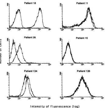
[image:4.612.315.556.605.685.2]plete panel of vaginal strains (data not shown). The same serotype was generally isolated from consecutive episodes of vaginitis, with the exception of three cases, in which we iden-tified a change in the serotype of the infecting strain (for example, Fig. 3, patient 18). In addition, comparison of the monoparametric histogram outputs for isolates from each of two consecutive episodes of vulvovaginal candidiasis present-ing the same serotype, revealed that their precise fluorescence patterns can sometimes be different (for example, Fig. 3, pa-tients 26 and 134). When these results were compared with the previously reported results of biotyping by the triplet scoring procedure (20) and genotype analysis by hybridization pattern with a C. albicans-specific probe (15), it was observed that fluorescence patterns were a reflection of strain lineage. Strains with identical fluorescence patterns presented similar biotypes and genotypes, while isolates of the same serotype, exhibiting differences in their fluorescence patterns, also pos-sessed different biotypes and genotypes.
DISCUSSION
Phenotyping procedures are still widely used to characterize clinical isolates of C. albicans on the basis of their distinctive assimilation, resistance, or antigenic properties. Although sub-division of C. albicans in two subgroups may seem of limited relevance, the serotyping procedure still contributes to our
ever-growing knowledge of the epidemiology of C. albicans infections. However, the value of serotyping as an epidemio-logic tool was recently questioned following the publication of widely divergent results concerning serotype distributions among patients with candidiasis (21). This lack of interlabora-tory reproducibility was attributed to the subjectivity involved in result interpretation and the reliance of investigators on widely divergent procedures and reagents (2). In an effort to circumvent these setbacks, we evaluated a novel serotyping procedure that relies on flow cytometric analysis.
The use of monoparametric histograms generated by flow cytometric analysis prevents subjective interpretations of re-sults. Subjective interpretations may lead to incorrect serotype assignment when agglutination or slide immunofluorescence assays are used. Autoagglutination, known to generate high levels of false positives in agglutination assays with IF6 (2), had no adverse effect on the flow cytometric procedure, allowing serotyping of all strains examined.
[image:5.612.137.482.65.418.2]In order to further evaluate the reliability of the flow cyto-metric procedure, 94 strains isolated from patients presenting vulvovaginal candidiasis were analyzed in parallel by a slide immunofluorescence serotyping assay with the preadsorbed serotype A-specific antiserum Ca\A-B. In this comparative analysis, the flow cytometric procedure appeared more precise than the slide immunofluorescence assay. In fact, the flow
FIG. 3. Representative examples of serotyping results obtained with C. albicans isolates from patients with recurrent vaginitis. The relationship between isolates from consecutive episodes of vaginitis was evaluated by flow cytometry following labelling of yeast cells with IF6 antiserum. The curves in each panel represent the fluorescence patterns observed for both isolates from the same patient. The negative control was omitted in each panel in order to facilitate the comparison of curves.
on May 15, 2020 by guest
http://jcm.asm.org/
cytometric method allowed us to detect the presence of two different strains in a supposedly pure culture and enabled the serotyping of four strains whose serotypes could not be deter-mined by slide immunofluorescence. However, the serotypes of four other isolates remained questionable since results from both techniques were contradictory. Further analyses of these strains, using previously established correlations between se-rotype and other phenotypic or genotypic traits, suggest that flow cytometry is more reliable than slide immunofluorescence for serotype assignment.
The results of previous epidemiological studies of C.
albi-cans have been questioned essentially because of reliability
problems associated with serotyping procedures. We thus ap-plied our new flow cytometric procedure as an alternative method to clarify the distribution of serotypes in two well-defined groups of C. albicans strains (15, 25). This study re-vealed that serotype distributions are similar (56.4% serotype A and 43.6% serotype B) in patients with single or recurrent episodes of vulvovaginal candidiasis. Accordingly, previous studies of vulvovaginal candidiasis reported that serotype B strains are more prevalent in North America than in Europe, where serotype A strains account for approximately 90% of vulvovaginal isolates (19). Our second group of strains con-sisted of clinical isolates from the oropharynxes of HIV-in-fected individuals. The distribution of serotypes in this group of patients remains controversial. Indeed, data were presented showing that serotype B isolates were more likely to be isolated from immunocompromised hosts while in other instances, strains of serotype A were shown to be predominant (4, 11, 21, 30). Our results tend to support these latter reports; however, they must be taken with caution since only a limited number of strains was studied.
Another application of the flow cytometric procedure was suggested by a closer look at multiple fluorescence patterns which revealed differences among strains of the same serotype. The observation that similar fluorescence patterns occur in numerous isolates from patients with consecutive episodes of vulvovaginal candidiasis suggests a close relationship among strains from the same patient. The value of the flow cytometric method was strongly supported when the serotyping results were compared with the previously established phenotypes and genotypes of these strains. Differences in fluorescence intensity patterns were observed only when isolates were evaluated as different by genotypic typing methods. The essentially identical fluorescence patterns observed in identical strains from the same patient were also further evidence supporting the reli-ability of the flow cytometric procedure. Differences in the exact patterns of fluorescence observed among different strains of the same serotype were likely to reflect differences in the expression levels of antigens at the surfaces of the cells. The capacity of the flow cytometric procedure to finely discriminate among strains would potentially be helpful, since the origin of the infecting strain is often questioned when clinicians encoun-ter patients with repeated episodes of Candida infections. Moreover, in clinical settings, when a cytometer is available, the procedure is probably more cost efficient than a commer-cially available kit for serotype determination per se.
In conclusion, the flow cytometric procedure coupled to the widely used IF6 antiserum could certainly provide investigators with a reproducible, accurate and reliable tool to determine the serotype of C. albicans strains. The technique was used with success and with consistent results by three different in-vestigators on our team. The technique was also performed in another center with a different cytometer (a FACScan from Becton Dickinson) with similar results. However, further eval-uations of the interlaboratory reproducibility of this method
will be necessary before it can be routinely applied to answer questions about discrepancies observed between studies con-cerned with serotype distribution.
ACKNOWLEDGMENTS
We thank E. Drouhet, G. St-Germain, M. McCullough, S. Riggsby, and J. Kwon-Chung for providing Candida strains; J. Joly, S. Poirier, and H. Boucher for technical assistance; and R. Marchand and L. de Repentigny for reviewing the manuscript.
This work was supported by a grant from the Fonds de la Recherche en Sante´ du Que´bec (FRSQ). S. Mercure was the recipient of a studentship from the FRSQ and the Medical Research Council of Canada, and G. Lemay is the recipient of a Chercheur boursier award from the FRSQ.
REFERENCES
1. Auger, P., C. Dumas, and J. Joly. 1979. A study of 666 strains of Candida
albicans: correlation between serotype and susceptibility to 5-fluorocytosine.
J. Infect. Dis. 139:590–594.
2. Brawner, D. L. 1991. Comparison between methods for serotyping of
Can-dida albicans produces discrepancies in results. J. Clin. Microbiol. 29:1020–
1025.
3. Brawner, D. L., G. L. Anderson, and K. Y. Yuen. 1992. Serotype prevalence of Candida albicans from blood culture isolates. J. Clin. Microbiol. 30:149– 153.
4. Brawner, D. L., and J. E. Cutler. 1989. Oral Candida albicans isolates from nonhospitalized normal carriers, immunocompetent hospitalized patients, and immunocompromised patients with or without acquired immunodefi-ciency syndrome. J. Clin. Microbiol. 27:1335–1341.
5. Chaffin, W. L., L. Ringler, and H. S. Larsen. 1988. Interactions of mono-specific antisera with cell surface determinants of Candida albicans. Infect. Immun. 56:3294–3296.
6. Coudert, J., T. Kien-Truong, P. Ambroise-Thomas, C. Douchet, and M. A.
Pothier. 1967. Septice´mies a` Candida albicans. E´ tudes se´rologiques par immuno-fluorescence, agglutination et immuno-e´lectrophore`se. Bull. Soc. Pathol. Exot. 60:497–503.
7. Drouhet, E., L. Mercier-Soucy, and S. Montplaisir. 1975. Sensibilite´ et re´sistance des levures pathoge`nes aux 5-fluoropyrimidines. I. Relation entre les phe´notypes de re´sistance a` la 5-fluorocytosine, le se´rotype de Candida
albicans et l’e´cologie de diffe´rentes espe`ces de Candida d’origine humaine. Ann. Microbiol. (Paris) 126B:25–39.
8. Guinet, R. M. F., and S. M. Gabriel. 1980. Candida albicans group A-specific soluble antigens demonstrated by quantitative immunoelectrophoresis. In-fect. Immun. 29:853–858.
9. Hasenclever, H. F., and W. O. Mitchell. 1961. Antigenic studies of Candida. I. Observation of two antigenic groups in Candida albicans. J. Bacteriol.
82:570–573.
10. Hasenclever, H. F., and W. O. Mitchell. 1963. Antigenic studies of Candida. IV. The relationship of the antigenic groups of Candida albicans to their isolation from various clinical specimens. Sabouraudia 2:201–204. 11. Imbert-Bernard, C., A. Valentin, J. Reynes, M. Mallie´, and J. M. Bastide.
1994. Relationship between fluconazole sensitivity of Candida albicans iso-lates from HIV positive patients and serotype, adherence and CD41 lym-phocyte count. Eur. J. Clin. Microbiol. Infect. Dis. 13:711–716.
12. Kanno, T., and J. Suzuki. 1972. A new method for the rapid identification of medically important yeasts, p. 289–293. In Proceedings of the Second Inter-national Symposium on Yeasts.
13. Kobayashi, H., N. Shibata, and S. Suzuki. 1992. Evidences for oligomanno-syl residues containing bothb-1,2 anda-1,2 linkages as a serotype A-specific epitope(s) in mannans of Candida albicans. Infect. Immun. 60:2106–2109. 14. Mercure, S., S. Montplaisir, and G. Lemay. 1993. Correlation between the
presence of a self-splicing intron in the 25S rDNA of C. albicans and strains susceptibility to 5-fluorocytosine. Nucleic Acids Res. 21:6020–6027. 15. Mercure, S., S. Poirier, G. Lemay, P. Auger, S. Montplaisir, and L. de
Repentigny.1993. Application of biotyping and DNA typing of Candida
albicans to the epidemiology of recurrent vulvovaginal candidiasis. J. Infect.
Dis. 168:502–507.
16. Mercure, S., S. Se´ne´chal, N. Rougeau, G. Lemay, and S. Montplaisir. 1994.
Candida albicans serotype analysis by flow cytometry, abstr. P01.19, p. D57. In Program & abstracts: XII Congress of the International Society for
Hu-man and Animal Mycology.
17. Merz, W. G. 1990. Candida albicans strain delineation. Clin. Microbiol. Rev.
3:321–334.
18. Miyakawa, Y., T. Kuribayashi, K. Kagaya, M. Suzuki, T. Nakase, and Y.
Fukazawa.1992. Role of specific determinants in mannan of Candida
albi-cans serotype A in adherence to human buccal epithelial cells. Infect.
Im-mun. 60:2493–2499.
19. Odds, F. C. 1988. Candida and candidosis, 2nd ed. Balliere Tindall, London. 20. Odds, F. C., and A. B. Abbott. 1980. A simple system for presumptive
VOL. 34, 1996 C. ALBICANS SEROTYPE ANALYSIS BY FLOW CYTOMETRY 2111
on May 15, 2020 by guest
http://jcm.asm.org/
identification of Candida albicans and differentiation of strains within the species. Sabouraudia 18:301–317.
21. Odds, F. C., J. Schmid, and D. R. Soll. 1990. Epidemiology of Candida infections in AIDS, p. 67–74. In H. Van den Bosshe, D. W. R. Mackenzie, G. Cauwenberger, J. VanCutsem, and E. Drouhet (ed.), Mycoses in AIDS patients. Plenum Press, New York.
22. Perrot, M. O., R. Grillot, and P. Ambroise-Thomas. 1977. Re´partition dans l’organisme et sensibilite´ a` la 5-fluorocytosine des deux se´rotypes de Candida
albicans. E´ tude de 680 souches. Bull. Soc. Fr. Mycol. Med. 6:305–309. 23. Poulain, D., V. Hopwood, and A. Vernes. 1985. Antigenic variability of
Candida albicans. Crit. Rev. Microbiol. 12:223–270.
24. Reiss, E., L. de Repentigny, R. J. Kuykendall, A. W. Carter, R. Galindo, P.
Auger, S. L. Bragg, and L. Kaufman.1986. Monoclonal antibodies against
Candida tropicalis mannan: antigen detection by enzyme immunoassay and
immunofluorescence. J. Clin. Microbiol. 24:796–802.
25. St-Germain, G., C. Dion, A. Espinel-Ingroff, J. Ratelle, and L. de
Repen-tigny.1995. Ketoconazole and itraconazole susceptibility of Candida albicans
isolated from patients infected with HIV. J. Antimicrob. Chemother. 36:109– 118.
26. Stiller, R. L., J. E. Bennett, H. J. Scholer, M. Wall, A. Polak, and D. A.
Stevens.1982. Susceptibility to 5-fluorocytosine and prevalence of serotype in 402 Candida albicans isolates from the United States. Antimicrob. Agents Chemother. 22:482–487.
27. Sullivan, D. J., T. J. Westerneng, K. A. Haynes, D. E. Bennett, and D. C.
Coleman.1995. Candida dubliniensis sp. nov.: phenotypic and molecular characterization of a novel species associated with oral candidosis in HIV-infected individuals. Microbiology 141:1507–1521.
28. Suzuki, M., and Y. Fukazawa. 1982. Immunochemical characterization of
Candida albicans cell wall antigens: specific determinant of Candida albicans
serotype A mannan. Microbiol. Immunol. 26:387–402.
29. Tsuchiya, T., Y. Fukazawa, M. Taguchi, T. Nakase, and T. Shinoda. 1974. Serologic aspects of yeast classification. Mycopathol. Mycol. Appl. 53:77–92. 30. Velegraki, A. 1995. In vitro susceptibility to itraconazole and fluconazole of switch phenotypes of Candida albicans serotypes A and B, isolated from immunocompromised patients. J. Med. Vet. Mycol. 33:83–85.