10.1261/rna.7282205 Access the most recent version at doi:
2005 11: 885-896 RNA
SHAOHUA XIAO, JEREMY J. DAY-STORMS, CHATCHAWAN SRISAWAT, et al.
processing
RNase P RNA reveals roles in holoenzyme assembly and tRNA
Characterization of conserved sequence elements in eukaryotic
References
http://rnajournal.cshlp.org/content/11/6/885.full.html#related-urls Article cited in:
http://rnajournal.cshlp.org/content/11/6/885.full.html#ref-list-1 This article cites 47 articles, 30 of which can be accessed free at:
service
Email alerting
click here top right corner of the article or
Receive free email alerts when new articles cite this article - sign up in the box at the
http://rnajournal.cshlp.org/subscriptions go to:
RNA To subscribe to
Characterization of conserved sequence elements
in eukaryotic RNase P RNA reveals roles in holoenzyme
assembly and tRNA processing
SHAOHUA XIAO,1JEREMY J. DAY-STORMS,1,2CHATCHAWAN SRISAWAT,1,3CAROL A. FIERKE,1,2 and DAVID R. ENGELKE1
1Department of Biological Chemistry and2Department of Chemistry, University of Michigan, Ann Arbor, Michigan 48109-0606, USA
ABSTRACT
RNase P is a ubiquitous endoribonuclease responsible for cleavage of the 50leader of precursor tRNAs (pre-tRNAs). Although the protein composition of RNase P holoenzymes varies significantly among Bacteria, Archaea, and Eukarya, the holoenzymes have essential RNA subunits with several sequences and structural features that are common to all three kingdoms of life. Additional structural elements of the RNA subunits have been found that are conserved in eukaryotes, but not in bacteria, and might have functions specifically required by the more complex eukaryotic holoenzymes. In this study, we have mutated four eukaryotic-specific conserved regions inSaccharomyces cerevisiaenuclear RNase P RNA and characterized the effects of the mutations on cell growth, enzyme function, and biogenesis of RNase P. RNase P with mutations in each of the four regions tested is sufficiently functional to support life although growth of the resulting yeast strains was compromised to varying extents. Further analysis revealed that mutations in three different regions cause differential defects in holoenzyme assembly, localization, and pre-tRNA processing in vivo and in vitro. These data suggest that most, but not all, eukaryotic-specific conserved regions of RNase P RNA are important for the maturation and function of the holoenzyme.
Keywords: nuclear RNase P;RPR1; tRNA maturation; ribonucleoprotein; RNA affinity tag; kinetics
INTRODUCTION
The best studied function of ribonuclease P (RNase P) is the removal of the 50leader of precursor tRNAs (pre-tRNAs), in which the enzyme catalyzes the hydrolysis of a specific phos-phodiester bond, leaving a phosphate at the 50 end of the mature tRNA and a hydroxyl group at the 30 end of the leader. The RNase P activity is found in all living cells, as well as mitochondria and chloroplasts (Frank and Pace 1998; Xiao et al. 2002). Most forms of RNase P holoenzymes are ribonucleoprotein complexes, i.e., they consist of RNA and protein subunits (Frank and Pace 1998; Xiao et al. 2002). Bacterial RNase P has an essential RNA moiety and a small, basic protein subunit. The RNA subunit by itself is able to catalyze the pre-tRNA cleavage reaction in vitro, and is one of
the first identified catalytic RNAs or ‘‘ribozymes’’ (Guerrier-Takada et al. 1983).
Although eukaryotic nuclear RNase P contains an essential RNA subunit, the protein content is far more complex com-pared to the bacterial enzyme. The yeastSaccharomyces cere-visiae and human nuclear enzymes have 9 and 10 tightly associated protein subunits, respectively (Chamberlain et al. 1998; Jarrous 2002). The nomenclature for the nine yeast protein subunits is Pop1p, Pop3p, Pop4p, Pop5p, Pop6p, Pop7p, Pop8p, Rpp1p, and Rpr2p, and the RNA subunit is encoded by the RPR1gene (Chamberlain et al. 1998). The
RPR1 RNA is synthesized as a precursor form, pre-RPR1
RNA, which contains an 84-nt 50 leader, where an internal
promoter element for RNA polymerase III resides, and a short 30-trailing sequence (Lee et al. 1991a). In actively grow-ing yeast culture, the pre-RPR1RNA appears to be converted to the mature form by removal of the 50-leader and 30-trailing sequences after seven of the nine protein subunits have asso-ciated with the RNA (Srisawat et al. 2002), and the ratio of mature to precursorRPR1RNA is5:1.
In eukaryotes, RNase P is structurally related to another ribonucleoprotein enzyme, RNase MRP, which is involved in the nucleolar maturation of ribosomal RNAs (Lindahl and Reprint requests to:David R. Engelke, Department of Biological
Chem-istry, University of Michigan, 1150 W. Medical Center Dr., 3200 MSRB III, Ann Arbor, Michigan 48109-0606, USA; e-mail: [email protected]; fax: (734) 763-7799.
3Present address: Department of Biochemistry, Faculty of
Medicine-Siriraj Hospital, Mahidol University, Bangkok 10700, Thailand
Article published online ahead of print. Article and publication date are at http://www.rnajournal.org/cgi/doi/10.1261/rna.7282205.
Zengel 1995; Reilly and Schmitt 1995; Tollervey 1995) and the turnover of selected messenger RNAs (Gill et al. 2004). Yeast RNase P and RNase MRP share eight protein subunits (Pop1p, Pop3p, Pop4p, Pop5p, Pop6p, Pop7p, Pop8p, and Rpp1p), but each enzyme has at least one unique protein subunit (Schmitt and Clayton 1994; Chamberlain et al. 1998). The RNA subunits of RNases P and MRP conform to a similar fold although the RNA sequences retain only limited identity between the two enzymes (Frank et al. 2000; Li et al. 2002). Eukaryotic RNase P and RNase MRP seem to have evolved from a common ancestor, but have diverged to carry out their distinct cellular functions.
In spite of the varied protein compositions in RNase P from different organisms, the RNA subunit remains indispens-able for activity. Phylogenetic-comparative studies have iden-tified a core structure that contains five
critical regions (CR I–V, Fig. 1) conserved in bacterial, archaeal, and eukaryotic nuclear RNase P RNAs (Chen and Pace 1997; Frank et al. 2000). The base pairing of CR I with CR V results in helix P4, which is postulated to be the catalytic core of the bacterial ribozyme (Nolan et al. 1993; Harris and Pace 1995; Kazantsev and Pace 1998; Christian et al. 2000; Crary et al. 2002). Structural elements have also been found that are conserved in eukaryotic RNase P RNA, but absent from the bacte-rial RNAs (Frank et al. 2000). One of the obvious eukaryotic-specific conserved regions is the internal loop of the P3 stem (Fig. 1). Nucleotide identities of the eukaryotic P3 loops are particularly well conserved between RNase P and RNase MRP RNA subunits in the same organism, suggesting that the sequences might have co-evolved with one or more protein subunits that bind to the P3 loop of both enzymes. Mutational and biochemical analyses have shown that this is the case. Mutation of the most conserved residues in the P3 internal loop of the yeastRPR1
RNA leads to a loss of specific binding by the largest protein subunit, Pop1p (Ziehler et al. 2001). In the case of human RNase P, the P3 region has also been shown to interact with an autoimmune To/Th protein antigen (Liu et al. 1994).
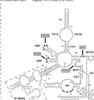
Several other eukaryotic-specific con-served elements in RNase P RNAs have been found by comparison of the eukary-otic and bacterial consensus structures (Frank et al. 2000). These regions include the eP8 hairpin, eP9 stem-loops, and the junction between P4 and P7 (designated
[image:3.669.231.549.273.611.2]as jP4/7; Fig. 1). Although in bacterial RNase P RNAs, P8 and P9 stem-loops seem to occupy positions similar to those of eP8 and eP9, their functional equivalence remains to be con-firmed. The fungal eP8 tetraloops (or pentaloops in some species) always have the sequence NUGA, while most of the fungal eP9 tetraloops have GNAA sequence (Frank et al. 2000). The jP4/7 loop, which is absent in bacterial RNase P, is found in fungal and human RNase P RNAs, although the loops are variable in size with low sequence conservation (Frank et al. 2000). In addition to the above conserved regions, two uridines are always found in an unpaired loop at the junction of the P7 stem and the eP15 stem in fungal RNase P RNAs (Fig. 1, jP7/15), whereas two adenosines occupy similar positions in bacterial RNAs (Tranguch and Engelke 1993; Frank et al. 2000).
FIGURE 1. Eukaryotic-specific conserved elements in the S1-RPR1 RNA. (P) Base-pair region. (eP) Stem structure in eukaryotic RNase P RNA that does not seem to have an obvious bacterial homolog (Frank et al. 2000). Loop regions between two stems are denoted with ‘jP’. Five critical regions (CR I–V) are conserved in bacterial, archaeal, and eukaryotic RNase P RNAs. Elements in quotation marks, P3, eP8, eP9, jP4/7, and jP7/15, are conserved features either unique to eukaryotic RNase P RNAs or with different sequence conservation in eukaryotic RNAs than in bacterial RNAs (Frank et al. 2000). Nucleotides in gray circles are most conserved in the corresponding eukaryotic conserved regions among yeast RNase P RNAs (Tranguch and Engelke 1993; Frank et al. 2000; Ziehler et al. 2001). Four conserved regions were mutated in this study where the underlined nucleotides were changed to the boxed sequences indicated with arrows. An S1 affinity tag with a short linker was inserted
between nucleotides 133 and 140 of the wild-typeRPR1RNA as indicated within dashed lines.
Little is known about the roles of the eukaryote-specific sequences and structural features of RNase P RNA. Their possible functions include (1) providing binding sites for protein subunits of RNase P, as is the case for the P3 internal loop; (2) binding to other molecules in the nucleus to provide needed eukaryotic-specific functions, such as proper localization and transport of the enzyme; and (3) interacting with pre-tRNA substrates to form eukaryote-specific contacts, for example, recognition of the 30end of pre-tRNAs by eukaryotic RNase Ps is different than by the bacterial enzymes (Ziehler et al. 2000).
In this study, we have made mutations in the yeastRPR1
RNA at the loop regions of eP8 hairpin (eP8m), eP9 hairpin (eP9m), the junction of P4/P7 (jP4/7m), and the junction of P7/eP15 (jP7/15m). Mutations in these conserved regions have varying effects on RNase P assembly, localization, and activity. The mutation of jP7/15m does not affect cell growth, tRNA processing, or RNase P maturation. In contrast, mutants in eP8m, eP9m, and jP4/7m display pronounced growth defects and pre-tRNA processing defects in vivo. Assembly of the RNase P ribonucleoprotein complexes har-boring each of these three mutations is affected, although each mutation has a distinctive effect. Mutations of eP9m and jP4/7m also result in significantly different subcellular distribution of RNase P, while mutation of eP8m has no obvious effect on RNase P localization. Steady-state kinetic studies of the pre-tRNA cleavage reaction reveal changes in
KMand/orkcatfor the reaction catalyzed by the three mutated
enzymes.
RESULTS
Growth phenotypes of the S1-RPR1mutants
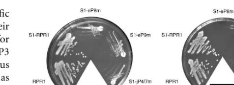
The mutated sequences of the fourRPR1mutations, eP8m, eP9m, jP4/7m, and jP7/15m, are indicated in Figure 1. A streptavidin affinity tag, S1, was inserted between nucleotides 133 and 140 of theRPR1RNA to facilitate characterization and purification of the enzymes (Srisawat and Engelke 2001). A secondary structure of the resulting S1-RPR1 RNA is shown in Figure 1.
Strains carrying wild-type or various mutatedRPR1RNAs were tested for growth at 30C, 37C, and 16C. The
wild-type and the S1 taggedRPR1strains show indistinguishable growth phenotypes at 30C and 37C, confirming that the
S1 affinity tag does not interfere with growth. In contrast, mutant strains S1-eP8m, S1-eP9m, and S1-jP4/7m grow rela-tively slowly at 30C, and the growth defect is more severe at
37C (Fig. 2). Although several strains grow slowly at both 16C (data not shown) and 30C, no notable cold-sensitive growth defects are observed. The mutation of jP7/15m, which converted the conserved UU to AA, had no effect on growth, tRNA processing, or RNase P maturation (data not shown), and was not characterized further.
Pre-tRNA and Pre-rRNA processing in the S1-RPR1 mutants
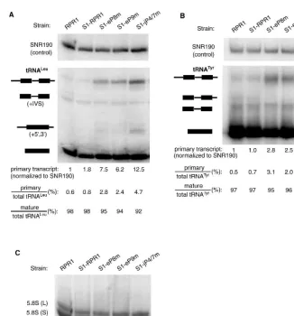
Effects of theRPR1mutations on RNase P function in vivo were examined by analyzing the accumulation of precur-sors for two tRNA species, tRNALeuand tRNATyr. Process-ing of both of these pre-tRNAs by RNase P had been studied previously (Lee et al. 1997; Ziehler et al. 2000), and both were tested because pre-tRNATyris more rapidly cleaved by RNase P than pre-tRNALeu.
In the maturation of pre-tRNALeuand most other yeast pre-tRNAs, 50cleavage precedes 30processing, in part because the structure formed between the 50 leader and 30 trailer blocks access to cleavage of the 30 trailer (Lee et al. 1997). Three major tRNALeuspecies are normally detected in North-ern blots (Lee et al. 1991b)—i.e., the primary transcript, an intermediate with mature 50and 30ends containing the 32-nt
intervening sequence (‘‘+IVS’’ in Fig. 3), and the mature tRNA. In wild-type strains, the majority of the tRNALeuexists as the mature form, whereas the primary transcript and the +IVS species are low in abundance. This processing pattern is observed in the untagged wild-type strain FSY1/RPR1 and strain FSY1/S1-RPR1(Fig. 3A). Although the abundance of pre-tRNALeu (but not pre-tRNATyr) increases slightly when the RNase P carries the S1 affinity tag, the defect is apparently not severe enough to slow growth significantly (Figs. 2, 3).
[image:4.669.318.556.76.163.2]In contrast, the amount of primary transcript increases significantly in mutant strains S1-eP8m (7.5-fold), S1-eP9m (6.2-fold), and S1-jP4/7m (12.5-fold) (Fig. 3A). Consistent with this, the percentage of the primary transcript relative to the total tRNALeuspecies also increases in the mutants, with 2.4–4.7% in different mutant strains versus 0.8% in the S1-RPR1strain (Fig. 3A). However, the ratio of the mature to the total tRNALeu remains relatively constant in all the strains tested (Fig. 3A). An additional processing intermedi-ate with the intron removed, but still containing the 50and 30 extensions (denoted ‘‘+5, 3’’ in Fig. 3) normally appears when RNase P processing is severely slowed in vivo, allowing splicing to precede terminal processing (Lee et al. 1991b). This intermediate appears most prominently in the S1-jP4/ 7m strain (Fig. 3A), but also accumulates at a lower level in the S1-eP8m and S1-eP9m strains (darker exposure not shown).
FIGURE 2. Growth of strains containing mutated S1-RPR1RNAs on
YPD medium. The plates were incubated for 2 d at 30C and 37C,
To make sure that the tRNA processing defects observed were not specific to tRNALeu, we also examined the proces-sing of tRNATyr in the S1-RPR1 mutant strains. The pri-mary transcript of tRNATyr, which contains a 12-nt 50 leader, an intron, and a 30trailing sequence, is accumulated modestly in all three mutant strains (2.5- to 3.3-fold accu-mulation; Fig. 3B). The ratio of the primary transcript to the total tRNATyrspecies is increased in the mutants, which is similar to what is observed for tRNALeu (Fig. 3A,B). These data suggest that theRPR1 RNA mutations cause a general defect in the 50-end processing of pre-tRNAs.
Previously, it was observed that a severe mutation in the CR IV domain of RPR1 RNA (Fig. 1) mildly affected processing of 5.8S rRNA for unknown reasons, giving a 30 extended form of about 7S rRNA in size (Chamberlain et al. 1996). We do not see the appear-ance of such aberrant bands in probing the 5.8S rRNA sequence in the RNase P mutants examined here (Fig. 3C). We also do not observe any change in the ratio of 5.8S (L) to 5.8S (S) forms, which would have indicated some per-turbation in the function of RNase MRP (Fig. 3C). Although this lack of effect on 5.8S rRNA is not surprising, we tested this as a control to demon-strate that RNA processing in general is not slowed in these mutant strains.
Maturation of the S1-RPR1RNA
To further understand why the RPR1
mutations cause defective RNase P activ-ity, we tested the effect of the mutations on maturation of the RPR1 RNA. We have previously found that mutations that either compromise the structure of theRPR1RNA or prevent protein bind-ing also block maturation of the precur-sor RPR1 RNA subunit (Pagan-Ramos et al. 1996a,b; Ziehler et al. 2001).
The maturation profiles of the mutated S1-RPR1RNAs expressed from plasmids were examined in strains containing the untagged wild-type RPR1 RNA on the chromosome. The S1-RPR1 precursor and mature RNAs were detected by Northern blotting with a probe comple-mentary to the S1 tag sequence. No bands are detected in the total RNA isolated from a control strain containing the vector alone, demonstrating that the probe does not hybridize to the endogen-ous untaggedRPR1RNA (Fig. 4, lane 1). The pre-RPR1RNA generally migrates as a slightly diffuse band on a Northern blot because the 30end is heterogeneous (Lee et al. 1991b). When
[image:5.669.49.375.75.425.2]the S1-RPR1strain is grown to mid-log phase, the ratio of mature to precursorRPR1RNA is5 (Fig. 4), which is normal for the maturation of wild-typeRPR1 RNA. However, the ratio decreases twofold in strains S1-eP9m and S1-jP4/7m (ratio of 2.3 and 2.2, respectively), with an intermediate ratio in strain S1-eP8m (ratio of 3.3; Fig. 4). Moreover, the steady-state levels of the precursor and matureRPR1RNAs in the mutant strains are consistently lower when normalized to the FIGURE 3. Pre-tRNA and 5.8S rRNA processing in S1-RPR1mutant strains. (A) Northern
blot analysis on pre-tRNALeu. The four major tRNALeuspecies detected on a Northern blot are
the primary transcript, an intermediate with the intervening sequence (+IVS), an intermediate
with 50and 30extensions (+5, 3), and the mature tRNALeu
(Lee et al. 1991b). (B) Northern blot
analysis on pre-tRNATyr. Identities of the four major tRNATyr species on the blot are the
primary transcript, an intermediate with the intron and a 30extension, the intermediate with
an intron, and the mature tRNATyr(Kufel et al. 2002). Names and schematic diagrams of tRNA
species are shown on theleftof the blots. Black bars represent mature tRNA sequences whereas
lines represent the 50 leader, the intron, and the 30 trailer sequences. Signal of the primary
transcript was normalized to that of the loading control,SNR190RNA. The amount of the
primary transcript in the RPR1 wild-type strain was arbitrarily set at 1, and was used for
comparison with data from the mutant strains. The relative amounts of primary transcript and mature tRNA are also listed as a percentage of the total tRNA species detected in the
North-erns. (C) Northern blot analysis on the 5.8S rRNA processing. The same blot shown inAwas
reprobed for the 5.8S rRNA. Ratio of the long to short forms of 5.8S in each strain is listed.
U6 snRNA (Fig. 4), suggesting that the mutatedRPR1RNAs might not be as stable.
The relative accumulation of theRPR1precursor implies a defect in the assembly or stability of RNase P holoen-zyme. Since the pre-RPR1RNA binds at least seven of nine protein subunits before maturation (Pop1p, Pop4p, Pop5p, Pop6p, Pop7p, Pop8p, and Rpp1p) (Srisawat et al. 2002), defects in the RNA structure could alter the rate of pre-cursor processing either directly or indirectly through blocking some essential RNP formation. Depletion of any of the seven protein subunits in the precursor complex, or compromised RNA structure or protein binding, will block maturation (Lygerou et al. 1994; Pagan-Ramos et al. 1996a,b; Chu et al. 1997; Stolc and Altman 1997; Chamberlain et al. 1998; Ziehler et al. 2001). In contrast, point mutations at CR II and CR III residues, which are primarily involved in pre-tRNA substrate interactions, allow pre-RPR1 maturation (Pagan-Ramos et al. 1996a). The relative accumulation of pre-RPR1RNA is therefore a likely indication that either the RNA structure is altered or protein association is blocked, or both, thus hampering maturation. It is also possible that the stability of the mature RNase P particles is impaired, making them more
susceptible to degradation, which could alter the ratio of the two species.
Association of the mutated S1-RPR1RNA with RNase P protein subunits
To address directly whether the mutations on the RPR1
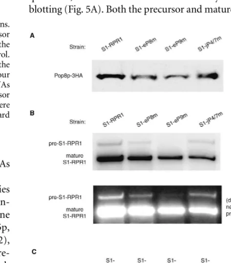
RNA affect protein association, we performed co-immuno-precipitation of RNA with one of the early associating protein subunits. For unknown reasons, epitope tagging of most protein subunits is synthetically lethal in the pres-ence of the S1 affinity tag on the RNA subunit. However, we were able to place three hemagglutinin (3HA) epitopes at the C terminus of Pop8p without negative effects on growth. Therefore, immunoprecipitation of Pop8p-3HA was performed, following which the S1-RPR1 RNA and Pop8p-3HA in the precipitate were examined by Northern blotting and Western blotting, respectively.
[image:6.669.80.271.78.321.2]Pop8p-3HA is found in all the pull-downs in roughly equal quantities, as detected with anti-HA antibody in the Western blotting (Fig. 5A). Both the precursor and mature forms of all FIGURE 4. Processing of the S1-RPR1 RNA in the mutant strains.
(A) Northern blotting of the S1-RPR1 RNA. Both the precursor
(pre-S1-RPR1) and the mature S1-RPR1RNA were detected on the
Northern blot. The U6 RNA was probed as a loading control.
(B) Quantitation of the S1-RPR1 RNA. Ratio of the mature to the
precursor S1-RPR1RNAs is listed with standard deviation from four
Northern blots. Signals of the mature and precursor S1-RPR1RNAs
were also normalized to that of the U6 RNA. Amounts of the precursor
and mature RNAs from the S1-RPR1strain were set at 100% and were
used for comparison with data from the mutant strains. Standard deviations obtained from four Northern blot analyses are provided.
FIGURE 5. Association of the mutated S1-RPR1RNA with
Pop8p-3HA in vivo. (A) Western blotting of Pop8p-3HA protein in the HA
immunoprecipitates. (B) Northern blotting of the S1-RPR1 RNA
copurified with Pop8p-3HA. Positions of the precursor and mature
S1-RPR1 RNA on the blot are indicated. (C) Quantitation of the
amount of Pop8p-3HA and S1-RPR1 in the immunoprecipitates.
Signals of the precursor and mature S1-RPR1RNA on the Northern
blot were normalized to the signals of Pop8p-3HA on the Western blot to account for the difference in the immunoprecipitation
effi-ciency among samples. The resulting ratio in the S1-RPR1 control
[image:6.669.317.554.340.610.2]mutated S1-RPR1RNAs are associated with Pop8p-3HA in the control and mutant strains, but the RNA-to-protein ratio changes significantly (Fig. 5B). Compared to the control, less precursor S1-eP8m RNA (38%) associates with the protein subunit (Fig. 5C). However, the binding of mature S1-eP8m RNA to Pop8p-3HA does not change significantly (Fig. 5C). This result suggests that the protein might be added more slowly to the precursor S1-eP8m RNA, but that the association is likely stable once the RNA is processed to the mature form because the matureRPR1-to-protein ratio is normal. The S1-eP9m mutation produces a more severe version of this, with a more than 10-fold decrease in the amount of bound precursor RNA, but only a twofold decrease in the amount of bound matureRPR1RNA (Fig. 5C). The S1-jP4/7m RNA presents a different case. Although both the precursor and matureRPR1
RNAs associate less with Pop8p-3HA, the mutation affects the interaction with the mature RNA more significantly (Fig. 5C). The result suggests that all three of these regions contribute to some extent to the efficiency of pre-RPR1 association with
proteins, whereas jP4/7 contributes to the stability of the mature RNase P complex once formed.
Localization of the mutated S1-RPR1RNA
Early steps of the tRNA biogenesis pathway, including cleav-age by RNase P, have been shown to occur in the nucleolus in yeast, and both the precursor and mature forms ofRPR1
RNAs are primarily nucleolar (Bertrand et al. 1998; Srisawat et al. 2002). If the RNase P holoenzyme is assembled slowly or misassembled, then the nucleolar localization of theRPR1
RNA might be compromised. To test this hypothesis, we examined the subcellular distribution of the mutated S1-RPR1 RNA in strains FSY1/S1-eP8m, FSY1/S1-eP9m, and FSY1/S1-jP4/7m.
In the FSY1/S1-RPR1control strain, the S1-RPR1RNA signal substantially overlaps with the nucleolar U14 snoRNA marker (Fig. 6), although there is also nucleoplasmic signal as seen in earlier work (Bertrand et al. 1998). The signal for S1-eP8m RNA is not strikingly different from that in the S1-RPR1strain, although examination of large numbers of cells could not rule out minor differences. This is consistent with the relatively small defect in RPR1 maturation in this strain (Fig. 5). In contrast, signals for S1-eP9m RNA and S1-jP4/7m RNA are repro-ducibly more diffuse, with considerably less overlap with the nucleolar U14 signal (Fig. 6). Thus, there is a correlation between holoenzyme assembly and locali-zation, although it is not possible to deter-mine causality between these events.
Effects of the S1-RPR1mutations on RNase P activity in vitro
To determine the effects of the RPR1
mutations on RNase P catalytic activity, we measured the steady-state turnover for cleavage of S. cerevisiae precursor tRNATyrwith a 12-nt 50leader catalyzed by RNase P mutants under initial velocity conditions. For each mutant the position of pre-tRNA cleavage appears normal (data not shown). We measured the activity of RNase P as a function of sub-strate concentration to determine the steady-state kinetic parameters,kcat,kcat/
KM and KM. The presence of the S1
affinity tag does not affect the value of
kcat, as compared to the previously
[image:7.669.55.376.316.678.2]determined nontagged wild-type holoen-zyme (1.2¯60.1 and 1.3¯60.1 sec1, FIGURE 6. Localization of the S1-RPR1RNA in the mutant strains. The signal of S1-RPR1
RNA is shown in red and the signal of the nucleolar U14 RNA is shown in green. The blue
DAPI staining indicates the nucleus.Rightpanels are the merged images of the corresponding
leftandmiddlepictures. Overlap of the red and green signals is shown in yellow. Images shown are representatives of large numbers of cells.
Fig 6. live 4/c
respectively; Fig. 7, Table 1) under identical conditions (Ziehler et al. 2000). The presence of the S1 tag decreases the observedKMfrom 55¯610 nM in the nontagged enzyme
to 20¯68 nM in the S1-RPR1 enzyme. The S1 tag also increases the value of kcat/KM two- to threefold, moving it
closer to the value of 13108 M1sec1 observed for wild-type RNase P cleavage of a pre-tRNA substrate containing no trailer sequence (Ziehler et al. 2000). Previous data suggest thatkcatmost likely describes the rate constant for product
release and kcat/KM nearly reflects the rate constant for
substrate association under the conditions studied (Ziehler et al. 2000). For this kinetic mechanism, the value of KM
cannot equal the substrate dissociation constant (Kd) but rather describes the apparent dissociation constant for the formation of all bound enzyme intermediates (Fersht 1985). These data suggest that the presence of the S1 affinity tag increases the apparent substrate association rate constant, likely by altering the partitioning between substrate dissocia-tion and substrate cleavage.
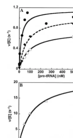
The steady-state kinetic data for the reactions catalyzed by the RPR1 mutants S1-eP8m, S1-eP9m, and S1-jP4/7m are depicted in Figure 7 and summarized in Table 1. Muta-tions in S1-eP9m and S1-jP4/7m decrease the observedkcat
slightly (1.0¯60.1 and 0.74¯60.03 sec1, respectively) as compared to wild type (1.2¯60.1 sec1) and increase the
KM to 70¯620 nM and 127¯616 nM, respectively.
Conse-quently, the specificity constant,kcat/KM, for the
S1-eP9m-catalyzed reaction is 1.5¯60.43107M1sec1, and for the S1-ejP4/7m is 0.58¯60.083107M1sec1. These values are decreased fourfold and 10-fold, respectively, as compared to the S1-RNase P-catalyzed reaction (Table 1). For pre-tRNA cleavage catalyzed by S1-eP8m, both kcat (22¯63
sec1) and KM (130¯640 nM) increase. Surprisingly, this
leads to a threefold increase in the specificity constant (Table 1; Fig. 7) to a value that is slightly higher than the
kcat/KMobserved for wild-type RNase P cleavage of a
pre-tRNA substrate containing no trailer sequence (Ziehler et al. 2000). In the case of the S1-eP8m mutant, the reac-tion is likely diffusion controlled since the specificity con-stant is 1.73108M1sec1. This increased value ofkcat/KM
indicates that every pre-tRNA that encounters RNase P is rapidly cleaved. This behavior may decrease the substrate selectivity of RNase P if cleavage of all pre-tRNA substrates is diffusion controlled.
DISCUSSION
[image:8.669.120.238.76.299.2]Characterization of RPR1 mutations at four eukaryotic-specific conserved regions has revealed different roles of FIGURE 7. Processing ofS. cerevisiaepre-tRNATyrin vitro. (A) The
cleavage of a pre-tRNA substrate with a 12-nt leader catalyzed by
either S1-RPR1(), S1-eP9m (¤) or S1-jP4/7m (*) in 10 mM HEPES
(pH 8), 100 mM KCl, and 10 mM MgCl2 was determined under
steady-state conditions where substrate concentration exceeds enzyme
concentration. (B) The cleavage of pre-tRNATyrby S1-eP8m (~) was
measured under identical conditions as listed inA. The data for the
S1-RPR1-catalyzed reaction () fromAis included for comparison.
The Michaelis–Menten equation is fit to the data to determine the
values forkcat,kcat/KM, andKM(listed in Table 1).
TABLE 1. Summary of the characterizations of theRPR1mutations
S1-RPR1 S1-eP8m S1-eP9m S1-jP4/7m
Mutated sequence S1 insertion between nucleotides 133 and 140
158 ACUG 161 178 CGUU 181 99 UCCG 102
Growth Normal Slow Slow Slow
Pre-tRNA processing Normal Deficient Deficient Deficient
Holoenzyme assembly Normal Mildly impaired precursor assembly
Severely impaired precursor assembly
Impaired assembly of precursor and mature complexes Localization Predominantly nucleolar Nucleolar Diffused, more
nucleoplasmic
Diffused, more nucleoplasmic
Kinetics
kcat(sec1) 1.2¯60.1 22¯63 1.0¯60.1 0.74¯60.03
KM(nM) 20¯68 130¯640 70¯620 127¯616
[image:8.669.62.559.577.729.2]the conserved elements in eukaryotic RNase P activity and maturation. Replacing the UU at the jP7/15 region with AA does not notably affect cell growth, pre-tRNA processing, or the maturation of RNase P RNA. However, mutations at the other three conserved sequences—i.e., eP8, eP9, and jP4/7 regions—lead to pronounced slow growth pheno-types and pre-tRNA maturation defects, although none of the mutations is lethal. Taking advantage of an RNA affi-nity tag, we are able to further demonstrate that the defec-tive RNase P enzymes have problems in holoenzyme assembly, localization, and in vitro pre-tRNA cleavage to varying degrees. The mutations of eP8m and eP9m reduce the binding of the protein subunit to pre-RPR1RNA more significantly than to the mature RPR1 RNA, with eP9m showing more severe defective phenotypes (Fig. 5). In contrast, jP4/7m mutation appears to impair the associa-tion of the protein subunit with the mature RPR1 RNA more than the precursor RNA (Fig. 5). Consistent with the defects in holoenzyme assembly, the localization of eP9m and jP4/7m mutated RNAs is diffuse compared to the wild-type RNA, and is less associated with the nucleolus (Fig. 6). Kinetic analyses show that ep9m and jP4/7m mutations decrease the catalytic efficiency (kcat/KM) of the pre-tRNA
cleavage reaction by fourfold and 10-fold, respectively, whereas mutation of eP8m increases the kcat/KM of the
reaction in vitro (Table 1). The apparent discrepancy between the increased efficiency of the eP8m enzyme in vitro and the pre-tRNA processing defect caused by the eP8m mutation in vivo will be discussed below. A summary of the phenotypes of the individual mutation is listed in Table 1. It is worth noting that the presence of the S1 affinity tag in the RPR1 RNA constructs in this study might contribute to the observed phenotypes, for example, by weakening the stability of RNA structures. However, since the S1-tagged wild-type RPR1 RNA is used as a control, the defective phenotypes must also reflect a con-tribution from the mutated conserved sequences.
Region jP4/7 is conserved in the RNase P RNA from fungi and vertebrates, but is not found in bacterial or archaeal RNase P RNA (Frank et al. 2000). Mutation at jP4/7 impairs the association between protein subunits and the RPR1 RNA, especially in the mature form, suggesting defects in holoen-zyme assembly and, possibly, the stability of the RNP com-plexes (Fig. 5). The observation is consistent with previous structure-sensitive footprinting results indicating that jP4/7 is protected from nuclease and chemical attacks in the holoen-zyme (Tranguch et al. 1994). It is not known, however, whether the jP4/7 loop contacts protein subunits directly.
Mutation at the jP4/7 region causes the most severe defect in pre-tRNA processing in vivo (Fig. 3). Moreover, this is accom-panied by a significant decrease (10-fold decrease) in the enzyme efficiency (kcat/KM) in vitro (Fig. 7; Table 1). Deficient
RNase P cleavage in vivo might be mainly attributed to desta-bilization of either the RNA subunit or the holoenzyme. How-ever, it is also plausible that jP4/7 could participate more
directly in pre-tRNA binding or catalysis since this region is right next to P4, the postulated catalytic core of RNase P. A recent study on the yeast RNase MRP, a sibling enzyme of yeast RNase P, suggested that a bulged-loop structure equivalent to jP4/7 might exist in the RNA subunit (Walker and Avis 2004). It will be intriguing to see whether the loop region plays similar roles in assembly and catalysis of RNase MRP.
Most fungal eP9 hairpins contain tetraloops of the GNRA type (Fig. 1). Presence of a tetraloop structure could increase the stability of RNA duplexes or participate in RNA–RNA interactions or RNA–protein interactions (Cheong et al. 1990; Gluck et al. 1992; Jaeger et al. 1994). Structure footprinting analysis of the yeast RNase P has indicated that the eP9 tetraloop is protected in the presence of protein subunits (Tranguch et al. 1994). In this study, the tetraloop of eP9 appears to be crucial for the assembly of precursor RNase P complex, since mutation at this region drastically reduces the association of pre-RPR1
RNA with protein subunits (Fig. 5). A recently refined consensus secondary structure of eukaryotic RNase P RNA has suggested that eP9 might be structurally homol-ogous to the bacterial P9 stem-loop (Frank et al. 2000). The P9 tetraloop in both Escherichia coli and Bacillus subtilis
RNase P RNAs was shown to be adjacent to the P1 stem of the RNA by crosslinking experiments (Harris et al. 1997; Chen et al. 1998). Whether eukaryotic eP9 is similarly positioned in the holoenzyme awaits further investigations. Crystal structures of the specificity domains of two bac-terial RNase P RNAs have shown that the P9, P10, and P11 stems form an opening structure that contributes to pre-tRNA recognition (Krasilnikov et al. 2003, 2004). A single nucleotide change in the tetraloop of the bacterial P9 mildly increases theKMandkcatof the pre-tRNA cleavage
reaction catalyzed by the holoenzyme (Pomeranz Krummel and Altman 1999). Consistent with the studies on the bacterial enzyme, mutation in the tetraloop of eP9 in the yeastRPR1RNA increases theKMof pre-tRNA processing
in vitro without obviously changing thekcatof the reaction
(Table 1). It is possible that eP9, like the bacterial P9, contributes to substrate recognition by eukaryotic RNase P, probably via formation of the substrate-binding site with P10/11 and potentially with protein subunits. However, with the increased protein content in eukaryotic RNase P and the diverged structural elements in the RNA subunit, eP9 might have different functions than the bacterial P9.
Mutation in the eP8 tetraloop only mildly reduces the association of protein subunits with precursor RPR1RNA, and has little effect on the stability of the mature holoenzyme (Fig. 5). Consistent with this, there is not a major perturbation in the association of theRPR1RNA with the nucleolus (Fig. 6). This mutation, however, causes a significant increase inKM
and kcat/KM in the in vitro pre-tRNA cleavage reaction
(Table 1). Therefore, this mutation leads to enhanced catalytic efficiency in vitro either by enhancing the association rate constant or changing the partitioning ratio between substrate
dissociation and cleavage. If the rate constant for substrate association/dissociation is normally limited by a conforma-tional change in wild-type RNase P, a less stable holoenzyme or one lacking one or more protein components could increase the rate of this conformational change.
The overall increase in catalytic efficiency (kcat/KM)
caused by the eP8m mutation seems to be contradictory to the accumulation of pre-tRNA species with a 50 exten-sion in vivo (Fig. 3). While there might be a nonobvious problem in nuclear localization or protein association, it is possible that the key change in the eP8m mutation is that it has become less specific for binding pre-tRNA substrates, since the reaction catalyzed by this mutated enzyme in vitro is diffusion controlled. In this case, the mutated enzyme might be more susceptible to competitive inhibition by other RNAs within the nucleoplasm or nucleolus. Consis-tent with this hypothesis, titration with total yeast RNAs as competitors shows that the apparent KI for the pre-tRNA
cleavage reaction catalyzed by eP8m is 0.02 mg/mL, 50-fold less than that for the reaction catalyzed by the wild-type enzyme (1.0mg/mL; data not shown).
Phylogenetic studies have demonstrated that the eukary-otic eP8 hairpin and the bacterial P8 might be homologs because they occupy similar positions in the four-way junction of RNase P RNA (Frank et al. 2000). Intriguingly, in the crystal structure of the specificity domain of an A-type RNase P RNA, P8 is involved in tertiary structural interactions to stabilize the conformation of the pre-tRNA recognition region (Krasilnikov et al. 2004). It is possible that eukaryotic eP8 might also stabilize the substrate recog-nition site, and that the mutated tetraloop could broaden the substrate specificity of the enzyme.
The studies presented here identify contributions of the eukaryotic-specificRPR1sequences to holoenzyme assembly and pre-tRNA cleavage, but it is entirely possible that there are additional consequences of mutations at these sites. For example, it is possible that, like the bacterial RNase P, the nuclear enzyme has additional substrates that have not yet been identified and that require some contribution from these sequences. Alternatively, the RNase P holoenzyme might need to interact with some proteins that are associated with substrates in vivo, for instance, the Lsm (Sm-like) or La proteins bound to the 30ends of pre-tRNA transcripts (Kufel
et al. 2002), or some other cellular component that facilitates substrate acquisition.
MATERIALS AND METHODS
Plasmids and mutagenesis
Mutation at eP8, eP9, or the junction of P4/P7 was individually introduced into the low-copy plasmid construct pRS315-S1-RPR1, which contained theRPR1gene under its own promoter and aLEU2 selectable marker. PCR-based mutagenesis was performed as described (Srisawat and Engelke 2001). Each of the mutatedRPR1
sequences, including a S1-RPR1‘‘wild-type’’ control, contained an ‘‘S1’’ RNA affinity tag that allowed the RNA to bind tightly to streptavidin, but to be released in the presence of biotin (Srisawat and Engelke 2001). A strain containing YCp50-RPR1 (Lee et al. 1991b), with the untagged wild-type gene on a low-copy plasmid, was used as a control for consequences of the S1 tag insertion.
Strains
Effects of the mutations on maturation of theRPR1 RNA were examined in the haploidS. cerevisiaestrain W3031A (MATa ade2-1 his3-ade2-1ade2-1, ade2-15 leu2-3, ade2-1ade2-12 trpade2-1-ade2-1 ura3-ade2-1 canade2-1-ade2-100) with mutated S1-RPR1RNAs expressed on pRS315 plasmids. The resulting strains were W3031A/S1-RPR1, W3031A/S1-eP8m, W3031A/S1-eP9m, and W3031A/S1-jP4-7m. These strains also had a chromosomal wild-typeRPR1gene intact.
Mutated S1-RPR1RNAs on pRS315 plasmids were transformed into strain FSY1 (MATaade2-1 his3-11, 15 leu2-3, 112 trp1-1 ura3-1 canura3-1-ura3-100 RPRura3-1łkan) containing a chromosomal deletion of the RPR1gene, but a copy of the wild-type gene on a counterselectable plasmid, YCp50-RPR1. Transformants containing the mutated RPR1genes were selected for the loss of YCp50-RPR1on medium with 5-fluoroorotic acid, resulting in strains FSY1/S1-RPR1, FSY1/ S1-eP8m, FSY1/S1-eP9m, and FSY1/jP4–7m. These strains, which contain only the mutated RPR1RNA, were used to test growth phenotypes, pre-tRNA and pre-rRNA processing profiles, and loca-lization of S1-RPR1 RNA.
To address whether the mutations on the RPR1 RNA cause assembly defects of the holoenzyme, a strain containing the chro-mosomalPOP8gene tagged with 3xHA sequences at the 30end was
used (Srisawat et al. 2002). The pRS315 plasmids with mutated S1-RPR1RNA genes were the only source of RPR1RNA in the strain. HA-immunoprecipitation of RNase P was performed to examine the association of Pop8p-3HA with mutatedRPR1RNAs.
Pre-tRNA and pre-rRNA processing
Strains with pRS315-S1-RPR1or its mutant derivatives in the FSY1 strain background were grown in YPD medium at 30C to an OD
600
of 1. Total cellular RNA was extracted with hot acidic phenol and precipitated with ethanol as described (Kohrer and Domdey 1991). Concentration of total RNA was determined by absorbance at 260 nm. Ten micrograms of isolated RNA were electrophoresed on an 8% denaturing polyacrylamide gel and transferred onto a nytran membrane. The blot was then probed with32P-labeled DNA oligo-nucleotides complementary to tRNALeu, tRNATyr, and to 5.8S rRNA to assess the in vivo activities of RNase P and MRP. The tRNALeu
probe was 50-TGCTAAGAGATTCGAACTCTTGCA-30, the tRNATyr probe was 50-AGTCGAACGCCCGATCTCAAGATT-30, and the 5.8S
rRNA probe was 50-CGCATTTCGCTGCGTTCTTCATCG-30. The SNR190RNA, a small nucleolar RNA that is not affected by RNase P mutations, was probed on the same blot as an internal normalization control (probe: 50-ATGGTCGAATCGGACGAG-30). Signals were
Maturation of theRPR1RNA
W3031A strains containing plasmid-borne S1-RPR1, S1-eP8m, S1-eP9m, S1-jP4/7m RNAs, or the empty vector were grown in SDC-Leu medium at 30C to an OD600of0.7. Ten micrograms
of total RNA were separated on a denaturing 6% polyacrylamide gel and transferred onto a nytran membrane. The blot was probed with radiolabeled DNA oligonucleotides complementary to the S1 affinity tag that detected the mutatedRPR1RNA from the plas-mid, but not the endogenous wild-typeRPR1RNA. Sequence of the S1 probe was 50-GCATGATTCTGGTCGGTCGACTCCC-30.
As an internal loading control, U6 RNA was probed on the same blot with radiolabeled DNA oligonucleotide with sequence 50-CTGATCATCTCTGTATTG-30. The hybridized RNAs were detected and quantitated as above. The ratio of mature to pre-cursor S1-RPR1 RNA listed in Figure 4 is the average of four Northern blotting analyses with standard deviation less than 0.3.
HA-immunoprecipitations and Western blotting
Strains containing Pop8p-3HA and pRS315 expressing S1-RPR1 or the mutated RNAs were grown in SDC-Leu medium at 30C to an OD600of1. Cells were washed with water and Buffer A (50 mM
Tris-HCl at pH 7.5, 150 mM NaCl, 5 mM EDTA, 0.1% Triton-X-100, 10% glycerol, 1 mM DTT, 1 mM PMSF). About 23109cells were resuspended in 400 mL of Buffer A and were broken by vortexing with glass beads (425–600 mm diameter). Total cell extracts were centrifuged at 16,000gfor 2 min at 4C. The
super-natant was centrifuged again at 16,000gfor 15 min at 4C. Protein content of the extract was determined with Micro Bicinchoninic Acid Assays (Pierce). FSY1 strain with wild-type Pop8p and S1-RPR1RNA was included as a negative control for HA-dependent immunoprecipitation.
Protein A-agarose (BioRad Affi-Gel) was incubated with rabbit anti-mouse IgG (2 mL per 200mL of packed beads, AffiniPure Rabbit Anti-Mouse IgG (H+L); Jackson ImmunoResearch) at 4C for 1 h, with gentle rotation. Anti-HA antibody 12CA5 (10mg per 200mL packed beads; Roche) was added to the coupled protein A-agarose-IgG complex, and the mixture was incubated at 4C for
2 h. The conjugates were washed six times with Buffer A. Total yeast extract with 3 mg of protein was bound to 200mL protein A–agarose–IgG–antiHA complex at 4C for 1 h, and then
washed with 1 mL of Buffer A five times. The volume of the bead slurry was brought to 300mL with Buffer A. Portions of the slurry were saved for Western blotting of Pop8p-3HA (80 mL) and Northern blotting of S1-RPR1RNA (220mL).
Eighty microliters of the bead slurry were spun down at 1500 rpm for 2 min. Proteins bound to the beads were extracted with 40mL of sample buffer with 6 M urea (60 mM Tris-HCl at pH 6.8, 2% SDS, 15% glycerol, 0.025% bromophenol blue, 5% b-mercaptoethanol, 6 M urea) at 95C for 5 min. The supernatant
sample was electrophoresed on a 12% polyacrylamide-Tris-HCl gel in SDS running buffer (BioRad) and transferred to a PVDF membrane. The blot was probed with anti-HA (12CA5; Roche) and goat anti-mouse HRP antibodies (Chemicon AP308) and was subjected to chemiluminescent substrate (ECL Plus Western Blot-ting Detection System; Amersham Biosciences). To detect the signals, the blot was exposed to an X-ray film (Kodak BIOMAX MR), and was also scanned with a Typhoon scanner (model 9400)
with fluorescence detection settings (Amersham application note: Fluorescent Western Blotting).
Northern blotting of S1-RPR1RNA in anti-HA immunoprecipitation
RNA bound to the 220 mL of Pop8p-3HA bead slurry was extracted with an equal volume of 5% SDS, 10% b-mercaptoetha-nol at 95C for 5 min. Eluted RNAs were extracted with phenol-chloroform, and ethanol precipitated with glycogen carrier. The RNA pellet was washed with 70% ethanol and resuspended in 23 formamide/EDTA/SDS loading buffer (95% formamide, 20 mM EDTA, 0.1% SDS, 0.05% bromophenol blue, 0.05% xylene cya-nol). RNA samples were separated on a denaturing 6% polyacryl-amide gel and transferred to a nytran membrane. The blot was probed with radiolabeled DNA oligonucleotide complementary to the RPR1 RNA (50-GCTGGAACAGCAGCAGTAATCGGTA-30).
The signals were detected and quantitated as above.
To examine the association of the mutated S1-RPR1RNAs with Pop8p-3HA, the amount of the S1-RPR1RNA in the precipitates was then normalized to that of Pop8p-3HA to account for the difference in immunoprecipitation efficiency among samples. Data displayed in Figure 5 are representations of Western and Northern analyses of three independent experiments, which gave quantitatively similar results within expected error.
RNase P enzyme assays
Strains containing wild-type or the mutated S1-RPR1 RNA expressed on the pRS315 plasmid in the FSY1 strain background were grown in YPD medium at 30C to an OD
600 of1. Yeast
extracts from 1.531010 cells were prepared as described by
Srisawat and Engelke (2001). RNase P complexes associated with the S1-RPR1RNA were purified by streptavidin affinity chroma-tography from yeast extract containing 50 mg of total protein as described (Srisawat and Engelke 2001). Concentrations of the eluted RNase P enzymes were determined by Northern blotting of the S1-RPR1 RNA in the biotin eluate in comparison to known amounts of in vitro transcribed S1-RPR1RNA.
The eluted RNase P enzymes were tested for catalysis of pre-tRNA cleavage in vitro under steady-state conditions as described (Ziehler et al. 2000) in a reaction buffer containing 10 mM HEPES (pH 7.9) 100 mM KCl, and 10 mM MgCl2. The 50radiolabeled
pre-tRNATyrsubstrate containing a 12-nt leader and a 30 polyur-idine trailer was incubated in 10 mM Tris HCl (pH 8), 0.1 mM EDTA at 95C for 3 min prior to the addition of 23 reaction buffer and subsequent incubation at 37C for 15 min. The enzyme
assay was initiated by the addition of RNase P (0.04– 16.3 pM). At various times, an aliquot of the reaction mixture was diluted into an equal volume of stop solution (200 mM EDTA at pH 8.0, 8 M urea, 0.05% bromophenol blue, and 0.05% xylene cyanol). Reac-tion times were chosen so that <10% of substrate was cleaved (initial velocity conditions). The substrate and 50leader product were separated on a 6% polyacrylamide 7 M urea gel. Quantita-tion was performed using a PhosphorImager with ImageQuant software (Molecular Dynamics). The Michaelis–Menten equation was fit to the data using KaleidaGraph software (Synergy Soft-ware) to determine the kinetic parameterskcat,KM, andkcat/KM.
Localization of S1-RPR1RNA in the mutants
Subcellular locations of the RNase P RNA in FSY1/S1-RPR1, FSY1/ S1-eP8m, FSY1/S1-eP9m, and FSY1/S1-jP4-7m were detected by fluorescent in situ hybridization (FISH). Yeast strains were grown in SDC-Leu medium at 30C to an OD
600of0.4. Cells were fixed,
hybridized with fluorescently labeled oligodeoxynucleotides, then stained with DAPI as described previously (Bertrand et al. 1998; Thompson et al. 2003), except that cells were spheroplasted with 0.2 mg/mL Zymolyase at 37C for 40 min. The S1-RPR1 RNA was probed with two fluorescent oligonucleotides, the anti-S1 fluores-cent probe (anti-S1-f) 50-GACT*ATCTTACGCACT*TGCAT GATTCT*GGTCGGTCGACT*CCC-30 and anti-RPR1 fluorescent
probe (anti-RPR1-f) 50-GACCT*AGGCCGAACTCCGT*GAATTT CTGAT*AACAACGGTCGGT*A-30with Cy3 (Amersham) attached
to the dT positions denoted with asterisks. The U14 snoRNA was probed as a nucleolar marker with oligonucleotides 50-CGAT* GGGTTCGTAAGCGT*ACTCCTACCGT*GG-30, with asterisks
indicating dTs labeled with Oregon Green 488 (Molecular Probes).
ACKNOWLEDGMENTS
We are grateful to F. Houser-Scott, P. Good, S. Walker, D. Coughlin, and members of the Engelke laboratory for helpful discussions. We thank J. Hsieh and M. Tsoi for technical assis-tance. This work was supported by National Institutes of Health (NIH) Grant GM34869 (D.R.E.) and GM55387 (C.A.F.). Partial support was provided by NIH Training Grant GM08270 (J.J.D.), and the H.H. Rackham Graduate School at the University of Michigan (S.X.).
Received December 21, 2004; accepted March 2, 2005.
REFERENCES
Bertrand, E., Houser-Scott, F., Kendall, A., Singer, R.H., and Engelke,
D.R. 1998. Nucleolar localization of early tRNA processing.Genes
&Dev.12:2463–2468.
Chamberlain, J.R., Pagan, R., Kindelberger, D.W., and Engelke, D.R. 1996. An RNase P RNA subunit mutation affects ribosomal RNA
processing.Nucleic Acids Res.24:3158–3166.
Chamberlain, J.R., Lee, Y., Lane, W.S., and Engelke, D.R. 1998. Purification and characterization of the nuclear RNase P holo-enzyme complex reveals extensive subunit overlap with RNase MRP.Genes&Dev.12:1678–1690.
Chen, J.L. and Pace, N.R. 1997. Identification of the universally
conserved core of ribonuclease P RNA.RNA3:557–560.
Chen, J.L., Nolan, J.M., Harris, M.E., and Pace, N.R. 1998. Comparative
photocross-linking analysis of the tertiary structures ofEscherichia
coliandBacillus subtilisRNase P RNAs.EMBO J.17:1515–1525. Cheong, C., Varani, G., and Tinoco Jr., I. 1990. Solution structure of
an unusually stable RNA hairpin, 50GGAC(UUCG)GUCC.Nature
346:680–682.
Christian, E.L., Kaye, N.M., and Harris, M.E. 2000. Helix P4 is a divalent metal ion binding site in the conserved core of the
ribonuclease P ribozyme.RNA6:511–519.
Chu, S., Zengel, J.M., and Lindahl, L. 1997. A novel protein shared by
RNase MRP and RNase P.RNA3:382–391.
Crary, S.M., Kurz, J.C., and Fierke, C.A. 2002. Specific
phosphor-othioate substitutions probe the active site of Bacillus subtilis
ribonuclease P.RNA8:933–947.
Fersht, A. 1985. Enzyme structure and mechanism. W.H. Freeman,
New York.
Frank, D.N. and Pace, N.R. 1998. Ribonuclease P: Unity and diversity
in a tRNA processing ribozyme.Annu. Rev. Biochem.67:153–180.
Frank, D.N., Adamidi, C., Ehringer, M.A., Pitulle, C., and Pace, N.R. 2000. Phylogenetic-comparative analysis of the eukaryal
ribonu-clease P RNA.RNA6:1895–1904.
Gill, T., Cai, T., Aulds, J., Wierzbicki, S., and Schmitt, M.E. 2004. RNase MRP cleaves the CLB2 mRNA to promote cell cycle
progression: Novel method of mRNA degradation. Mol. Cell.
Biol.24:945–953.
Gluck, A., Endo, Y., and Wool, I.G. 1992. Ribosomal RNA identity elements for ricin A-chain recognition and catalysis. Analysis with
tetraloop mutants.J. Mol. Biol.226:411–424.
Guerrier-Takada, C., Gardiner, K., Marsh, T., Pace, N., and Altman, S. 1983. The RNA moiety of ribonuclease P is the catalytic subunit of
the enzyme.Cell35:849–857.
Harris, M.E. and Pace, N.R. 1995. Identification of phosphates involved
in catalysis by the ribozyme RNase P RNA.RNA1:210–218.
Harris, M.E., Kazantsev, A.V., Chen, J.L., and Pace, N.R. 1997. Analysis of the tertiary structure of the ribonuclease P ribozyme-substrate
complex by site-specific photoaffinity crosslinking.RNA3:561–576.
Jaeger, L., Michel, F., and Westhof, E. 1994. Involvement of a GNRA
tetraloop in long-range RNA tertiary interactions.J. Mol. Biol.236:
1271–1276.
Jarrous, N. 2002. Human ribonuclease P: Subunits, function, and
intranuclear localization.RNA8:1–7.
Kazantsev, A.V. and Pace, N.R. 1998. Identification by
modification-interference of purine N-7 and ribose 20-OH groups critical for
catalysis by bacterial ribonuclease P.RNA4:937–947.
Kohrer, K. and Domdey, H. 1991. Preparation of high molecular
weight RNA.Methods Enzymol.194:398–405.
Krasilnikov, A.S., Yang, X., Pan, T., and Mondragon, A. 2003. Crystal
structure of the specificity domain of ribonuclease P.Nature421:
760–764.
Krasilnikov, A.S., Xiao, Y., Pan, T., and Mondragon, A. 2004. Basis for
structural diversity in homologous RNAs.Science306:104–107.
Kufel, J., Allmang, C., Verdone, L., Beggs, J.D., and Tollervey, D. 2002. Lsm proteins are required for normal processing of pre-tRNAs and their efficient association with La-homologous protein
Lhp1p.Mol. Cell. Biol.22:5248–5256.
Lee, J.Y., Evans, C.F., and Engelke, D.R., 1991a. Expression of RNase P
RNA inSaccharomyces cerevisiaeis controlled by an unusual RNA
polymerase III promoter.Proc. Natl. Acad. Sci.88:6986–6990.
Lee, J.Y., Rohlman, C.E., Molony, L.A., and Engelke, D.R. 1991b. Characterization of RPR1, an essential gene encoding the RNA
component ofSaccharomyces cerevisiaenuclear RNase P.Mol. Cell.
Biol.11:721–730.
Lee, Y., Kindelberger, D.W., Lee, J.Y., McClennen, S., Chamberlain, J., and Engelke, D.R. 1997. Nuclear pre-tRNA terminal structure and
RNase P recognition.RNA3:175–185.
Li, X., Frank, D.N., Pace, N., Zengel, J.M., and Lindahl, L. 2002. Phylogenetic analysis of the structure of RNase MRP RNA in
yeasts.RNA8:740–751.
Lindahl, L. and Zengel, J.M. 1995. RNase MRP and rRNA processing.
Mol. Biol. Rep.22:69–73.
Liu, M.H., Yuan, Y., and Reddy, R. 1994. Human RNaseP RNA and nucleolar 7–2 RNA share conserved ‘To’ antigen-binding
domains.Mol. Cell. Biochem.130:75–82.
Lygerou, Z., Mitchell, P., Petfalski, E., Seraphin, B., and Tollervey, D. 1994. The POP1 gene encodes a protein component common to the RNase
MRP and RNase P ribonucleoproteins.Genes&Dev.8:1423–1433.
Nolan, J.M., Burke, D.H., and Pace, N.R. 1993. Circularly permuted tRNAs as specific photoaffinity probes of ribonuclease P RNA
structure.Science261:762–765.
Pagan-Ramos, E., Lee, Y., and Engelke, D.R. 1996a. A conserved RNA motif involved in divalent cation utilization by nuclear RNase P.
RNA2:1100–1109.
———. 1996b. Mutational analysis of Saccharomyces cerevisiae
nuclear RNase P: Randomization of universally conserved
posi-tions in the RNA subunit.RNA2:441–451.
tetraloop motif in a catalytic RNA. Proc. Natl. Acad. Sci. 96: 11200–11205.
Reilly, T.H. and Schmitt, M.E. 1995. The yeast,Saccharomyces
cerevi-siae, RNase P/MRP ribonucleoprotein endoribonuclease family.
Mol. Biol. Rep.22:87–93.
Schmitt, M.E. and Clayton, D.A., 1994. Characterization of a unique protein component of yeast RNase MRP: An RNA-binding protein
with a zinc-cluster domain.Genes&Dev.8:2617–2628.
Srisawat, C. and Engelke, D.R. 2001. Streptavidin aptamers: Affinity
tags for the study of RNAs and ribonucleoproteins. RNA 7:
632–641.
Srisawat, C., Houser-Scott, F., Bertrand, E., Xiao, S., Singer, R.H., and Engelke, D.R. 2002. An active precursor in assembly of yeast
nuclear ribonuclease P.RNA8:1348–1360.
Stolc, V. and Altman, S. 1997. Rpp1, an essential protein subunit of nuclear RNase P required for processing of precursor tRNA and
35S precursor rRNA inSaccharomyces cerevisiae. Genes&Dev.11:
2414–2425. Erratum11:2926–2937.
Thompson, M., Haeusler, R.A., Good, P.D., and Engelke, D.R. 2003.
Nucleolar clustering of dispersed tRNA genes.Science302:1399–1401.
Tollervey, D. 1995. Genetic and biochemical analyses of yeast RNase MRP.Mol. Biol. Rep.22:75–79.
Tranguch, A.J. and Engelke, D.R. 1993. Comparative structural
ana-lysis of nuclear RNase P RNAs from yeast. J. Biol. Chem. 268:
14045–14055.
Tranguch, A.J., Kindelberger, D.W., Rohlman, C.E., Lee, J.Y., and Engelke, D.R. 1994. Structure-sensitive RNA footprinting of
yeast nuclear ribonuclease P.Biochemistry33:1778–1787.
Walker, S.C. and Avis, J.M. 2004. A conserved element in the yeast RNase MRP RNA subunit can participate in a long-range
base-pairing interaction.J. Mol. Biol.341:375–388.
Xiao, S., Scott, F., Fierke, C.A., and Engelke, D.R. 2002. Eukaryotic
ribonuclease P: A plurality of ribonucleoprotein enzymes.Annu.
Rev. Biochem.71:165–189.
Ziehler, W.A., Day, J.J., Fierke, C.A., and Engelke, D.R. 2000. Effects
of 50 leader and 30 trailer structures on pre-tRNA processing by
nuclear RNase P.Biochemistry39:9909–9916.
Ziehler, W.A., Morris, J., Scott, F.H., Millikin, C., and Engelke, D.R. 2001. An essential protein-binding domain of nuclear RNase P
RNA.RNA7:565–575.