BIROn - Birkbeck Institutional Research Online
Rizi, K. and Murdan, S. and Danquah, Cynthia A. and Nicklin, Jane and
Bhakta, Sanjib (2015) Development of a rapid, reliable and quantitative
method – “SPOTi” for testing antifungal efficacy. Journal of Microbiological
Methods 117 , pp. 36-40. ISSN 0167-7012.
Downloaded from:
Usage Guidelines:
Please refer to usage guidelines at
or alternatively
Development of a rapid, reliable and quantitative method
— “
SPOTi
”
for
testing antifungal ef
fi
cacy
Khalida Rizi
a,b, Sudaxshina Murdan
a, Cynthia A. Danquah
b, Jane Faull
b, Sanjib Bhakta
b,⁎
aDepartment of Pharmaceutics, UCL School of Pharmacy, 29-39 Brunswick Square, London WC1N 1AX, UK
bDepartment of Biological Sciences, Institute of Structural and Molecular Biology, Birkbeck, University of London, Malet Street, London WC1E 7HX, UK
a b s t r a c t
a r t i c l e i n f o
Article history:
Received 7 June 2015
Received in revised form 2 July 2015 Accepted 2 July 2015
Available online 13 July 2015
Keywords:
SPOTi Antifungals Dermatophytes Screening Drug discovery
A reference method for the antimicrobial susceptibility testing of common fungal pathogens such as dermato-phytes, is currently lacking. In this study, we report the successful adaptation of solid agar-based spot culture growth inhibition assay (SPOTi) for dermatophytes, currently being used as a gold-standard in the anti-tubercular drug discoveryfield. The fungal-SPOTi assay correlated with the disc-diffusion method, and is validat-ed using mycelial plugs. We propose the fungal-SPOTi as a high-throughput alternative to the disc-diffusion and broth micro-dilution anti-fungal assays to screen novel anti-fungals.
© 2015 The Authors. Published by Elsevier B.V. This is an open access article under the CC BY license (http://creativecommons.org/licenses/by/4.0/).
1. Introduction
Dermatophytes, a group of fungi, are responsible for causing infec-tion of the hair, skin and nails in one-fifth of the world's population (Marques et al., 2000). Treatment of fungal infections is not always straight forward, for example, in onychomycosis, up to 50% of patients in clinical practice are estimated to be treatment failures (Arrese and Pierard, 2003) and 47% suffer a relapse, possibly as a result of incorrect treatment and strain heterogeneity (Bradley et al., 1999). An immediate solution to improve treatment outcomes is a rapid, straightforward and accurate means to profile the drug susceptibility of these fungal patho-gens. This will allow clinicians to make an informed choice when pre-scribing a therapy. While several drug susceptibility screening methods for bacteria (Wiegand et al., 2008) and fungi such as yeasts andfilamentous fungi have existed for many years (Rex et al., 1993), dermatophytes still lack an undisputable quantitative assay. An increase in research activity related to anti-fungal susceptibility testing of der-matophytes has led to the development of a number of broth micro-and macro-dilution methods (Araujo et al., 2009), agar-based methods using disc diffusion (Nweze et al., 2010; Macura, 1993); and E-Test (Mendez et al., 2008), colorimetric modifications (Castro et al., 2007), bioluminescence assays to enhance MIC determinations (Yoshida et al., 1997). A limitation of many of these reported methods is their
long incubation times (up to 15 days) (Macura, 1993; Karaca and Koc, 2004). In addition, solid agar-based methods for dermatophytes often report the minimum inhibitory concentration (MIC) of a drug as the concentration at which the fungal growth showed a relative growth in-hibition in percent, which can give rise to discrepancies owing to operator-subjectivity (Ghannoum et al., 2006).
A rapid, but reliable method that is universally reproducible is need-ed in both the clinical setting, where determination of antibiotic resis-tance profile is the goal, and the academic and/or industrial drug-discovery wet-laboratories where irrefutable identification of novel lead molecules takes precedence. This calls for a stewardship engaged in the development of assay techniques that are objective, gold-standard and feasible.
We originally developed and previously reported a high-throughput, solid agar-based assay conducted in multi-well (six-well to ninety-six well) plates to test inhibitors of bacterial growth and via-bility, known as the SPOTi assay (Evangelopoulos and Bhakta, 2010; Gupta and Bhakta, 2012; Guzman et al., 2013). The latter has been inde-pendently used in a clinical setting for antibacterial drug susceptibility testing (Hall et al., 2012).
In this paper, we report the successful adaptation and application of this methodology, which can be used to test anti-fungal drug suscepti-bility as well as screening novel inhibitor libraries against fungal patho-gens. At thefirst instance, we tested four antifungal agents; amorolfine, terbinafine, ciclopirox olamine and nystatin against four dermatophyte strains;Trichophyton rubrum,Trichophyton tonsurans,Trichophyton
⁎ Corresponding author.
E-mail addresses:[email protected],[email protected](S. Bhakta).
http://dx.doi.org/10.1016/j.mimet.2015.07.005
0167-7012/© 2015 The Authors. Published by Elsevier B.V. This is an open access article under the CC BY license (http://creativecommons.org/licenses/by/4.0/). Contents lists available atScienceDirect
Journal of Microbiological Methods
equinumandTrichophyton mentagrophytes, and validated the assay using disc diffusion and mycelial plug methods.
2. Materials and methods
2.1. Materials
Four antifungal agents; amorolfine HCl (Ranbaxy Research Laborato-ries, India), terbinafine HCl (AK Scientific, USA) and ciclopirox olamine (Insight Biotechnology Ltd, UK) and nystatin (Sigma Aldrich Ltd, UK) were used. Sabouraud's dextrose agar (SDA), Sabouraud's dextrose broth (SDB) and dimethyl sulfoxide DMSO were purchased from Sigma-Aldrich, UK.
2.2. Isolates
Four dermatophytes strainsT. rubrum(CBS 118892),T.tonsurans
(CBS 112818),T.equinum(CBS 127.97) andT.mentagrophytes(MUCL 9823) were used.
2.3. Methods
2.3.1. Preparation of inocula
Stock inoculum suspensions of the dermatophytes were prepared from 14-day old cultures grown on Sabouraud's dextrose agar (SDA) slants at 25 °C. The fungal colonies were covered with 2 mL of Sabouraud's dextrose broth (SDB), and suspensions were obtained by gently probing the surface of the agar with the tip of a sterile Pasteur pi-pette, generating a mixture of conidial and hyphal fragments. The ob-tained suspensions were thenfiltered through four layers of sterile muslin, which retains hyphal fragments but permits the passage of der-matophyte micro-conidia. Afiltration process was used as this has been shown to provide a greater reproducibility and reliability of susceptibil-ity testing.
The number of conidia per milli-litre (mL) of the inocula were deter-mined by: 1) counting the number of conidia using a haemocytometer
and 2) determining colony forming units per mL (CFU mL−1) with
plate dilution assays and are shown inTable 1. For each isolate the num-bers of conidia were determined infive viewingfields and averaged. With the CFU determination, the results shown are the average of three plates from three independent experiments in which strains were harvested and counted.
2.3.2. High throughput SPOTi assay
The methodology, with required modification from previously published method (Guzman et al., 2013; Evangelopoulos and Bhakta, 2010; Gupta and Bhakta, 2012), is schematically shown in
Fig. 1. Serial two fold dilutions of drug were prepared from a stock solution in DMSO. TwoμL of each dilution were dispensed into the individual wells of a 96-well plate followed by the addition of 200μL of SDA media using a Multidrop™Combi multi-dispenser (Thermo Scientific, UK). Thefinal drug concentration range in the agar ranged between 10 and 0.005μg/mL for ciclopirox olamine and nystatin and 0.15 and 0.00007μg/mL for terbinafine HCl and amorolfine HCl. The wells were then spotted using a multichannel pipette with 5μL of dermatophyte spore suspension. This was equiv-alent to approximately 2000 dermatophyte spores per well.
SDA media was used as it was found to be one of the most favoured for both soil and clinical dermatophytes isolates (Sharma and Sharma, 2011). An incubation temperature of 32 °C was used as these dermato-phytes often infect keratinous tissue (Weitzman and Summerbell, 1995) such as skin, whose temperature is 32 °C. Furthermore, tempera-tures of 30 to 35 °C have been shown to result in optimal fungal growth and sporulation forT.rubrumandT.mentagrophytes(Sharma et al., 2012). Dermatophyte growth was assessed visually over a period of 6 days (Fig. 1).
2.3.3. Disc diffusion susceptibility testing
[image:3.595.41.294.76.129.2]Discs loaded with ciclopirox olamine, nystatin, amorolfine HCl and terbinafine HCl were prepared according to the potency in the Neo-Sensitabs Susceptibility Testing Catalogue (Rosco Diagnostica Ltd, Table 1
The average conidial count and CFU of the dermatophytes.
Fungi Count (spores/ml) CFU (spores/ml)
T. rubrum 50 × 104
46 × 104
T.equinum 61 × 104
43 × 104
T.tonsurans 47 × 104
26 × 104
[image:3.595.309.562.77.142.2]T.mentagrophytes 38 × 104 19 × 104
Fig. 1.Schematic explanation of the spot culture growth inhibition (SPOTi) assay. Table 2
MICs as determined from the SPOTi assay. (n = 3. Sd = 0 for all).
Fungi MIC (μg/ml)
Amorolfine Terbinafine Ciclopirox Nystatin
T.rubrum 0.01 0.0006 2.5 1.2
T.equinum 0.005 0.0012 2.5 1.2
T.tonsurans 0.01 0.0012 1.2 0.6
[image:3.595.104.502.544.730.2]2007/2008). The drug load per disc, hence the amounts available for drug diffusion into the agar gel were 50μg for ciclopirox olamine and nystatin, and 5μg for terbinafine and amorolfine. 100μl ofT.rubrum in-ocula was spread on the surface of SDA plates then the diffusion discs were placed in the centre of the plates. The plates were then incubated at 32 °C for 6 days. The diameters of ZOI were measured, using Image J software. Results were averaged for 3 plates for each antifungal agent.
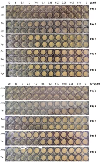
Fig. 2.Susceptibility ofT.rubrumto antifungal drugs using the 96-well plate SPOTi assay. The assay was performed in triplicates. Cic, ciclopirox; Nys, nystatin; Amo, amorolfine; Ter, terbinafine.
Table 3
ZOIs of ciclopirox, nystatin, amorolfine and terbinafine againstT.rubrum. Mean ± standard deviation are shown (n= 3).
Antifungal agents Zone of inhibition (cm)
Ciclopirox 5.39 ± 0.31
Nystatin 2.65 ± 0.37
Amorolfine 9.00 ± 0.00
[image:4.595.51.267.692.744.2]2.3.4. Validation of the SPOTi assay using mycelial plugs
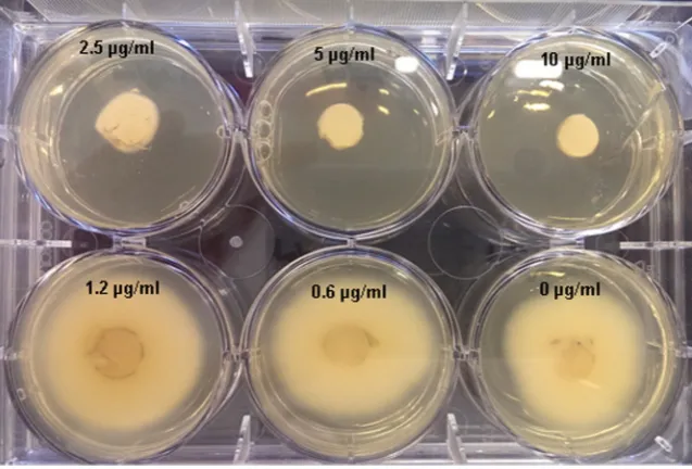
Using ciclopirox as a model drug, the SPOTi assay described above in 96-well plates was validated using a 6-well plate and mycelial plugs of 5 mm diameter. Drug solutions were diluted in 5 ml SDA in each well to produce drug concentrations of: 10, 5, 2.5, 1.2 and 0.6μg/mL. In the last well, the pure solvent (DMSO) was used to ensure that it does not negatively affect fungal growth.T.rubrummycelial plugs of 0.6 mm di-ameter were then placed in the centre of each well and incubated at 32 °C. This experiment was performed in triplicates. An additional 6-well plate was used as a growth control where no antifungals or sol-vents were mixed with SDA.
3. Results and discussion
3.1. MICs determined by SPOTi, ZOI and mycelial plugs
With the modified SPOTi assay, dermatophyte growth was assessed over a 6 day period after inoculation the MICs (Table 2.) of the antifun-gal drugs could be reliably determined at day 3, as shown by photo-graphic images shown inFig. 2forT.rubrum. Compared to the disc-diffusion and broth micro-dilution methods, where a 7 day incubation period is recommended (Norris et al., 1999), SPOTi offers a significant advantage and ease of performance.
Encouragingly, the results from the SPOTi, quantitative in nature, agreed with the qualitative ones obtained from the disc-diffusion tests (a qualitative analysis) that were performed alongside to validate the former approach, i.e. a higher potency of amorolfine and terbinafine which gave larger ZOI at lower concentrations, compared to ciclopirox and nystatin (Table 3).
Taking ciclopirox as a model drug, the SPOTi assay was compared with the traditional method of inoculation using mycelial plugs (Wright et al., 1983) (Fig. 3), where the MIC of ciclopirox was found to be 2.5μg/mL (Fig. 3). This is the same as that determined with SPOTi (Table 2) and establishes the fact that, in spite of using only the conidia of the fungi in the SPOTi assay, the MIC determination by SPOTi is representative of all forms of the pathogen-hyphae and spores, thereby displaying the robustness and reproducibility of the assay.
In the absence of a dermatophytes' susceptibility assessment stan-dard, disc-diffusion method is still widely used primarily due to its con-venience. The major critique of this method arises from the fact that when a disc containing a given amount of an antifungal is placed on
an agar plate, there follows a slow release of the drug out of the disc, into the agar, followed by its diffusion within the agar over time. This is likely to form a concentration gradient with higher drug concentra-tions found near the drug-loaded disc. Thus dermatophytes further away from the disc will be exposed to a lower drug concentration. Fur-thermore, the interpretation of zone of inhibition (ZOI) can often be subjective, with definitions of measured areas ranging from“area of no growth”to“areas up to normal growth”. Finally, ZOI does not allow the direct comparison of various agents in terms of MIC; and is only qualitative even when areas of inhibitions are reported, as these values cannot be used to determine MIC. For example, an area of more than 20 mm is indicative of susceptibility when testing terbinafine but only an area of 15 mm is indicative of susceptibility with nystatin (Rosco Diagnostica Ltd, 2007/2008). Nonetheless, comparison of this method with the broth micro-dilution assay has resulted in favourable correla-tion with some antifungal agents, where an increase of MIC is accompa-nied by a decrease of inhibition zone diameters (National Committee for Clinical Laboratory Standards, 1997; National Committee for Clinical Laboratory Standards, 2002), as is also seen through this work (Tables 2 and 3).
With the SPOTi assay, more quantitative results are obtained through direct observation. Drug concentrations resulting in‘no fungal growth’are identified as the MIC. The main advantage of this assay, com-pared to the disc diffusion method, is that the concentration of the drugs that fungal colonies are exposed to is uniform in each well as the drug is mixed thoroughly in the agar. Hence, SPOTi allows for quantitative mea-surement of MICs.
4. Conclusions
[image:5.595.143.462.54.270.2]We have shown SPOTi to be a rapid, reliable and quantitative sus-ceptibility assay for dermatophytes. The encouraging results attest to the use of this assay on other fungi, for antifungal susceptibility testing as well as for the screening of novel antifungal agents and it may prove to be a gold-standard susceptibility assay for fungi. SPOTi has al-ready proven to be a success story in the identification of anti-mycobacterials from varied sources such as natural, synthetic and has recently been instrumental in repurposing drugs (Guzman et al., 2013). We expect this success to translate effortlessly when using fungi, and endeavour to adapt this convenient, effective technique for other types of fungal pathogens.
Funding
This project was partly funded by EPSRC, grant code EP/I009221/1.
Competingfinancial interests
The authors declare no competingfinancial interests.
Acknowledgments
This work was conducted during the efficacy testing of anti-fungal nail patches, and we acknowledge Prof A Kinloch, Dr M Charalambides and Dr Idris Mohammed, from Imperial College London, who worked on the mechanical properties of nail patches. Marie-josee Maugueret-Minerve, Microbiology Research Unit manager, is also thanked for her technical help.
References
Araujo, C.R., Miranda, K.C., Fernandes, O.D.L., Soares, A.J., Silva, M.D.R., 2009.In vitro sus-ceptibility testing of dermatophytes isolated in Goiania, Brazil, againstfive antifungal agents by broth microdilution method. Rev. Inst. Med. Trop. Sao Paulo 51, 9–12.
Arrese, J.E., Pierard, G.E., 2003.Treatment failures and relapses in onychomycosis: a stub-born clinical problem. Dermatology 207, 255–260.
Bradley, M.C., Leidich, S., Isham, N., Elewski, B.E., Ghannoum, M.A., 1999.Antifungal sus-ceptibilities and genetic relatedness of serialTrichophyton rubrumisolates from pa-tients with onychomycosis of the toenail. Mycoses 42 (Suppl. 2), 105–110.
Castro, C., Serrano, M., Valverde, A., Pemán, J., Almeida, C., Martín-Mazuelos, E., 2007.
Comparison of the Sensititre YeastOne colorimetric antifungal panel with the modi-fied Clinical and Laboratory Standards Institute broth microdilution (M38-A) method for antifungal susceptibility testing of dermatophytes. Chemotherapy 54, 427–430.
Evangelopoulos, D., Bhakta, S., 2010.Rapid methods for testing inhibitors of mycobacteri-al growth. Methods Mol. Biol. 642, 193–201.
Ghannoum, M.A., Arthington-Skaggs, B., Chaturvedi, V., Espinel-Ingroff, A., Pfaller, M.A., Rennie, R., Rinaldi, M.G., Walsh, T.J., 2006.Interlaboratory study of quality control iso-lates for a broth microdilution method (modified CLSI M38-A) for testing susceptibil-ities of dermatophytes to antifungals. J. Clin. Microbiol. 44, 4353–4356.
Gupta, A., Bhakta, S., 2012.An integrated surrogate model for screening of drugs against
Mycobacterium tuberculosis. J. Antimicrob. Chemother. 67, 1380–1391.
Guzman, J.D., Evangelopoulos, D., Gupta, A., Birchall, K., Mwaigwisya, S., Saxty, B., Mchugh, T.D., Gibbons, S., Malkinson, J., Bhakta, S., 2013.Antitubercular specific activity of ibu-profen and the other 2-arylpropanoic acids using the HT-SPOTi whole-cell phenotyp-ic assay. BMJ Open 3.
Hall, L., Jude, K.P., Clark, S.L., Dionne, K., Merson, R., Boyer, A., Parrish, N.M., Wengenack, N.L., 2012.Evaluation of the Sensititre MycoTB plate for susceptibility testing of the
Mycobacterium tuberculosiscomplex againstfirst- and second-line agents. J. Clin. Microbiol. 50, 3732–3734.
Karaca, N., Koc, A.N., 2004.In vitro susceptibility testing of dermatophytes: comparison of disk diffusion and reference broth dilution methods. Diagn. Microbiol. Infect. Dis. 48, 259–264.
Macura, A.B., 1993.In vitro susceptibility of dermatophytes to antifungal drugs: a compar-ison of two methods. Int. J. Dermatol. 32, 533–536.
Marques, S.A., Robles, A.M., Tortorano, A.M., Tuculet, M.A., Negroni, R., Mendes, R.P., 2000.
Mycoses associated with AIDS in the Third World. Med. Mycol. 38 (Suppl. 1), 269–279.
Mendez, C.C., Serrano, M.C., Valverde, A., Peman, J., Almeida, C., Martin-Mazuelos, E., 2008.
Comparison of E-Test, disk diffusion and a modified CLSI broth microdilution (M 38-A) method for in vitro testing of itraconazole,fluconazole and voriconazole against dermatophytes. Med. Mycol. 46, 119–123.
National Committee For Clinical Laboratory Standards, 1997.Reference method for broth dilution antifungal susceptibility testing Of yeasts. Approved Standard NCCLS Docu-ment M27-A. Wayne: National Committee for Clinical Laboratory Standards.
National Committee For Clinical Laboratory Standards, 2002.Reference method for broth dilution antifungal susceptibility testing offilamentous fungi. Approved Standard CLSI Document M38-A. Wayne: Committee for Clinical Laboratory Standards.
Norris, H.A., Elewski, B.E., Ghannoum, M.A., 1999.Optimal growth conditions for the de-termination of the antifungal susceptibility of three species of dermatophytes with the use of a microdilution method. J. Am. Acad. Dermatol. 40, S9–S13.
Nweze, E.I., Mukherjee, P.K., Ghannoum, M.A., 2010.Agar-based disk diffusion assay for susceptibility testing of dermatophytes. J. Clin. Microbiol. 48, 3750–3752.
Rex, J.H., Pfaller, M.A., Rinaldi, M.G., Polak, A., Galgiani, J.N., 1993.Antifungal susceptibility testing. Clin. Microbiol. Rev. 6, 367–381.
Rosco Diagnostica Ltd, 2007/2008.User's Guide NEO-SENSITABS Susceptibility Testing. 19th ed Rosco Diagnostica Ltd, Denmark.
Sharma, M., Sharma, M., 2011.Influence of culture media on mycelial growth and sporulatin of some soil dermatophytes compared to their clinical isolates. J. Microbiol. Antimicrob. 3, 196–200.
Sharma, M., Sharma, M., Chandra, S., 2012.Influence of temperature and relative humid-ity on growth and sporulation of some common dermatophytes. Indian J. Fundam. Appl. Life Sci. 2, 1–6.
Weitzman, I., Summerbell, R.C., 1995.The dermatophytes. Clin. Microbiol. Rev. 8, 240–259.
Wiegand, I., Hilpert, K., Hancock, R.E., 2008.Agar and broth dilution methods to deter-mine the minimal inhibitory concentration (MIC) of antimicrobial substances. Nat. Protoc. 3, 163–175.
Wright, L., Scott, E., Gorman, S., 1983.The sensitivity of mycelium, arthrospores and microconidia ofTrichophyton mentagrophytesto imidazoles determined by in-vitro tests. J. Antimicrob. Chemother. 12, 317–327.