Transforming Growth Factor+ in Psoriasis
Pathogenesis and Therapy
JAMES
T.
ELDER, LARRY R. ELLINGSWORTH,‘ GARY J. FISHER, AND JOHN J. VOORHEESDepartment of Dermatology University of Michigan Ann Arbor, Michigan 48109
“Immunology Laboratory Connective Tissue Research Laboratories
Collagen Corporation Palo Alto, California 94303
INTRODUCTION
Psoriasis is a common skin disease, affecting 1 to 2 % of the U.S. population at a cost of some 1.5 billion dollars per year.’ Psoriasis is characterized by marked in- creases in keratinocyte proliferation? abnormal patterns of keratinocyte differentia- tion? prominent alterations in dermal capillary vasculature~ and the presence of dermal and epidermal T cells, monocytehacrophages, and polymorphonuclear leukocytes?.6 We have recently shown that expression of transforming growth factor-a (TGF-a) is markedly increased in psoriatic lesions: This result, coupled with the fluctuating clin- ical course of psoriasis, suggests that the molecular pathogenesis of psoriasis may in- volve unstable or altered regulation of TGF-a and possibly other epidermally derived cytokines, such as interleukin-6 (IL-6).8
Transforming growth factor-p (TCF-P) acts synergistically with TCF-a to promote anchorage-independent growth of certain cell types? However, it has potent antiprolifer- ative effects upon a variety of epithelial cell types in vitro,’O including keratinocytes.“ In human keratinocytes, TGF-P causes a reversible inhibition of proliferation, predom- inantly in the GI phase of the cell cycle.I2 On the basis of these results, it has been hypothesized that TGF-p functions as a negative growth regulator in normal skin.ll,12 Given the marked keratinocyte hyperproliferation characteristic of psoriasis, it is pos- sible that psoriatic keratinocytes could be deficient in either the production of or re- sponsiveness to TGF-p, resulting in a deficient state of growth regulation. We have tested this hypothesis in several different ways with respect to TGF-P1, the first member of the TGF-0 gene family to be molecularly cloned and biochemically characterized. First, we have compared the expression of TGF-01 mRNA in normal and psoriatic epidermis by Northern blotting. These results confirm those already reported using a slot blot procedure? Second, we have compared the responsiveness of keratinocytes cultured from the skin of normal individuals and from psoriatic lesions to purified
TCF-pl. The responses studied include TGF-p1-induced alterations in c-myc and plas- minogen activator inhibitor-1 (PAI-1) mRNA levels as well as the proliferative response.
ELDER er 01.: TGFO IN PSORIASIS 219
Finally, we have characterized the keratinocyte TGF-pl receptor by Scatchard analysis and V3F-pl crosslinking, and compared the receptors of normal and psoriatic ker- atinocytes. The results indicate that TGF-PI is capable of acting directly on psoriatic as well as normal keratinocytes, and suggest that TGF-P could be an effective anti- psoriatic agent.
MATERIALS AND METHODS
Bone-derived KF-pl was purified from the noncollagenous, guanidine-HCI-soluble proteins of demineralized bovine bone as previously de~cribed!~ For binding and receptor crosslinking studies, TGF-pl was radiolabeled with 1 mCi of Nalz51 (15 mCi/pg, Amersham) as previously de~cribed!~
Keratinocyte Culture
Primary cultures of human keratinocytes were prepared by trypsinization of kera- tome or punch biopsies of skin removed from normal or psoriatic volunteers under lidocaine anesthesia. The procedures used ha e been described previou~Iy.'s-~~ Cul- tures were propagated in Keratinocyte Grow ti! Medium (KGM, Clonetics, Boulder, C O ) , a modification of MCDB 153 defined medium,'6 optimized for high density ker- atinocyte growth, and used in the second to fifth passage. For assays of keratinocyte proliferation, cells were seeded in KGM at lo5 cells/60-mm dish (Falcon) and allowed to attach overnight. TGF-pl (1 ng/ml) or diluent (0.1 M HCI, 1 mg/ml BSA) was then added and cells were trypsinized and counted at the indicated intervals using a hemacytometer.
R NA Isolation and Analysis
Keratinocytes (2-4 X lo6 cells per 100-mm dish) were treated with TGF-Pl(1 ng/ml) or recombinant human y-interferon (y-IFN, Genentech, 100 U/ml) 24 h after feeding with KGM. After 3 h, cells were lysed with 4 M guanidinium isothiocyanate, 5 mM sodium citrate, pH 7.0, 100 mM beta-mercaptoethanol, 0.5 % sodium sarcosinate, and RNA was isolated by overnight centrifugation at 100,000 x g over 5.7 M CsCI, 100 mM EIYIA, pH
7.0.17
For extraction of RNA from keratome biopsies of normal and psoriatic epidermis, biopsies were immediately frozen in liquid nitrogen after removal and stored at -70°C. The frozen biopsies were finely pulverized in liquid nitrogen prior to addition of guanidinium isothiocyanate buffer, then homogenized in a Polytron tissue grinder (Brinkmann). RNAs were quantitated by absorbance at 260 nm, and equal quantities of total RNA were size-fractionated on 1 % formaldehyde-agarose gels.'s RNAs were blotted onto derivatized nylon membranes (Zeta-probe, Bio-Rad) using220 ANNALS NEW YORK ACADEMY OF SCIENCES
hydrogenase cDNA plasmids were obtained through the American Type Culture Col- lection. A lipocortin I1 cDNA probe was used for normalization of normal and psori- atic epidermal RNA loading and inta~tness?~
ZGF-Pl Binding and Receptor Crosslinking Assays
Specific binding of radioiodinated TGF-pl to cultured keratinocytes was determined as previously described?',22 Briefly, cell monolayers were rinsed several times in ice- cold binding buffer (Dulbecco's modified Eagle's medium containing 25
m M
HEPES, pH 7.4, 0.1% BSA) followed by incubation with the indicated concentrations of 1251- labeled TGF-PI at 4°C for 3 to 4 h with gentle agitation. Cells were rinsed four times in ice-cold Hanks buffered salt solution containing 0.1% BSA, and solubilized in 1% Triton X-100, 10% glycerol, 0.01 % BSA, 20 mM HEPES, pH 7.4. Bound and unbound TGF-01 was then determined using a gamma counter. Nonspecific binding, determined by addition of 10 nM unlabeled TGF-PI, was subtracted from the total bound radioactivity. 1251-labeled TGF-PI was crosslinked to receptors on keratinocyte monolayers using disuccinimidyl suberate (DSS, Pierce) exactly as previously describedJ2TOTAL
RNA
[image:3.432.77.355.267.539.2]NORMAL
I
UNINV 1 PSORIATICELDER et al.: TGFO IN PSORIASIS 221
RESULTS
[image:4.432.97.347.277.546.2]In FIGURE 1, TGF-Pl steady-state mRNA levels in normal epidermis as well as in normal-appearing (uninvolved) and lesional psoriatic epidermis have been compared by RNA blot hybridization. A predominant band of approximately 2.4 kilobases (kb) is detected in all three sample types. Moreover, as reported previously using a slot- blotting technique! the intensity of the 2.4 kb band is similar in all three groups, al- though some variation within each group is seen (upper panel). The lipocortin I1 con- trol gene probe detects a 1.6 kb transcript at comparable intensities in all three groups (lower panel).
FIGURE 2 depicts the responses of several keratinocyte mRNA transcripts to treat- ment of cultures derived from normal epidermis or active psoriatic lesions with puri- fied TGF-Pl (1 ngiml; 40 pM). For a comparison, parallel dishes were treated with another cytokine known to inhibit keratinocyte proliferation, y-IFN (100 U/ml). In each instance, TGF-Pl caused substantial decreases in c-myc mRNA 3 h after treatment (top panel). These results could not be accounted for by a nonspecific toxic effect, since re-hybridization of the same blots with the PAL1 probe revealed markedly increased PAL1 mRNA levels (second panel). Repeated hybridizations with the TGF-PI probe
NORMAL KC
PSORIATIC
KC
p
= TGFP1, lng/rnl, 3hrsY = Y IFN, 1000 U/ml, 3hrs
222 ANNALS NEW YORK ACADEMY OF SCIENCES
nlm
NORMAL (N=6)
0 9 .
PSORIATIC (N=6)
T
F-13
( 1
n n / m l \ /0
1
2
3
4
5
6
[image:5.432.78.344.60.292.2]DAY
FIGURE 3. Proliferative responses of normal and psoriatic keratinocytes to 'IGF-pl treatment.
(third panel) and the GAD probe (bottom panel) revealed no effects on mRNA tran- script levels, confirming the normalization of mRNA loading and demonstrating a lack of TGF-P autoinduction. No induction of PAI-1 mRNA was seen 3 h after IFN-y treat- ment, a!though there was a trend towards reduction of c-myc mRNA transcript levels in response to IFN-y in both normal and psoriatic groups.
The autoradiograms depicted in FIGURE 2 were quantitated by laser densitometry. TGF-gl reduced c-myc mRNA levels to 27.9 & 3.9% of control levels in the normal group, and 21.4% of control in the psoriatic group. In contrast, PAI-1 mRNA levels were increased to 1,133% of control levels in the normal group, and 786% of normal in the psoriatic group. In neither case was the difference between the normal and psori- atic groups statistically significant, as judged by Student's t test using a two-tailed hy- pothesis (p
>
0.1).FIGURE 3 compares the proliferative responses of keratinocyte cultures derived from six normal individuals and lesional epidermis of six psoriatic patients to purified TGF- pl (1 nglml, 40 pM). Proliferation was assayed by cell counting rather than [3H]thymi- dine incorporation, avoiding potential problems of interpretation due to keratinocyte catabolism of thymidine?* TGF-PI caused a marked (four to fivefold) and significant (p
<
0.01) inhibition of proliferation was observed 6 days after treatment in both the normal and psoriatic keratinocytes. However, there was no evident or statistically sig- nificant (p>
.Ol) difference between the antiproliferative responses of the two groups (compare solid lines, FIG. 3).ELDER et 01.: TGF-p IN PSORIASIS 223
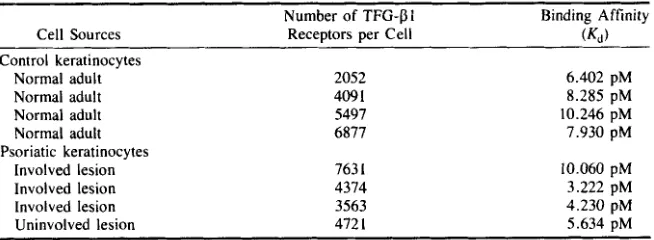
dinated TGF-f!l by Scatchard analysis. FIGURE 4 displays the binding data obtained from representative individual cultures of keratinocytes derived from normal (upper panel), uninvolved psoriatic (middle panel), and involved psoriatic epidermis (lower panel). Similar data were used to calculate binding constants
(Kd)
and the number of sites per cell for an additional three normal and two involved psoriatic keratinocyte cultures. These results are summarized in TABLE 1. The data indicate that normal, uninvolved, and lesional keratinocytes are similar in terms of binding affinities and number of binding sites per cell for TGF-f!l.Three forms of the TGF-f!l receptor that differ in molecular weight have been de- tected in fibroblasts and other cell types by crosslinking 1251-labeled ‘TGF-f! to the receptor with disuccinimidyl suberate (DSS) We have applied this technique to keratino- cytes to determine whether normal and psoriatic keratinocytes differ in terms of receptor molecular weight profiles (FIG. 5). Normal and psoriatic keratinocytes displayed an unusual receptor profile, displaying a closely spaced triplet of bands corresponding to receptor-TGF-pl complexes in the 65 K molecular weight range (lanes 1 and 3, FIG. 5). In comparison, 3T3 fibroblasts displayed bands corresponding to receptor com- plexes at 65, 90, and 280 K (lane 5) as previously reported?1,22 All bands were effec- tively competed by addition of excess (20 nM) unlabeled TGF-f!l to the crosslinking reaction (lanes 2 , 4 , and 6). However, there was no clear difference between the receptor crosslinking patterns of normal and psoriatic keratinocytes. Increasing concentrations of unlabeled TGF-pl reduced labeling of all three bands of the triplet to a similar ex- tent, indicating that each molecular weight form of the keratinocyte receptor bound TGF-01 with comparable efficiency (FIG. 6).
DISCUSSION
Increased keratinocyte proliferation in psoriasis2 could be due either to increased production of or sensitivity to factor(s) that stimulate growth, or to decreased produc- tion of or sensitivity to factor(s) that inhibit growth. We have shown markedly increased expression of TGF-a in psoriatic lesions, while expression of TGF-f!l mRNA was similar in normal epidermis and psoriatic lesions? Recently, Mansbridge and coworkers have reported focally increased expression of TGF-Pl in uninvolved and lesional psoriatic epidermis by immunocytochemical techniques?4 Therefore, we have extended our pre- viously reported studies to confirm by Northern blot analysis the similarity of TGF-Pl
expression in normal and psoriatic skin (FIG. 1) and cultured keratinocytes (FIG. 2, third panel). Taken together, these results indicate that the increased TGF-f!l protein ob- served in psoriatic skin by Mansbridge and coworkers may be due to deposition of TGF-01 synthesized elsewhere, differential activation of latent epidermally derived TGF-f!l precursor^?^ localized variations in TGF-f!l RNA not detectable by our tech- niques, or altered regulation of TGF-f!l translation in psoriatic skin. Moreover, it re- mains possible that other members of the TGF-f! gene family26-28 may be abnormally expressed in psoriatic skin.
224 ANNALS NEW YORK ACADEMY OF SCIENCES
ELDER et al.: TGF-p IN PSORIASIS 225
TABLE 1. Transforming Growth Factor-f3l Receptor Binding and Affinity on Normal and Psoriatic (Lesionall Keratinocvtes
Number of TFG-PI Binding Affinity
Cell Sources Receptors per Cell (KI)
Control keratinocytes Normal adult Normal adult Normal adult Normal adult
Involved lesion Involved lesion Involved lesion Uninvolved lesion Psoriatic keratinocytes
2052 409 1
5497 6877
763 1
4374 3563 472 I 6.402 pM 8.285 pM 10.246 pM 7.930 pM 10.060 pM 3.222 pM 4.230 pM 5.634 pM
to markedly induce the expression of PAI-1 mRNA in human carcinoma, glioblastoma, and fibroblast cell line^?^.^' However, this is the first report of such a response in a human primary culture system (FIG. 2, second panel). Neither this response nor the reduction of c-myc mRNA after WF-Pl treatment was significantly different in normal and psoriatic keratinocytes, indicating that both cell types are comparably responsive to TGF-Bl at the concentration tested (1 ng/ml, 40 pM).
As shown by Wilke, Pittelkow, and coworkers?Z TGF-P induces a reversible state of growth arrest without induction of terminal differentiation in cultured neonatal human keratinocytes. We have extended these results to adult human keratinocytes derived from normal and psoriatic epidermis (FIG. 3), and find that normal and psoriatic cells do not differ in antiproliferative responsiveness to TGF-Pl (1 ng/ml, 40 pM).
Consistent with these findings, Scatchard analysis of TGF-f31 binding revealed a comparable range of values for binding constants (Kd) and numbers of binding sites per cell for normal and psoriatic keratinocytes (FIG. 4, TABLE 1). The Kd values are substantially higher and the numbers of sites per cell are substantially lower than those reported for neonatal foreskin keratinocytes by Shipley and coworkers (428 pM, 35,000 sitedcell).' I However, these differences may be accounted for by the fact that the latter
experiments were performed at room temperature, while our experiments were per- formed at 4°C.
Receptor crosslinking studies using Iz5I-labeled WF-Pl and DSS yielded the unex- pected result that both normal and psoriatic keratinocytes displayed a prominent triplet of bands at approximately 65 kD, and very little signal at 90 and 280 kD as observed in 3T3 fibroblasts (FIG. 5). Unlabeled TGF-Pl competed equally well for crosslinking of all three bands of the 65 kD triplet (FIG. 6 ) . suggesting that each form of the 53 kD receptor bind TGF-Pl with comparable affinity. Our finding that keratinocytes ex- press the low molecular weight form(s) of the receptor is consistent with its recent identification as the receptor conferring growth inhibition in mink lung epithelial ~ e l l s . 3 ~ This form of the TGF-P receptor has 10- to 30-fold higher affinity for TGF-01 than for TCF-P2.34 Thus, it is puzzling that cultured keratinocytes and mink lung epithelial cells display very similar growth inhibitory responses to TGF-PI and -/32 (M. Pittelkow, personal comm~nication)?~
[image:8.432.54.381.81.201.2]226 ANNALS NEW YORK ACADEMY OF SCIENCES
[image:9.432.80.356.57.476.2]BINDING
PROTEIN
FIGURE 5. Receptor crosslinking analysis of keratinocyte and fibroblast E F - p l receptors. Light- colored line at 94 kD is an artifact.
sive to TGF-PI as are normal keratinocytes, and that TGF-pl can have a potent and direct antiproliferative effect on psoriatic keratinocytes. These results imply that T3F-p
ELDER et 01.: TGFP IN PSORIASIS 221
Unlabeled
TGF-P1
(nM)
kD
205
-
116
94
67
43
29
[image:10.432.75.366.59.386.2]0
.1
.25
.5 .75
1
2.5
5
7.5
10
FIGURE 6. Competition of TGF-PI receptor crosslinking with unlabeled TGF-PI.
cated by the potential for pleiotypic effects on a variety of cell types, and by its rapid hepatic clearance.)6 Further work on the use of penetration enhancers to promote epidermal delivery of TGF-P is clearly warranted.
Finally, it is very important that the proliferative response of psoriatic epidermis may be intimately related to the presence of epidermal and dermal T cells, monocytes, and macrophages in psoriatic lesions?.6 The dramatic response of psoriasis to low doses of cyclosporin A (CsA) underscores the potential role of T cells in this process, since CsA inhibits T cell function at doses far lower than those required to suppress keratinocyte proliferation directly in vitro?7 Thus, CsA inhibits T cell release of y- IFN, which we have recently shown to be capable of inducing
TGF-a
in keratinocytes." TGF-P is a potent inhibitor of IL-1-independent thymocyte p r o l i f e r a t i ~ n ~ ' . ~ ~ and IL-228 ANNALS NEW YORK ACADEMY OF SCIENCES
[NOTE ADDED IN PROOF: A 2kb PAI-1 transcript was detected in normal and psoriatic epidermal RNA by blot hybridization. PAIN mRNA levels were not significantly increased or decreased in psoriatic lesions. Since PAI-I is strongly induced by X F - P l in keratinocytes in vifro, these results suggest that TGF-Dl activity is not increased or decreased in psoriatic epidermis.]
REFERENCES
I . KRUEGER, G . G . , P. R. BERGSTRESSER, N. LOWE, J. J. VOORHEES & G. D. WEINSTEIN. 1984. Psoriasis. J. Amer. Acad. Dermatol. 11: 937-947.
2. WEINSTEIN, G. D. & E. J. VAN SCOTT. 1965. Autoradiographic analysis of turnover times of normal and psoriatic epidermis. J. Invest. Dermatol. 45: 257.
3. BERNARD, B. A,, D. ASSELINEAU, L. SCHAFFAR-DESHAYES& M. Y. DARMON. 1988. Abnormal sequence of expression of differentiation markers in psoriatic epidermis: inversion of two steps in the differentiation program? J. Invest. Dermatol. 90: 801-805.
4 . BRAVERMAN, I . M., & J. SIBLEY. 1982. Role ofthe microcirculation in the treatment and patho- genesis of psoriasis. J. Invest. Dermatol. 78: 12-17,
5. BAKER, B. S., A. F. S W A I N , H. VALDIMARSSON & L. FRY. 1984. T-cell subpopulations in the blood and skin of patients with psoriasis. Br. J. Dermatol. 110: 37-44.
6. H A M M A R , H., S . - Q . Gu, A. JOHANNESSON, K.-G. SUNDKVIST & P. BIBERFELD. 1984. Sub- populations of mononuclear cells in microscopic lesions of psoriatic patients. Selective ac- cumulation of suppressorkytotoxic T cells in epidermis during the evolution of the lesion. J. Invest. Dermatol. 83: 416-420.
7. ELDER, J. T., G. J . FISHER, P. B. LINDQUIST, G. L. BENNETT, M. R. PITTELKOW, R. J. COFFEY, L. ELLINGSWORTH, R. DERYNCK & J . J. VOORHEES. 1989. Overexpression of transforming growth factor a in psoriatic epidermis. Science 243: 811-814.
8. GROSSMAN, R. M., J . KRUEGER, D. YOURISH, A. GRANELLI-PIPERNO, D. P. MURPHY, L. T . MAY, T. S. KUPPER, P. B. SEHCAL & A. B. GOTTLIEB. 1989. Interleukin 6 is expressed in high levels in psoriatic skin and stimulates proliferation of cultured human keratinocytes. Proc. Natl. Acad. Sci. USA 86: 6367-6371.
9. SPORN, M. B., A. B. ROBERTS, L. M. WAKEFIELD& R. K. ASSOIAN. 1986. Transforminggrowth factor-6: Biological function and chemical structure. Science 233: 532-534.
10. KESKI-OJA, J. & H. L. MOSES. 1987. Growth inhibitory polypeptides in the regulation of cell proliferation. Med. Biol. 65: 13-20.
I I . SHIPLEY, G. D., M. R. PITTELKOW, J. J. WILLE, R. E. SCOTT& H. L. MOSES. 1986. Reversible inhibition of normal human prokeratinocyte proliferation by type
6
transforming growth factor- growth inhibitor in serum-free medium. Cancer Res. 46: 2068-2071.12. WILKE, M. S., B. M. Hsu, J . J. WILLE, M. R. PITTELKOW & R. E. SCOTT. 1988. Biologic mechanisms for the regulation of normal human keratinocyte proliferation and differentiation. Am. J. Pathol. 131: 171-181.
13. ELLINGWORTH, L. R., J. E. BRENNAN, K. FOK, D. M. ROSEN, H. BENTZ, K. A. PIEZ & S. M. SEYEDIN. 1986. Antibodies to the N-terminal portion of cartilage-inducing factor A and trans- forming growth factor 6. J. Biol. Chem. 261: 12362-12367.
FROLIK, C . , L. WAKEFIELD, D. SMITH & M. SPORN. 1984. Characterization of a membrane receptor for transforming growth factor$ in normal rat kidney fibroblasts. J. Biol. Chem.
15. WILLE, J . J., M. R. PITTELKOW, G. R. S H I P L E Y & R. E. SCOTT. 1984. Integrated Control of growth and differentiation of normal human prokeratinocytes cultured in serum-free medium: clonal analyses, growth kinetics. and cell cycle studies. J. Cell. Physiol. 121: 31-44. 16. BOYCE, S. T. & R. G. HAM. Normal human epidermal keratinocytes. In In Vitro Models for
Cancer Research. Vol. 111. M. M. Webber & L. I. Sekely, Eds.: 245-274. CRC Press. Boca Raton, FL.
17. NICKOLOFF. B. J., R. S. MITRA, J . T. ELDER, G. J . FISHER & J . J. VOORHEES. 1989. Decreased growth inhibition by recombinant gamma interferon is associated with increased transforming growth factor-a production in keratinocytes cultured from psoriatic lesions. Br. J. Dermatol. 14.
259: 10995-11000.
ELDER et al.: TGFD IN PSORIASIS 229 18. 19. 20. 21. 22. 23. 24. 25. 26. 27. 28. 29. 30. 31. 32. 33. 34. 35. 36. 37. 38.
ELDER, J . T., A. K. GUPTA, G . J. FISHER & J. J. VOORHEES. 1988. Cyclosporine inhibits or- nithine decarboxylase gene expression and acute inflammation in response to phorbol ester treatment of hairless mouse skin. Transplant Proc. 20: 95-104.
ROBERTS, M. B. SPORN & D. V. GOEDDEL. 1985. Human transforming growth factor-p com- plementary DNA sequence and expression in normal and transformed cells. Nature 316 701-705.
K. DANO, R. V. LEBO & T. D. GELEHRTER. cDNA cloning of human plasminogen activator- inhibitor from endothelial cells. J Clin Invest 78: 1673-1680.
ELLINCSWORTH, L. R., D. N A K A Y A M A , P. SEGARINI, J. DASCH, P. C A R R I L L O & W. WAECELL. Transforming growth factor-es are equipotent growth inhibitors of interleukin-I-induced thymo- cyte proliferation. Cell. Immun. 114: 41-54.
KIMCHI, A., X.-F. WANG, R. A. WEINBERG, S. CHEIFETZ & J. MASSAGUB. 1988. Absence of TGF-b receptors and growth inhibitory responses in retinoblastoma cells. Science 240:
SCHWARTZ, P. M., L. C. KUCELMAN, Y. COIFMAN, L. M. HOUGH & L. M . MILSTONE. 1988. Human keratinocytes catabolize thymidine. J. Invest. Dermatol. 90: 8-12.
KANE, C. J. M., P. C. HANAWALT, A. M. KNAPP& J. N. MANSBRIDGE. 1989. Immunohistochem- ical localization of transforming growth factor$ protein in psoriasis. J. Invest. Dermatol. 92A: 455.
WAKEFIELD, L. M., D. M. SMtTH, K. C. FLANDERS & M. B. SPORN. 1988. Latent transforming growth factor-6 froin human platelets. J. Biol. Chem. 263: 7646-7654.
MASSAGUE, J. 1987. The TGF-0 family of growth and differentiation factors. Cell 49: 437-438. DERYNCK, R., P. B. LINDQUIST, A. LEE, D. WEN, 1. T A M M , J. L. GRAYCAR, L. RHEE, A . J .
MASON, D. A. MILLER, R. 1. COFFEY, H. L. MOSES & E. Y. C H E N . 1988. A new type of
transforming growth factor-p, TGF-p3. EMBO J. 7: 3737-3743.
JAKOWLEW, S . B., P. J. DILLARD, M. B. SPOKN & A. B. ROBERTS. 1988. Complementary deox- yribonucleic acid cloning of a messenger ribonucleic acid encoding transforming growth factor
p4 from chicken embryo chondrocytes. Mol. Endo. 2: 1186-1195.
COFFEY, R. J., C. C. BASCOM, N. 1. SPIES, R. GRAVES-DEAL, B. E. WEISSMAN & H. L. MOSES. 1988. Selective inhibition of growth-related gene expression i n m u r k keratinocytes by trans- forming growth factor p. Mol. Cell Biol. 8: 3088-3093.
HELSETH, E . , A. DALEN, G. UNSGAARD, T . SKANDSEN, J. GRONDAHL-HANSEN & L. R. L U N D . 1988. Transforming growth factor-p1 is a potent inducer of plasminogen activator inhibitor type-1 in human glioblastoma and carcinoma cell lines. Acta Pathol. Microbiol. Immunol. Scand. 9 6 845-849.
KESKI-OJA, J., R. RAGHOW, M. SAWDEY, D. J. LOSKUTOFF, A. E. POSTLETHWAITE, A. H. KANG & H. L. MOSES. 1988. Regulation of mRNAs for type-I plasminogen activator inhib- itor, fibronectin, and type I procollagen by transforming growth factor-0. J. Biol. Chem. 263:
WiLKE, M. S., B. M. Hsu & R. E. SCOTT. 1988. Two subtypes ofreversible cell cycle restriction points exist in cultured normal human kertinocyte progenitor cells. Lab. Invest. 58: 660-666. ELDER, 1. T . , A. TAVAKKOL, S . B. KLEIN, M. E. ZEIGLER, M. WICHA & 1. 1. VOORHEES.
1990. Protooncogene expression in normal and psoriatic skin. J. Invest. Dermatol. 94: 19-25. BOYD, F. T . & J . MASSAGUE. 1989. Transforming growth factor-b inhibition of epithelial cell proliferation linked to the expression of a 53-kDa membrane receptor. J. Biol. Chem. 2 6 4
MUSTOE, T . A ., G. F. PIERCE, A. THOMASON, P. GRAMATES, M. B. SPORN & T. F. DEUEL. 1987. Accelerated healing of incisional wounds in rats induced by transforming growth factor- b. Science 237 1333-1336.
COFFEY, R. J., L. J. KOST, R. M. LYONS, H. L. MOSES & N. F. LARUSSO. 1987. Hepatic pro- cessing of transforming growth factor p in the rat. J. Clin. Invest. 80: 750-757.
FISHER,G. J., E. A. D U E L L , B . J. NICKOLOFF,T. M. ANNESLEY, J . K. KOWALKE, C. N. ELLIS
& J. J. VOORHEES. 1988. Levels of cyclosporin in epidermis of treated psoriasis patients differen- tially inhibit growth of keratinocytes cultured in serum free versus serum containing media. J. Invest. Dermatol. 91: 142-146.
WAHL, S. M . , D. A. H U N T , H. L. WONG, S. DOUGHERTY, N. MCCARTNEY-FRANCIS, L. M. DERYNCK, R., J. A. J A R R E T T , E. Y. C H E N , D. H. E A T O N & J . R. BELL, R. K. ASSOIAN, A. B.
GINSBURG, D., R. ZEHEB, A. Y. YANG, U. M. RAFFERTY, P. A. ANDREASEN, L. NIELSEN,
196-199.
3 I1 1-3 I 15.
230 ANNALS NEW YORK ACADEMY OF SCIENCES
W A H L , L. ELLINGSWORTH, J . A. SCHMIDT, G . HALL, A . B. ROBERTS& M . B. SPORN. Trans- forming growth factor-p is a potent immunosuppressive agent that inhibits IL-I-dependent lym- phocyte proliferation. J. Immunol. 140: 3026-3032.
DERYNCK, M. B. SPORN & A. S. F A U C I . 1986. Production of transforming growth factor 0 by human T lymphocytes and its potential role in the regulation of T cell growth. J. Exp. Med.
NICKOLOFF, B. J. & R. S. MITRA. 1988. Transforming growth factor-beta is a keratinocyte-derived
39. KEHRL, J . H . , L. M. WAKEFIELD, A . B. ROBERTS, S. JAKOWLEW, M. ALVAREZ-MON, R.
163: 1037-1050.
40.