EXPERIMENTALDERMATOLOGY
ISSN 0906-6705
Proprotein convertase expression and
localization in epidermis: evidence for
multiple roles and substrates
Pearton DJ, Nirunsuksiri W, Rehemtulla A, Lewis SP, Presland RB, Dale David J. Pearton1, BA. Proprotein convertase expression and localization in epidermis: evi- Wilas Nirunsuksiri1,3,
dence for multiple roles and substrates. Alnawaz Rehemtulla2,
Exp Dermatol 2001: 10: 193–203.CMunksgaard, 2001 S. Patrick Lewis1,
Richard B. Presland1and Abstract: Specific proteolysis plays an important role in the terminal
dif-Beverly A. Dale1 ferentiation of keratinocytes in the epidermis and several types of proteases
1Departments of Oral Biology, Periodontics, have been implicated in this process. The proprotein convertases (PCs)
Biochemistry, Medicine/Dermatology, University are a family of Ca2π-dependent serine proteases involved in processing and
of Washington, Seattle, WA 98195;2Department activation of several types of substrates. In this study we examined the
of Radiation Oncology, University of Michigan, expression and some potential substrates of PCs in epidermis. Four PCs Ann Arbor, MI;3Present address Dow are expressed in epidermis: furin, PACE4, PC5/6 and PC7/8. Furin is de- AgroSciences LLC, Indianapolis, IN tected in two forms, either with or without the transmembrane domain,
suggesting occurrence of post-translational cleavage to produce a soluble enzyme. In addition the furin active site has differential accessibility in the granular layer of the epidermis relative to the basal layer, whereas antibodies to the transmembrane domain stain both layers. These
find-ings suggest that furin has access to different types of substrates in granu- Key words: epidermal differentiation – lar cells as opposed to basal cells. PC7/8, in contrast, is detected throughout keratinocytes – furin – PACE4 – PC5/PC6 – the epidermis with antibodies to both the transmembrane and active site PC7/PC8 – profilaggrin – Notch-1 and no soluble form observed. A peptide PC inhibitor (dec-RVKR-CMK) Dr Beverly A. Dale, Dept of Oral Biology, inhibits cleavage of Notch-1, a receptor important in cell fate determi- University of Washington, Box 357132, Seattle, nation that is found throughout the epidermis. Profilaggrin, found in WA 98195 7132
the granular layer, is specifically cleaved by furin and PACE4in vitroat Tel.: 206 543 4393 Fax: 206 685 3162 a site between the amino terminus and the first filaggrin repeat. This
e-mail: bdale/u.washington.edu work suggests that the PCs play multiple roles during epidermal
differen-tiation. Accepted for publication 12 January 2001
Introduction
The sequential differentiation of keratinocytes in the epidermis is a complex, highly regulated series of events that results in the formation of the anu-cleate cornified cells of the stratum corneum from the relatively undifferentiated cells of the basal layer. This differentiation process is calcium de-pendent and requires extensive and tightly con-trolled changes in both protein expression and
en-zymatic activities resulting in characteristic
morphological changes of the cells. Proteolytic processing is an important mechanism for many of these events, including signaling, proprotein acti-vation, structural remodeling and desquamation. A number of different proteases have been
iden-tified that play a role in epidermal differentiation. These include calpain I (1), cathepsins C (2, 3), B, H and L (4), profilaggrin endopeptidase (PEP1) (5), the stratum corneum chymotryptic-like tease (SCCP) and the stratum corneum thiol pro-tease (SCTP) (6). The importance of propro-teases in the epidermis is highlighted by the finding that null mutations of cathepsin C result in palmoplantar keratoderma disorders, specifically Haim–Munk and Papillon–Lefevre syndromes (2, 7, 8).
A protease family that has been implicated in processing and differentiation in a number of tissues is the Proprotein Convertase (PC) family.
This is a family of Ca2π-dependent serine
iden-tified; furin (also known as PACE), PC2, PC1/3, PACE4, PC4, PC5/6 and PC7/8/LPC. Of these fur-in, PACE4, PC5/6 and PC7/8 are widely expressed whereas PC2 and PC1/3 are limited to neuro-endo-crine tissues and PC4 is restricted to the testis, sug-gesting both generalized and tissue-specific pat-terns of expression and function. The PC enzymes recognize basic motifs, cleaving after paired basic residues (PC2 and PC1/3) or after a canonical RX(R/K)R motif (furin and PACE4); in some sub-strates a P6 Arg can substitute for the Arg at the P4 position (13). Furin has been shown to process a wide variety of substrates including receptors, growth factors, hormones, plasma proteins, matrix metalloproteinases and extracellular matrix com-ponents. In addition PC family members are also involved in the processing of exogenous proteins such as some viral envelope glycoproteins and bac-terial endotoxins (12).
Several proteins relevant to keratinocyte devel-opment have been shown to be substrates for PC processing or contain potential PC cleavage sites. These include receptors such as Notch-1 (14, 15), matrix components such as collagen XVII (BP180) (16), matrix metalloproteinases, desmosomal com-ponents such as desmoplakin-1 and desmoglein,
integrins including integrins a3 and a6 (17) and
structural proteins such as profilaggrin.
Although PCs have diverse substrates and play a role in differentiation in some systems there has been no systematic study of PC expression in epi-dermis (2). We therefore investigated the express-ion and distributexpress-ion of the members of the PC family in epidermis and examined the processing of several potential substrates.
In this study, 4 members of the PC family (furin, PACE4, PC5/6 and PC7/8) were detected in epider-mis. Furin and PC7/8 were detected in distinct, al-though overlapping, patterns within the epidermis. In addition the active site of furin showed differen-tial accessibility during differentiation. Inhibition of the PC family in culture inhibited the processing
of Notch-1 andin vitroresults characterized a
fur-in/PACE4 processing site in the amino terminus of profilaggrin. The specific localization pattern, cal-cium dependence, and identification of possible substrates are consistent with a role for PCs in multiple events during epidermal differentiation.
Methods
Keratinocyte culture
Human foreskin keratinocytes were cultured using the methods described by Fleckman et al. (18), a modification of the Rheinwald–Green system. Cells were harvested for RNA or protein at either
∂60–70% of confluence, at 100% confluence or
several days post-confluence.
Detection of proprotein convertase expression in skin using degenerate primers
The sequences of the known human PC members (furin, PACE4, PC1, PC2, PC5/6, PC7/8) were ob-tained from Genbank using the ENTREZ server. The sequences were multiply aligned using Clus-talW (19, 20) and a highly conserved region of the catalytic domain was identified to which
degener-ate primers were designed. The sense primer (5ø
-CA(C/T) GG(C/G) AC(A/T/G/C) (A/C) G(A/T/G/
C) TGTGC (A/T/G) GG-3ø) was based on the
se-quence R194HGTRCAG201in furin which contains
the active site arginine (R194). This sequence is
100% conserved across the known human mem-bers of the PC family and highly conserved in
other species. The antisense primer (5ø-GG (T/G)
CCC CAG CT (T/G) (C/G) (A/C) (A/G) CTG
TA-3ø) was based on the sequence I249YSASWG255
which is also highly conserved across the family,
although in PC7/8 the alanine (A252) is replaced
by a cysteine. RT-PCR using these primers would amplify a 187 bp segment from each PC that could be distinguished on the basis of its nucleotide se-quence.
Total RNA was isolated from 5-day post-con-fluent cultured human keratinocytes using the Tri-zol reagent (Life Technologies, Gaithersburg, MA, USA) and the manufacturer’s protocol. After DNase I treatment the total RNA was reverse transcribed using the Superscript II RT system (Life Technologies) and the resulting cDNA was
PCR amplified (40 cycles at 95æC for 30 s; 60æC
for 1 min; 72æC for 1 min) using Taq plus Taq
extender (Promega, Madison, WI, USA). The re-sulting 187 bp product was gel purified and TA
cloned into the pCR3.1 vector (Invitrogen,
Carlsbad, CA, USA).
Northern analysis
RT-PCR analysis of PC5/6 and PC7/8
Oligoprimers specific for PC5/6 and PC7/8/LPC were made according to published sequences
(Gen-bank acc. NM–004716). For PC5/6 the sense
primer used comprised nucleotides 962–982 (5ø
-TCA GCA GCA CTG CAG AAA GC-3ø) and the
antisense primer, nucleotides 1409–1429 (5ø-TGG
GGT CTT CAA CTC GGC TA-3ø), giving a 447
bp product. For PC7/8 the sense primer 163–183
(5ø-GCT GGT AGG CTG TTT CAC CG-3ø) and
667–647 (5ø-GGA TTG CTG CCA GAG AAG
TC-3ø) were used giving a 504 bp product. Reverse
transcription was performed on the total RNA using the Superscript II kit (Life Technologies). PCR analysis was performed using 32 cycles of 94
æC, 1 min; 50æC, 45 s; 72æC, 1 min using Taq
poly-merase (Promega). PCR products were analyzed on a 1% agarose gel and their identity confirmed by sequence analysis.
PC protein expression in epidermis and keratinocytes
Protein from foreskin epidermis and cultured hu-man keratinocytes was extracted using a modified RIPA buffer (50mM HEPES, pH 7.5, 150 mM NaCl, 50 mM NaF, 1% NP-40, 0.5% deoxycholate, 1 mM sodium vanadate, 1 mM
nitrophenylphos-phate, 5 mM benzamidine, 2 mM PMSF, 10mg/ml
aprotonin, 1¿ complete protease inhibitor
(EDTA-free, Boehringer)) (24). The extracted pro-teins were separated on 7.5–12% SDS–PAGE gels
and blotted onto nitrocellulose for western
immunoblot detection using specific antibodies and ECL. Furin was detected using a polyclonal antibody directed against the C-terminal tail (c-fur) (a gift from Ruth Hogue Angeletti, New York University, USA) and monoclonal antibodies raised against catalytic domain (MON148) or cys-teine-rich domain (MON152) epitopes (Alexis Bio-chemicals, San Diego, CA, USA). PC7/8 was de-tected using polyclonal antibodies directed against the catalytic and C-terminal domains (gift from Dr W. J. M. Van de Ven, Katholieke Universiteit Leuv-en, Belgium). PACE4 was detected using an anti-body directed to the pro and catalytic domains (Alexis Biochemicals).
Localization of furin in epidermis
Immunofluorescence was performed on methanol fixed frozen or paraffin embedded methyl-Carnoy’s fixed human epidermis. Carnoy’s fixed tissues were first rehydrated and from then on all sections were treated the same. Antigen unmasking was per-formed on selected sections by incubation with
0.1% trypsin for a variable length of time (usually 3 min). After preblocking the sections with serum specific for the secondary antibodies used, the sec-tions were incubated with the primary antibodies
in TBS containing 1% BSA overnight at 4æC. The
primary antibodies were detected with either FITC-conjugated secondary antibodies (Vector, Eugene, OR, USA) or biotin-conjugated second-ary antibodies followed by streptavidin-conjugated
Texas Red (Vector). DNA was stained with 4ø
,6-diamidino-2-phenylindole dihydrochloride (DAPI,
0.001%). Sections were mounted in ProlongA
mounting medium (Molecular Probes, Eugene, OR, USA). Images were obtained using a CCD camera attached to an epifluorescence Nikon SA microphot microscope.
Inhibition of proprotein convertases in cultured keratinocytes
Mouse epidermal keratinocytes were cultured to confluence as described previously (25). The PC
in-hibitor
decanoyl-Arg-Val-Lys-Arg-chloromethyl-ketone (Bachem, Torrence, CA, USA) (26) was added to the medium at feeding. Cells shed into the medium were collected by centrifugation and extracted first in RIPA buffer (24) and the pellet was subsequently re-extracted in 8.8 M urea-Tris buffer (27). Equal amounts of protein were run on SDS–PAGE gels, blotted, probed with specific antibodies, including Notch-1 (a gift from L. Miln-er, Fred Hutchinson Cancer Research CentMiln-er, Se-attle, WA, USA) and visualized by ECL.
In vitro cleavage of profilaggrin by furin and
PACE4
Preparation of furin and PACE4 enzymes. Transi-ent transfectants of COS-1 or 293T cells with fur-in, mutant furfur-in, and PACE4 expression constructs and with the control vector were performed as de-scribed elsewhere (22). Conditioned media from cells expressing soluble furin (furin), mutant sol-uble furin with the active site altered from serine to alanine (furin-SA), PACE4, and pMT3 (a control vector) (22, 23, 28) were harvested and concen-trated 10-fold using Centricon 10 (Amicon, Bever-ly, MA, USA) in 0.2 M sodium acetate pH 5.5. The concentrated media were stored in aliquots at
ª80æC until use.
Construction and expression of the profilaggrin substrate
profilaggrin, including the entire amino-terminal
A π B domains, plus the truncated filaggrin
se-quence. Preparation of the construct is described elsewhere (27). The construct encodes the 8 amino acid FLAG epitope at its amino-terminus. The profilaggrin cDNA construct was cloned into
pcDNA3 (Invitrogen) for in vitro expression
studies. COS-7 cells were transiently transfected using LipofectAMINE (Life Technologies) with
5.5 mg of the profilaggrin cDNA constructs. Cells
were usually harvested 48 h after transfection and lysates prepared in 0.25 M Tris/HCl, pH 7.8, using three freeze/thaw cycles in dry ice-ethanol and 37
æC water baths. Solubilized cell extracts were
clari-fied by centrifugation at 12,000 r.p.m. for 10 min and stored frozen in aliquots until use.
Site-directed in vitromutagenesis
Site directed mutagenesis on double-stranded pFLAG467proF DNA was performed using the method based on the QuikChange mutagenesis kit (Stratagene, LaJolla, CA, USA) to render a non-conservative mutation, either at P1, P2, or P4 of the cleavage site, from Arg/Lys to Ala. The three mutagenic oligonucleotide primers (Life
Technolo-gies) were as follows (5ø to 3ø) (mutated bases are
underlined, and the amino acid changes produced at the cleavage site are shown in brackets):
P1/Ala,
GGACAAGAAAGCGTGCGGGATCCA GAGTTAGC (RKRA);
P2/Ala,
GGACAAGAAAGGCTAGGGGATCCA GAGTTAGC (RKAR);
P4/Ala,
GGAGTCCAGGA
CAGCAAAGCGTAGGGG (AKRR). The altered PC cleavage sites were confirmed by sequencing the mutated constructs.
Proprotein convertase cleavage assay and immunoblotting procedure
COS-7 cell extracts containing the substrate were incubated with conditioned or mock medium
(en-zyme or control prep) at 37æC for 16 h in a final
volume of 50ml of 0.1 M HEPES buffer (pH 7.0),
1 mM b-mercaptoethanol, and 5 mM CaCl2. The
[image:4.595.312.542.64.274.2]reaction was terminated by the addition of SDS– PAGE sample buffer and boiled before loading onto a 12% or 12–15% gradient SDS-polyacryl-amide gel. The gel was electroblotted to nitrocellu-lose and the amino-terminal domain of profilag-grin identified using the polyclonal anti-B1-peptide antibody described previously (specific for the B domain) (27).
Figure 1.Expression of PC family members in cultured human keratinocytes and epidermis. (A) Northern analysis of total RNA from subconfluent (S) and confluent (C) cultured human keratinocytes. Medullo blastoma cell line (UW) was used as a positive control for furin/PACE expression. RNA was fraction-ated on a 1% glyoxal gel, blotted, and hybridized with DNA probes specific for furin/PACE, PACE4, and profilaggrin. The blots were subsequently hybridized with glyceraldehyde-3-phos-phate dehydrogenase (GAPDH) to normalize the amount of RNA. Molecular weight markers are designated on the left. Both subconfluent and confluent (profilaggrin expressing) kera-tinocytes expressed a 5 kb furin message and a similar sized PACE4 transcript. The furin message was increased 3-fold in confluent vs subconfluent cells. (B) RT-PCR analysis of total RNA from foreskin epidermis (F) and from subconfluent (S), confluent (C), and postconfluent (P) keratinocytes cultures. Primers specific for PC5/6, PC8, and actin were used. Both cells and tissue had detectable levels of PC5/6 and PC7/8.
Results
Members of the proprotein convertase family are expressed in human keratinocytes
GAPDH indicates that the level increased approxi-mately 3-fold upon confluence. GAPDH express-ion levels are unchanged in pre vs postconfluent cells (29). Similar results were obtained with PACE4 expression (Fig. 1A). As a control for changes in keratinocyte gene expression with con-fluence, the level of human profilaggrin mRNA was evaluated. Profilaggrin expression increased dramatically in the confluent cultures (Fig. 1A), consistent with previous results (29). In addition, mRNAs for PC5/6 and PC7/8/LPC were detected by RT-PCR amplification using specific primers (Fig. 1B) both in cultured keratinocytes and fore-skin epidermis; the identity of these products was confirmed by sequencing. Probes corresponding to PC1/PC3, PC2, and PC4 were tested by Northern blotting but did not hybridize to keratinocyte RNA under the conditions used, nor were they de-tected in RT-PCR experiments (data not shown), in agreement with previous evidence suggesting that these PC family members are specific to other cell
Figure 2.Western blots of PC expression in extracts of foreskin epidermis and cultured keratinocytes. Foreskin (F) and subconfluent (S), confluent (C) and postconfluent (P) keratinocyte extracts were separated on 7.5–12% SDS–PAGE gels, transferred to nitrocellu-lose and probed with specific antibodies using the ECL system. The positions of molecular weight standards are indicated on the left. (A) Expression of furin. Antibody c-fur, specific for the C-terminal cytoplasmic domain, recognized a 97 kDa band in epidermal and keratinocyte extracts. Antibodies directed against the active site (MON148) and cysteine-rich domain (MON152) recognized 97, 75, and 120 kDa bands (arrows), although the 120 kDa band was not detected in epidermal extracts. The 97 kDa band is equivalent in size to the expressed furin cDNA construct from COS cells (lanefurin MON 148 and MON 152 blots), while the 75 kDa band is slightly smaller in size than the soluble furin construct lacking the transmembrane and C-terminal domain (lanesolin MON 148 and MON 152 blots). The 120 kDa band detected in keratinocytes may correspond to the proenzyme. (B) Diagram of furin indicating epitopes for the antibodies used in A. The transmembrane domain is shown in black and the position of the protein truncation to produce the soluble form (sol) is shown by the waved line. (C) Expression of PACE4. Bands of 95 kDa, 75 kDa and 70 kDa (arrows) were recognized by the PACE4 polyclonal antibody in control PACE4 expressed in 293T cells (PACE4) and subconfluent (S) and postconfluent (P) keratinocytes. (D) Expression of PC7/8. The active 75 kDa protein (arrow) is detected in both subconfluent and postconfluent cells using antibodies to both the C-terminus (KP1) and the N-terminus (MP1).
types or tissues (12). Thus, we have evidence for expression of four members of the PC family in epidermal keratinocytes.
Proprotein convertase protein expression in epidermis and keratinocytes
Antibodies to the catalytic domain and cytoplasmic domains of both furin and PC7/8 were used to further examine expression and localization of these proteins in epidermis (Figs 2, 3 & 4).
The 97 kDa mature furin was detected in fore-skin epidermal extracts and in subconfluent and postconfluent keratinocytes (Fig. 2A). The 97 kDa protein was recognized by antibodies to the C-ter-minal cytoplasmic domain (c-fur), catalytic
do-main (MON148) and the cys-rich domain
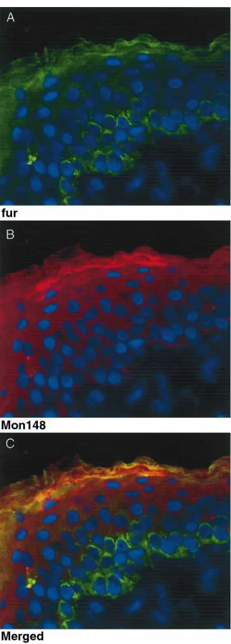
con-Figure 3.Double immunofluorescence of epidermis using the c-fur and MON148 antibodies. (A) c-c-fur reaction. (B) MON148 reaction. (C) Merged c-fur and MON148 reactions; the yellow color indicates where the green and red signals are superim-posed to give yellow fluorescence. Tissue sections were trypsin treated (0.01%, 1 min) and incubated overnight (4æC) with the rabbit polyclonal c-fur antibody and the mouse monoclonal MON148 antibody. The c-fur antibody was detected with a FITC-labeled anti-rabbit secondary antibody (green); the MON148 was detected using a biotinylated anti-mouse second-ary/Texas Red streptavidin ternary complex (red); DNA in nu-clei was stained with DAPI (blue). The C-terminal antibody (c-fur, A & C) stains both the basal and granular layer with significantly reduced staining in the spinous layer. The catalytic domain antibody (MON148, B & C) stains mainly the granular layer with lighter spinous and very little basal staining. In the granular layer the merged staining (C) shows both co-localiz-ation (yellow) as well as patches of differential localizco-localiz-ation of the C-terminus (green) and catalytic (red) domains.
struct. An additional 70–75 kDa band was ob-served in epidermis and keratinocytes that reacted with antibodies to the catalytic domain (MON148) and cys-rich domain (MON152), but not the C-terminal cytoplasmic domain (c-fur). This band is slightly smaller than soluble furin produced from a construct lacking the transmembrane and C-ter-minal domains, indicating cleavage and removal of the transmembrane domain in the tissue. A larger
band (∂120 kDa), possibly corresponding to the
proenzyme, was detected in keratinocytes but not in foreskin epidermal extracts. The specificity and identity of the bands recognized by the antibodies was confirmed by stripping representative blots and reprobing with an alternate antibody (data not shown).
Three bands (96 kDa, 75 kDa and 70 kDa) posi-tive for PACE4 were detected on western blots in sub- and post-confluent keratinocyte extracts (Fig. 2C). PC7/8 protein was also detected in pre- and post-confluent keratinocytes (Fig. 2D). The active
form at∂75 kDa was seen in both pre- and
post-confluent cell extracts with both antibodies.
Localization of proprotein convertases in epidermis
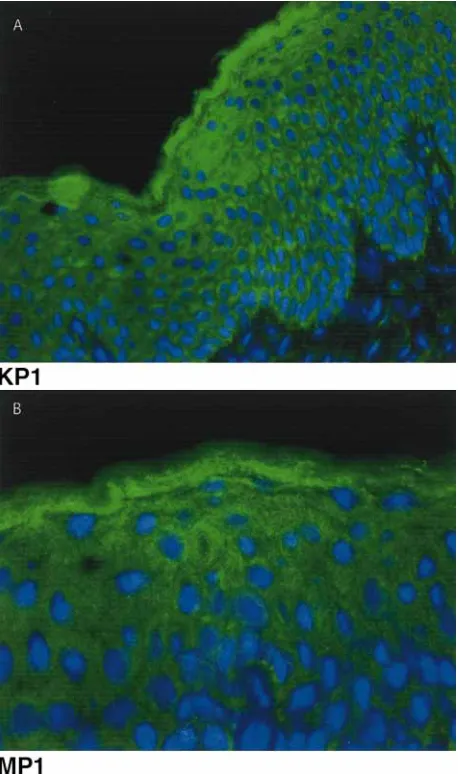
[image:6.595.64.293.59.698.2]yel-Figure 4. Immunofluorescence of epidermis using antibodies specific for PC7/8. (A) Staining with the antibody KP1 which is directed at an epitope in the C-terminal cytoplasmic domain. (B) Staining with antibody MP1 which recognizes the N-ter-minus of the catalytic domain. Tissue sections were incubated with the polyclonal KP1 or MP1 (1 h, RT) and detected using a FITC-labeled anti-rabbit secondary (green). Nuclei were stained with DAPI (blue). Both antibodies stained all layers of the epidermis and were confined to the cytoplasm of the cells. Note the slight increase of staining in the granular layer.
low in the granular layer indicate where the red MON148 staining and green c-fur staining are col-ocalized (merged). There are, however, significant areas where the two antibodies are not superim-posed (red and green staining in the granular layer). Strong furin staining was also seen in the glandular structures of the dermis (not shown).
[image:7.595.326.508.62.192.2]Antibodies specific for PC7/8 showed patchy staining throughout the epidermis (Fig. 4). This distribution was similar with both the antibody di-rected against the C-terminal cytoplasmic domain (KP1) (Fig. 4A) and that directed against the cata-lytic domain (MP1) (Fig. 4B).
Figure 5.Inhibition of Notch-1 processing in mouse keratino-cytes by decanocyl-RVKR-CMK. Mouse keratinokeratino-cytes were treated with the inhibitor or vehicle for 4 days. Cell extracts were separated on a 7.5–15% SDS–PAGE gel, blotted onto nitrocellulose and probed with a monoclonal Notch-1 anti-body directed to an epitope in the intracellular C-terminal do-main using the ECL system. The 120 kDa active Notch-1 amino-terminal domain (*) is detected in the pretreated cultures (day 2) as well as in the day 4 cultures without the PC inhibitor but is significantly reduced in cultures treated with 100mM and 250mM of the inhibitor. The position of molecular weight stan-dards is shown on the left. The Notch-1 pro-form is not de-tected due to its poor reactivity with the antibody (Milner, per-sonal communication).
Identification of potential PC substrates in epidermis
Two potential substrates, Notch-1 and profilag-grin, relevant in epidermal differentiation, were
in-vestigated using either in vivo or in vitro
ap-proaches.
Inhibition of Notch-1 processing by inhibition of proprotein convertase processing in culture
Notch-1 is a cell-surface receptor involved in cell fate determination and patterning. Processing of Notch-1 by furin at an RXRR site in the carboxy terminus during transport to the plasma mem-brane is required to produce the functional recep-tor (14, 30). A cell permeable PC inhibirecep-tor (de-canoyl-RVKR-CMK) (26) was added to mouse keratinocyte cultures to test the role of furin on Notch-1 processing in these cells. Inhibition of the processing of Notch-1 from the precursor form (220 kDa) to the functional 120 kDa form (Fig. 5) was seen with dramatic reduction of the 120 kDa
form using both 100 and 250 mM of the inhibitor.
The inhibitor did not exhibit toxic effects on the cells at these concentrations.
Furin and PACE4 specifically cleave the amino terminus of profilaggrin
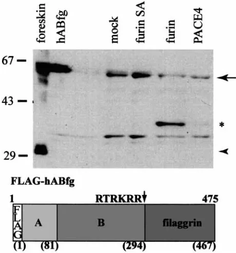
Profilaggrin is a large (⬎400 kDa) protein,
[image:7.595.55.284.64.451.2]unique N and C-termini, that is expressed in the granular layer of the epidermis (reviewed in 31). Normal keratinocyte culture systems can be in-duced to produce profilaggrin (18, 29, 32) but do not fully process it into filaggrin and the amino-terminal fragment. Therefore, to study potential PC processing of profilaggrin at the predicted
se-quence (R288TRKRR293) at the junction of the
amino terminus and the filaggrin domains, an in
vitro system was used. Extracts obtained from COS-7 cells transfected with the N-terminal
profi-laggrin expression construct, pFLAG467proF,
were used to mimic profilaggrin; profilaggrin itself is insoluble in aqueous buffers suitable for enzyme assays and therefore could not be used in these as-says. Media conditioned by COS-1 or 293T cells transfected with expression constructs encoding either a soluble form of furin, its inactive mutant derivative (furin-SA), or PACE4 were sources of the enzymes. Cleavage of the substrate was
de-Figure 6.Cleavage of profilaggrin (pFLAG467proF) by furin and PACE4. Extracts of COS-7 cells transfected with the pFLAG467proF were incubated with conditioned media con-taining expressed enzyme. The products were analyzed using antibody specific for the B domain of the profilaggrin amino-terminus. The position of uncleaved product is shown by the arrow. Note that a cleaved product (approx. 37 kDa*) is de-tected in the samples containing furin (strong) and PACE4 (weak), but not those containing an inactive mutant (furin-SA) or mock transfected media or the unreacted substrate (hABfg). The endogenous profilaggrin N-terminus (32 kDa) is visible in the foreskin extract (arrowhead). Molecular weight markers are indicated on the left. A diagram of the FLAG467proF con-struct is shown to illustrate the A and B domains and the posi-tion of the predicted cleavage site.
tected by the appearance of the processed fragment on western blots using antibodies specific for the B domain of the profilaggrin N-terminus. The antibody specific for the amino-terminus reacts with both the amino-terminal-profilaggrin sub-strate and the expected cleavage product (Fig. 6), whereas the antibody specific for filaggrin reacts only with the amino-terminal-profilaggrin sub-strate and not with the cleaved product (data not shown). As shown in Fig. 6, only treatment of the profilaggrin substrate with the soluble form of fur-in or with PACE4 but not with the mutant furfur-in (furin-SA) resulted in the appearance of a new band representing the proteolytic cleavage product with a size of approximately 37 kDa. This cleavage
is Ca2π-dependent and can be inhibited with
EDTA (data not shown). Control substrate (von Willebrand factor or CPA-PACE) was used to ver-ify that the enzyme preparations functioned as ex-pected (data not shown).
In order to confirm the specific site of PC
cleav-age, the proposed cleavage site (R288TRKRR293)
[image:8.595.62.292.334.580.2]was mutated to see if specific processing could be affected. Fig. 7 shows that substitution at either the P1 position (to RKRA) or the P4 position (to AKRR) virtually abolished the cleavage which produces the 37 kDa product in the normal con-struct, whereas the alteration of the P2 position (to RKAR) had little effect on the cleavage. The re-sults demonstrated that the predicted paired amino acid sequence is required as the primary target site
[image:8.595.311.542.454.588.2]for furin/PACE cleavagein vitro. Interestingly, mu-tagenesis of each of the critical residues (P1, P4) yielded another cleaved product migrating at about 40 kDa. This product could be either an in-termediate of a sequential cleavage or a product resulting from an alternative secondary site, poss-ibly the sequence GSRHPR present at residues 331–336.
Discussion
The dramatic remodeling of cytoplasm and cell membrane that occurs at the transition of the granular and cornified layers to form the protec-tive skin surface requires multiple proteolytic events, breakdown of nucleic acids and membrane-bounded cellular organelles, and restructuring of the keratin cytoskeleton and plasma membrane. The changes in keratinocytes during the basal to spinous layer transition are not as dramatic morphologically yet regulate the balance between proliferation and withdrawal from the cell cycle and differentiation. The results presented here show that members of the PC family are likely to play a role in these events, consistent with their involvement in differentiation of gut epithelia and other tissues (33).
We have shown the expression in epidermis of four PC family members; furin, PACE4, PC5/6 and PC7/8 at the mRNA level and detected protein for three of these proteases. Furin, in particular, is detected in two forms – the mature 97 kDa form and a smaller 75 kDa form that lacks reactivity with the c-fur antibody specific for the C-terminus. Based on size and antibody reactivity, the 75 kDa form is expected to lack the transmembrane do-main. This size difference may be associated with the change in the accessibility of furin in the differ-ent layers of the skin shown by immunocytochemi-stry using antibodies directed to specific epitopes. The cytoplasmic C-terminal domain is detected in both the basal (less differentiated) layer and the granular layer whereas the catalytic domain is most readily detected in the granular layer where cells are preparing to undergo terminal differen-tiation. This implies that the catalytic domain is more accessible to the antibody in the granular cells. As the cells of the granular layer are under-going significant remodeling, including the dissol-ution of membranous organelles and the remodel-ing of the golgi/transgolgi apparatus into lamellar granules (34–36), it is likely that there is some re-arrangement of the normal contents of these or-ganelles, including the normally trans-golgi resi-dent furin, which would allow it access to a wider range of substrates in these cells.
These results suggest that furin might play
multiple roles in epidermal differentiation depend-ing on the cell layer. Thus in the basal layer it would be expected to be involved in processing of secreted substrates including collagen XVII,
inte-grins a3 anda6, and Notch, which mature via the
golgi–transgolgi route. Our studies clearly show that inhibition of furin (and other PCs) in kera-tinocytes blocked the processing of Notch-1, a cell surface receptor that has been shown to be in-volved in cell fate determination and patterning (37–41) and integrin signaling (17). Notch is ex-pressed in all layers of the epidermis and has been suggested to have a role in stem cell fate and main-tenance in epidermis (15) and hair follicles (42, 43). The potential importance of the Notch pathway is emphasized by the expression of the fringe genes, modulators of Notch signaling, in specific patterns in epidermis (44). Furin over-expression has been
shown to inhibit differentiation in a-cells of
pan-creatic islets (45) and gastric mucosal cells (33). In contrast inhibition of furin in epidermal or-ganotypic cultures leads to premature differen-tiation of keratinocytes, possibly via its effects on the Notch pathway (Pearton et al., in preparation). Furin is not strongly expressed in the spinous layers, but is detectable in the granular layer with altered epitope accessibility correlated with detec-tion of the 75 kDa form of the enzyme. In this layer furin, and other PCs, would have access to a different range of substrates including cytoplasmic proteins such as profilaggrin. Both furin and
PACE4 are able to cleave profilaggrin in vitro to
release a fragment of the size predicted by cleavage at the junction with the filaggrin domains (27). PC5/6 and PC7/8 are also expressed in the epider-mis but it is not known whether they have similar cleavage specificity towards the profilaggrin sub-strate.
Profilaggrin is a highly phosphorylated insol-uble protein of keratohyalin. During terminal dif-ferentiation it is processed by phosphatases and proteases to release the amino-terminus and indi-vidual filaggrin domains which bind to and aggre-gate keratin filaments (46). Although several en-zymes involved in profilaggrin processing are known, the early events are not understood (re-viewed in 31). The amino-terminus containing the
S-100 Ca2π-binding domain (A domain) and a
cat-ionic B domain moves to the nucleus in transition cells while the filaggrin domains remain in the cytoplasm (47) (27, 48). A canonical PC cleavage
motif (R288TRKRR293) lies at the junction
domain-specific cleavage of profilaggrin in vitro (37 kDa product) is similar but not identical to that
de-tected in vivo (approx. 32 kDa) (Figs 6, 7). This
difference could be due in part to the presence of the FLAG epitope which was required to enhance solubility of this test substrate. However,
ad-ditional cleavages may also occur in vivo, and
further study will be required to determine the
in-volvement of PC in this processing pathwayin vivo.
Nevertheless, cleavage of the profilaggrin
amino-terminus is a critical event for altering the solubility of the protein for subsequent PEP1 cleavage of the filaggrin units (Resing et al., sub-mitted), as well as for generating the S-100-like do-main for nuclear translocation where it may have distinct functions in calcium-regulated events dur-ing terminal differentiation (47, 48, 50, 51). The profilaggrin domain-specific processing, the furin localization and its change in accessibility during differentiation, and the calcium-dependent pro-cessing, all suggest a functional role for PCs in
ter-minal differentiationin vivo. Our findings
empha-size the pivotal role of proteases in processing pro-filaggrin and adds furin/PACE to the list of proteases (i.e. profilaggrin endoproteinase 1 (5), calpain (52), plasmin and chymotrypsin-like pro-teases (53)) that are likely to have significant roles in terminal differentiation of epidermal cells.
In summary we present the first systematic analysis into the expression and localization of the PCs in epidermis. We have shown the localization of furin and PC7/8 using immunofluorescence and present evidence that furin undergoes a change of accessibility between the basal and granular layers that is related to cleavage to a soluble form that would have access to different types of substrates in these two distinct sites. We show that inhibition of PCs in keratinocytes inhibits processing of Notch-1, a known substrate with implications for cell fate determination and patterning. We also demonstrate that PCs are able to specifically cleave the amino-terminus of profilaggrin, a novel sub-strate found in the granular layer of the epidermis. This work indicates that the PCs may play multiple roles during the differentiation of cells within the epidermis.
Acknowledgements
We thank Dr Michael Bobola for the UW 228–1 cell line, Janet Kimball for her assistance with immunoblotting, Barbara Hag-er for hHag-er expHag-ertise in tissue culture, Dr Ruth Houge Angeletti (Albert Einstein College of Medicine, NY) for the gift of the anti-furin antibody, Dr W.J.M. Van de Ven (Katholieke Univer-siteit Leuven, Belgium) for the gift of the anti-PC7/8 antibody and Dr Lauri Milner (Fred Hutchinson Cancer Research Cent-er, Seattle, WA, USA) for the gift of the Notch-1 antibody. Tissue culture procedures were performed in the Keratinocyte
Culture Core Laboratory, Dr Philip Fleckman, Director, and immunostaining was photographed in the Morphology Core Laboratory, Dr Micheal Piepkorn, Director, Division of Der-matology with assistance from Mr Robert Underwood, part of the Dermatology Program Project, P01 AR21557. Autoradio-graphic analysis was carried out by the PhosphorImager facility of the Markey Molecular Medicine Center at the University of Washington. This work was supported by NIH grants R39 DE04660 and P01 AR21557 to BAD. R.B. Presland was the recipient of a Career Development Award from the Derma-tology Foundation.
References
1. Kam E, Resing K A, Lim S K, Dale B A. Identification of rat epidermal profilaggrin phosphatase as a member of the protein phosphatase 2A family. J Cell Sci 1993: 106: 219. 2. Hart T C, Hart P S, Bowden D W et al. Mutations of
the cathepsin C gene are responsible for Papillon–Lefevre syndrome. J Med Genet 1999: 36: 881.
3. Kim S Y, Bae C D. Calpain inhibitors reduce the cornified cell envelope formation by inhibiting proteolytic processing of transglutaminase 1. Exp Mol Med 1998: 30: 257. 4. Tanabe H, Kumagai N, Tsukahara T et al. Changes of
lys-osomal proteinase activities and their expression in rat cul-tured keratinocytes during differentiation. Biochim Bi-ophys Acta 1991: 1094: 281.
5. Resing K A, Johnson R S, Walsh K A. Characterization of protease processing sites during conversion of rat profilag-grin to filagprofilag-grin. Biochemistry 1993: 32: 10036.
6. Watkinson A. Stratum corneum thiol protease (SCTP) a novel cysteine protease of late epidermal differentiation. Arch Dermatol Res 1999: 291: 260.
7. Hart T C, Hart P S, Michalec M D et al. Haim–Munk syndrome and Papillon–Lefevre syndrome are allelic muta-tions in cathepsin C. J Med Genet 2000: 37: 88.
8. Toomes C, James J, Wood A J et al. Loss-of-function muta-tions in the cathepsin C gene result in periodontal disease and palmoplantar keratosis. Nat Genet 1999: 23: 421. 9. Creemers J W, Jackson R S, Hutton J C. Molecular and
cellular regulation of prohormone processing. Semin Cell Dev Biol 1998: 9: 3.
10. Gensberg K, Jan S, Matthews G M. Subtilisin-related ser-ine proteases in the mammalian constitutive secretory path-way. Semin Cell Dev Biol 1998: 9: 11.
11. Nakayama K. Furin: a mammalian subtilisin/Kex2p-like endoprotease involved in processing of a wide variety of precursor proteins. Biochem J 1997: 327 (Pt 3): 625. 12. Seidah N G, Chretien M. Pro-protein convertases of
subtili-sin/kexin family. Methods Enzymol 1994: 244: 175. 13. Krysan D J, Rockwell N C, Fuller R S. Quantitative
char-acterization of furin specificity: Energetics of substrate dis-crimination using an internally consistent set of hexapepti-dyl methylcoumarinamides. J Biol Chem 1999: 274: 23229. 14. Logeat F, Bessia C, Brou C et al. The Notch1 receptor is cleaved constitutively by a furin-like convertase. Proc Natl Acad Sci USA 1998: 95: 8108.
15. Lowell S, Jones P, Le Roux I, Dunne J, Watt F M. Stimula-tion of human epidermal differentiaStimula-tion by delta-notch sig-nalling at the boundaries of stem-cell clusters. Curr Biol 2000: 10: 491.
16. Schacke H, Schumann H, Hammami-Hauasli N, Raghun-ath M, Bruckner-Tuderman L. Two forms of collagen XVII in keratinocytes. A full-length transmembrane protein and a soluble ectodomain. J Biol Chem 1998: 273: 25937. 17. Berthet V, Rigot V, Champion S et al. Role of
Kera-tinocytes cultured from subjects with ichthyosis vulgaris are phenotypically abnormal. J Invest Dermatol 1987: 88: 640. 19. Thompson J D, Higgins D G, Gibson T J. CLUSTAL W: improving the sensitivity of progressive multiple sequence alignment through sequence weighting, position-specific gap penalties and weight matrix choice. Nucleic Acids Res 1994: 22: 4673.
20. Higgins D G, Thompson J D, Gibson T J. Using CLUS-TAL for multiple sequence alignments. Methods Enzymol 1996: 266: 383.
21. Chomczynski P, Sacchi N. Single-step method of RNA iso-lation by acid guanidinium thiocyanate- phenol-chloroform extraction. Anal Biochem 1987: 162: 156.
22. Rehemtulla A, Dorner A J, Kaufman R J. Regulation of PACE propeptide-processing activity: requirement for a post-endoplasmic reticulum compartment and autoproteo-lytic activation. Proc Natl Acad Sci U S A 1992: 89: 8235. 23. Rehemtulla A, Barr P J, Rhodes C J, Kaufman R J. PACE4 is a member of the mammalian propeptidase family that has overlapping but not identical substrate specificity to PACE. Biochemistry 1993: 32: 11586.
24. Aplin A E, Juliano R L. Integrin and cytoskeletal regula-tion of growth factor signaling to the MAP kinase pathway. J Cell Sci 1999: 112: 695.
25. Hager B, Bickenbach J R, Fleckman P. Long-term culture of murine epidermal keratinocytes. J Invest Dermatol 1999: 112: 971.
26. Garten W, Hallenberger S, Ortmann D et al. Processing of viral glycoproteins by the subtilisin-like endoprotease furin and its inhibition by specific peptidylchloroalkylketones. Biochimie 1994: 76: 217.
27. Presland R B, Kimball J R, Kautsky M B, Lewis S P, Lo C Y, Dale B A. Evidence for specific proteolytic cleavage of the N-terminal domain of human profilaggrin during epidermal differentiation. J Invest Dermatol 1997: 108: 170. 28. Rehemtulla A, Kaufman R J. Protein processing within the
secretory pathway. Curr Opin Biotechnol 1992: 3: 560. 29. Nirunsuksiri W, Presland R B, Brumbaugh S G, Dale B A,
Fleckman P. Decreased profilaggrin expression in ichthyosis vulgaris is a result of selectively impaired posttranscrip-tional control. J Biol Chem 1995: 270: 871.
30. Rand M D, Grimm L M, Artavanis-Tsakonas S et al. Cal-cium depletion dissociates and activates heterodimeric notch receptors. Mol Cell Biol 2000: 20: 1825.
31. Dale B A, Resing K A, Presland R B. Keratohyalin granule proteins. In: Leigh I, Lane B, Watt F, eds. The Keratinocyte Handbook. Cambridge.: Cambridge University Press, 1994: 323.
32. Haydock P V, Blomquist C, Brumbaugh S, Dale B A, Hol-brook K A, Fleckman P. Antisense profilaggrin RNA de-lays and decreases profilaggrin expression and altersin vitro
differentiation of rat epidermal keratinocytes. J Invest Dermatol 1993: 101: 118.
33. Konda Y, Yokota H, Kayo T et al. Proprotein-processing endoprotease furin controls the growth and differentiation of gastric surface mucous cells. J Clin Invest 1997: 99: 1842. 34. Madison K C, Sando G N, Howard E J et al. Lamellar granule biogenesis: a role for ceramide glucosyltransferase, lysosomal enzyme transport, and the Golgi. J Investig Dermatol Symp Proc 1998: 3: 80.
35. Elias P M, Cullander C, Mauro T et al. The secretory granular cell: the outermost granular cell as a specialized secretory cell. J Investig Dermatol Symp Proc 1998: 3: 87. 36. Madison K C, Howard E J. Ceramides are transported
through the Golgi apparatus in human keratinocytesin vi-tro. J Invest Dermatol 1996: 106: 1030.
37. Milner L A, Bigas A. Notch as a mediator of cell fate deter-mination in hematopoiesis: evidence and speculation. Blood 1999: 93: 2431.
38. Bigas A, Martin D I, Milner L A. Notch1 and Notch2 inhibit myeloid differentiation in response to different cyto-kines. Mol Cell Biol 1998: 18: 2324.
39. Milner L A, Bigas A, Kopan R, Brashem-Stein C, Bern-stein I D, Martin D I. Inhibition of granulocytic differen-tiation by Notch1. Proc Natl Acad Sci U S A 1996: 93: 13014.
40. Perron M, Harris W A. Determination of vertebrate retinal progenitor cell fate by the Notch pathway and basic helix– loop–helix transcription factors. Cell Mol Life Sci 2000: 57: 215.
41. Rogister B, Ben-Hur T, Dubois-Dalcq M. From neural stem cells to myelinating oligodendrocytes. Mol Cell Neu-rosci 1999: 14: 287.
42. Favier B, Fliniaux I, Thelu J et al. Localisation of members of the notch system and the differentiation of vibrissa hair follicles: Receptors, ligands, and fringe modulators. Dev Dyn 2000: 218: 426.
43. Lin M H, Leimeister C, Gessler M, Kopan R. Activation of the Notch pathway in the hair cortex leads to aberrant differentiation of the adjacent hair-shaft layers. Develop-ment 2000: 127: 2421.
44. Thelu J, Viallet J P, Dhouailly D. Differential expression pattern of the three Fringe genes is associated with epider-mal differentiation. J Invest Dermatol 1998: 111: 903. 45. Kayo T, Sawada Y, Suda M et al. Proprotein-processing
endoprotease furin controls growth of pancreatic beta-cells. Diabetes 1997: 46: 1296.
46. Dale B A, Lonsdale-Eccles J D, Holbrook K A. Stratum corneum basic protein: an interfilamentous matrix protein of epidermal keratin. Curr Probl Dermatol 1980: 10: 311. 47. Ishida-Yamamoto A, Takahashi H, Presland R B, Dale B
A, Iizuka H. Translocation of profilaggrin N-terminal do-main into keratinocyte nuclei with fragmented DNA in normal human skin and loricrin keratoderma. Lab Invest 1998: 78: 1245.
48. Presland R B, Bassuk J A, Kimball J R, Dale B A. Char-acterization of two distinct calcium-binding sites in the amino-terminus of human profilaggrin. J Invest Dermatol 1995: 104: 218.
49. Thulin C D, Walsh K A. Identification of the amino ter-minus of human filaggrin using differential LC/MS tech-niques: implications for profilaggrin processing. Biochemis-try 1995: 34: 8687.
50. Presland R B, Haydock P V, Fleckman P, Nirunsuksiri W, Dale B A. Characterization of the human epidermal profi-laggrin gene. Genomic organization and identification of an S-100-like calcium binding domain at the amino ter-minus. J Biol Chem 1992: 267: 23772.
51. Markova N G, Marekov L N, Chipev C C, Gan S Q, Idler W W, Steinert P M. Profilaggrin is a major epidermal cal-cium-binding protein. Mol Cell Biol 1993: 13: 613. 52. Miyachi Y, Yoshimura N, Suzuki S et al. Biochemical
dem-onstration and immunohistochemical localization of cal-pain in human skin. J Invest Dermatol 1986: 86: 346. 53. Suzuki Y, Nomura J, Koyama J, Horii I. The role of