http://dx.doi.org/10.4236/ajac.2013.412085
Development and Validation of Extraction Method for the
Determination of UC781 in Cervicovaginal Fluid
Naser L. Rezk1,2,3*, Zhiqing Qiao1, Majed Jeriasy3
1Eshelman School of Pharmacy, Division of Pharmacotherapy and Experimental Therapeutics,
University of North Carolina, Chapel Hill, USA
2Andor Labs, Durham, North Carolina, USA
3King Abuallah International Medical Research Center, Riyadh, KSA
Email: *[email protected], *[email protected], *[email protected]
Received October 25, 2013; revised November 26, 2013; accepted December 1, 2013
Copyright © 2013 Naser L. Rezk etal. This is an open access article distributed under the Creative Commons Attribution License, which permits unrestricted use, distribution, and reproduction in any medium, provided the original work is properly cited.
ABSTRACT
Liquid chromatography plays the important and critical role in the field of clinical pharmacology of evaluating drugs in biological matrices. Studying antiretroviral drugs in the female genital tract has important implications for using drugs as a vaginal microbicide for prevention of HIV-1 sexual transmission. Accurate measurement of drug levels is ex- tremely important in optimizing drug concentration in gel formulation. Extracting drugs from small volumes of viscous, proteinaceous substances like cervicovaginal fluid (CVF) is a practically challenging process. The proposed method was designed to introduce procedure for sample collection and drug extraction procedure for CVF matrix before the chromatographic separation. Based on this extraction method, we validated a reverse phase high performance liquid chromatography with electrospray ionization mass spectrometry assay in order to quantify UC781 in female genital tract compartment. The LC-MS method was validated based on a novel extraction technique which proved to be effi- cient in reducing analyte degradation with an average extraction efficiency of 72%. This method is accurate, demon- strating an average accuracy over three QC (n = 30) concentrations ranging from 99.9% to 106.1%. Average precision within-day and between-day ranged from 3.1% to 10.2% and 5.1% to 6.4%, respectively. We demonstrated that the analyte was able to maintain its stability under various conditions using this extraction method. The sample preparation, extraction, and the powerful liquid chromatography and mass spectrometry can readily be applied for accurate quantifi-cation of similar drugs in CVF.
Keywords: HIV-Prevention; Cervicovaginal Fluid; Extraction; LC-MS; Nonnucleoside Reverse Transcriptase Inhibitor
1. Introduction
Sexual transmission of HIV is the principal mode of spread of HIV throughout the world [1]. The majority of HIV-1 infections are acquired sexually, and interventions to prevent sexual transmission are urgently needed to curb the growth of the HIV pandemic [2].
Methods to reduce or prevent sexual transmission of HIV-1 are urgently needed to sharply reduce the global HIV-1 epidemic [3]. Understanding the pharmacokinet- ics of drugs in human body compartments, such as the female genital tract, is especially important [4]. Among several classes of HIV inhibitors, many drugs belong to non-nucleoside reverse transcriptase inhibitors (NNRTIs). The thiocarboxanilide ((N-[4-chloro-3-(3-methyl-2-bu-
tenyloxy) phenyl]-2-methyl-3-furancarbotthioamide) or UC-781 (Figure 1(a)) is ranked among the most potent NNRTI’s [5-8]. Currently, UC781 is being investigated as a prevention therapy [9,10]. The highly potent non- nucleoside reverse transcriptase inhibitor UC781 has been tested as a safe vaginal microbicide gel formulation for preventing the sexual transmission of HIV-1 virus [10]. To investigate whether UC781 retained anti-infective activity following exposure to the female genital tract, we developed the assay for the analysis UC781 levels and antiviral activity in cervicovaginal fluid (CVF). Based on our LC-ESI-MS method [11] for sensitive and accu- rate determination of UC781 in blood plasma, we modi- fied the extraction method to fit with the (CVF) matrix.
An optimal extraction method for female genital tract secretion must include the release of drug candidate from
(a)
(b)
Figure 1 (a) Chemical structure of UC781; (b) Chemical structure of F2591 (IS).
the viscous proteinaceous substances [12] like CVF. In this manuscript, the primary objective of this work is to develop and optimize an extraction method for a sample clean-up procedure to fit with CVF. The second objective is to validate the procedure to be a standard assay for quantification of drug concentration in CVF.
2. Materials and Methods
2.1. Chemicals
UC781 (purity 98.7%) was supplied by Regis Technolo- gies, Inc (Morton Grove, IL, USA). F2951 (purity 99.2%), used as internal standard, was supplied by Chemtura, Technology Center (Guelph, Ontario, Canada). Tetrahy- drofuran (purity 99.9%) was purchased from Aldrich (St. Louis, MO, USA). HPLC-grade reagents and chemicals were purchased from Fisher Scientific (Norcross, GA, USA). Purified compressed nitrogen gas was obtained from National Welders Supply (Charlotte, NC, USA).
2.2. Equipment
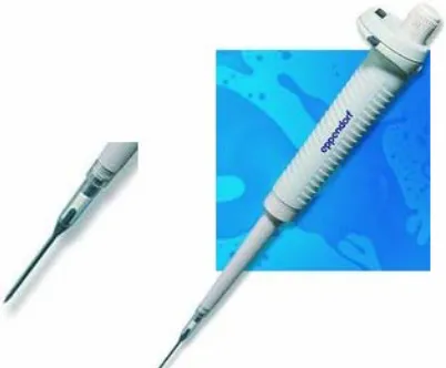
An Eppendorf® Positive Displacement contamination- free pipette (PDP), adjustable ranges 1 µl - 20 µl and Positive Displacement Tips obtained from (Eppendorf North America, Washington DC, USA). This pipette func- tions according to the positive-displacement principle in
[image:2.595.56.288.83.378.2]conjunction with the special Positive Displacement Tip (Figure 2). An Eppendorf 5415D centrifuge (Eppendorf AG, Hamburg, Germany) was used during sample prepa- ration. The high-performance liquid chromatography (HPLC) system consisted of an Agilent Technologies (Wilmington, DE, USA) HP1100 binary pump, degasser, and thermostatic auto sampler (programmed at 4˚C). The HPLC system was connected to a 1100 Series Mass Spec- trometer. Positive electrospray ionization was the mode used for analytical compounds. Data analysis was per- formed using HP ChemStation software (Version A.09.03) run on a Dell computer (Windows 2000 Professional operating system).
2.3. Blank Vaginal Fluid Collection and Preparation
The biological matrix, vaginal secretion fluid (CVF) was collected from five healthy subjects. The collection of CVF has been approved by the institutional review board (IRB) at the University of North Carolina at Chapel Hill (UNC) according the Federal-Wide Assurance (FWA) #4801, the IRB approval # (45 CFR 46 CFR 46.110), and all volunteers/patients gave informed consent prior to study participation.
Before spiking drug to the pooled CVF, the vaginal secretion was diluted 1:3 in normal saline. Because the volume of CVF varies from subject to subject, a similar approach was used on the patient sample directly after the collection of CVF secretion and before storage. Based on the amount of collected CVF, the PDP adjustable positive displacement pipette should be adjusted on 5 or 10 or 15 or 20, then pipette the possible volume, wipe the narrow tip. Then transfer the aspirate into a clean tube containing three times the volume of the aspirate.
2.4. Preparation of Standards
A total of 5.066 mg of UC781 (molecular weight 335.82)
[image:2.595.323.524.553.719.2]powder with a purity of 98.7% was accurately weighed and dissolved in 5 mL of methanol to produce a final concentration of 1 mg·mL−1. The master stock solution was prepared by diluting a stock of 1 mg·mL−1 to 500
μg·mL−1 in 50% HPLC-grade methanol/HPLC-grade wa- ter. This 500 μg·mL−1 master solution was used to pre- pare six working solutions in methanol/HPLC-grade wa- ter (1:1) at concentrations of 0.5 - 500 mg·mL−1 of UC781. CVF calibration samples (50, 100, 500, 1000, 5000, 10,000, 25,000 and 50,000 ng·mL−1 of UC781) were prepared by using a 1:10 dilution of the respective working solutions to blank CVF. From an additional 500 ng·mL−1 working stock solution, concentrations of 1.5
μg·mL−1, 15 μg·mL−1 and 150 μg·mL−1 of UC781 were prepared in methanol/HPLC-grade water (1:1). CVF qual- ity control samples at concentrations of 150, 1500, and 15,000 ng·mL−1 of UC781 were prepared using a 1:10 dilution of their respective working solutions to blank CVF.
2.5. Internal Standard (IS) Preparation
F2951 (Figure 1(b)) powder (5.187 mg; purity 99.2%) was dissolved in 3 mL methanol, then complete the vol- ume of methanol (5.0 mL) to achieve a final concentra- tion of 1.0 mg·mL−1 (stock solution). From this solution, an aliquot was diluted in HPLC-grade acetonitrile to a final concentration of 20.0 ng/mL (working solution). Small aliquots of working solution must be stored at
−20˚C.
2.6. Extraction Procedure
The extraction of UC781 from the CVF matrix was per- formed with liquid-liquid extraction, using diethylether. Prior to extraction, CVF protein was precipitated. The precipitation occurred by adding 10 µL aliquot of CVF blank, calibrators, and QCs into 2.0 mL tube containing 20 µL the internal standard made in acetonitrile. All ma- trix aliquots were pipette using positive displacement pipette with narrow end tip (Figure 2) which easy to wipe off the remaining outside sticky fluid for better ac- curacy. The tips are narrow, transparent and long enough to monitor the volume. The solutions were mixed via vortex-mixing for 30 seconds. After vortex-mixing, 10 µl of (0.1 N) NaOH was added to each tube, followed by 1.7 mL of the extraction liquid. All tubes were immedi- ately capped and gently mixed for 30 min at low speed. All tubes were placed in a dry ice/acetone bath for ap- proximately 1 minute; the aqueous portion of the sample was frozen and the organic layer was immediately trans- ferred to a centrifuge tube and evaporated until dry under a nitrogen stream at 35˚C for approximately 8 mints. Finally, the residue was reconstituted with 100 µL of methanol/water (50/50). The resulting solutions were
carefully vortexed for 30 s and centrifuged at 14,000 rcf for 3 mints. The supernatants were transferred to 200 µL HPLC micro-vials (Agilent Technologies), and 10 µL of each sample was injected for LC-MS analysis.
2.7. Chromatography Separation Conditions 1) High performance liquid chromatography condi- tions
Chromatographic separation was performed using gra- dient elution. Separation was conducted using an Allure C-18 (100 × 2.1 mm, 3.0 µm particle size, Restek, Belle- fonte, PA, USA) analytical column with an Allure C-18 (10 × 2.1 mm, 5.0 µm particle size, Restek) guard col- umn. Two mobile phase components were utilized throughout the study. Mobile phase A consisted of 10 mM of ammonium formate in water, and mobile phase B was composed of LC-MS-grade methanol containing 0.01% tetrahydrofuran (THF). A linear gradient was pro- grammed as 80% mobile phase B to 100% B over the first 5 minutes, followed by 0.5 minutes at 100% mobile phase B, then 6 minutes at 80% B, and finally 4 minutes of re-equilibration. The analysis was performed at 30˚C with a mobile phase flow rate of 0.3 mL·min−1.
2) Mass spectrometry detection conditions
Mass spectral analysis was performed on an Agilent Quadrupole 1100 Mass Spectrometer fitted with elec- trospray ionization (ESI) source and operated in the posi- tive ionization mode. The vaporizer was operated at 300˚C, the nebulizer gas pressure was set to 40 psi and the capillary voltage was set to 3000 V. The IS and UC781 were detected by their positive ion (m/z 326.0 and 336.1, respectively) using the single ion monitoring (SIM) mode.
2.8. Assessment of Performance Characteristics and Calculation
2.8.1. Linearity
An equal weighted regression was performed to assess linearity. The deviation in the mean calculated concen- trations over five runs was required to be within 15% of the nominal concentration for the non-zero calibration standards.
2.8.2. Accuracy and Precision
Accuracy was calculated as the percent deviation from the nominal concentrations. All intra- and inter-day pre- cision was required to be within a coefficient of variation (CV%) of 15% or less. Sample ranges included a low QC where the concentration was three times of the LOQ [13,14], a medium QC and a high QC.
2.8.3. Extraction Efficiency (%)
response of three pre-spiked QC levels (low, medium, and high in mobile phase) by the area response of ex- tracted blank plasma that was post-spiked with the same three QC concentrations.
2.8.4. Stability
The stability of UC781 during sample handling was veri- fied by subjecting samples to three freeze-thaw cycles and storage for 2 days in refrigerator 4˚C prior to analy- ses. An additional test was performed to verify drug sta- bility in the final extract for 48 hours in autosampler while tubes waiting for HPLC analysis. The samples were left at room temperature for 6 hours prior to analy- ses. Two concentrations medium and high (QC) samples were utilized in the stability tests.
2.8.5. Applying the Method on the Clinical Samples The patient samples will be diluted 1:3 before pipetting 10 µL using positive displacement pipette. An SOP was developed describing the sample collection procedure and submitted to participating clinical centers. After the measurement in order to obtain the final concentration calculated concentration will be multiplied*3.
3. Results
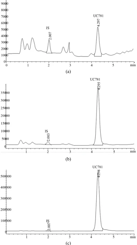
3.1. Chromatographic Separation and Selectivity The approximate retention times for UC781 and IS were 2.0 and 4.3 min, respectively. As depicted in Figures 3(a)-(c) (chromatograms of extracted drugs from CVF, with internal standard at low, medium and high QC’s respectively), none of the endogenous substances from the blank CVF extracts interfered with the analyte or internal standard.
3.2. Linearity and Limit of Quantification
The peak area of the UC781: IS ratio for calibration standards were proportional to the level of drug in CVF over the range of tested concentrations. The calibration curves were fitted by performing weighted least-squares linear regression. The linear regression data for the cali- bration curves obtained from this method (n = 5) consis- tently demonstrated a coefficient of determination ≥0.999. Using this method, we produced data that were linear from 50 - 50,000 ng·mL−1. The low limit of quantifica- tion of UC781 was 50 ng·mL−1. As shown in Table 1(a), this concentration demonstrated high accuracy and preci- sion. Linearity was also tested without the internal stan- dard to determine the direct proportionality of the UC781 peak area to the corresponding concentrations. The cal- culated regression coefficient (r2) of all calibration curves was ≥0.999.
(a)
(b)
(c)
Figure 3. (a) LC-MS chromatogram of the low QC (150 ng·mL−1); (b) LC-MS chromatogram of the high QC (1500
ng·mL−1); (c) LC-MS chromatogram of the high QC (15,000
ng·mL−1).
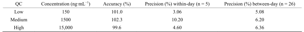
3.3. Accuracy and Precision
[image:4.595.307.540.91.509.2]Table 1. Summary of accuracy and precision (%) during method validation at low, medium and high QC concentration.
QC Concentration (ng·mL−1) Accuracy (%) Precision (%) within-day (n = 5) Precision (%) between-day (n = 26)
Low 150 101.0 3.06 5.08
Medium 1500 102.3 10.20 6.20
High 15,000 99.6 4.60 6.36
centrations, the intra-day precision was always lower than 10.2%. Overall, the mean inter-day precision was 6.4%, with mean RSDs ranging from 5.1% - 6.4%.
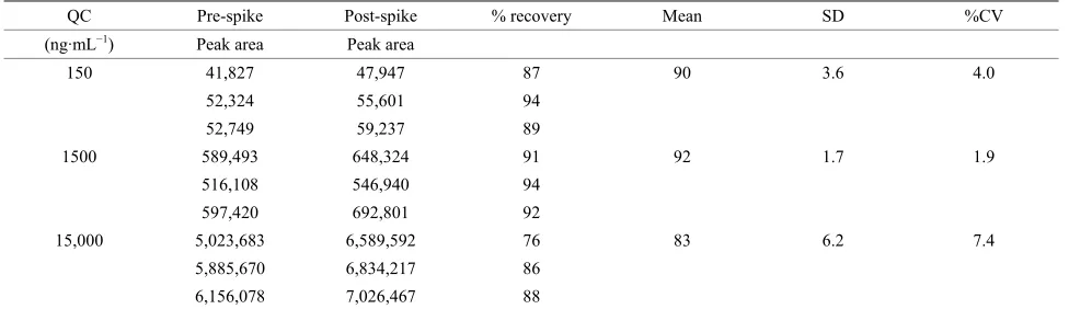
3.4. Extraction Efficiency (%)
The extraction efficiencies for UC781 and IS from CVF using the described liquid-liquid extraction method were calculated using the ratio of the concentration of analyte in CVF to the identical concentration of the analyte pre- pared in mobile phase without extraction. The absolute recovery of analyte from CVF using the diethyether (liq- uid-liquid extraction) procedure was investigated (Table 2). This extraction method reliably eliminated interfering material from CVF and demonstrated high recovery (88.3%) for three (QC) concentration levels.
3.5. Stability
Stability data for UC781 under various conditions are provided in Table 3. For all conditions tested, UC781 proved to be stable. All results were within the accep- tance criteria of ±15% deviation from the nominal con- centration.
4. Discussion
Liquid chromatography separation hyphenated to mass spectrometry (LC-MS) has been developed into an im- portant application in clinical pharmacology—not only for research purposes but also for routine use. At present, most important application fields are target analyses in drug measurements in a variety of biological matrices for therapeutic drug monitoring (TDM), metabolic disorders diagnosis, and many other applications. The essential strengths of LC-MS include a potentially high analytical specificity, a wide range of applicability to small or large molecules, and the opportunity to develop powerful as- says with a high degree of flexibility.
There has been more than 25 million people have al- ready died because of HIV since the discovery of the virus in 1981. This makes AIDS is one of the most disas- trous epidemics in human history [15,16]. In year 2008 2.7 million people got infected by the HIV virus, mainly due to hetero sexual HIV transmission [17,18]. Despite the enormous efforts made, the development of a pro- phylactic AIDS-vaccine will likely not be available in the recent years to come [19]. This leads researchers to focus more on the importance of microbicides (i.e. chemical
entities that can prevent or reduce the transmittance viral infection) which can applied locally (e.g. vaginally and rectally) as an alternative approach in preventing HIV- transmission [20-22].
Our research group at the University of North Carolina was first research team to focus on investigating antiret- roviral (ARV); protease inhibitors (IP), nucleoside re- verse transcriptase inhibitors (NRTI) and non-nucleoside reverse transcriptase inhibitors (NNRTI) in female geni- tal tract (FGT) [23,24]. It was the first method to quantify drugs in direct aspirates of cervicovaginal fluids (CVF). In that study we found large deference in CVF drugs pene- tration (from ≤10% to ≥100%) of blood plasma concen- tration [25]. Since then, we went over to the evaluation of many other ARV medications in FGT [4,26,27]. Most of these studies supported the usage of several ARV which could be an excellent pre-exposure/post-exposure prophylaxis (PrEP/PEP) candidate.
The nonnucleoside reverse transcriptase inhibitor UC781 proved to be a potential microbicide to prevent sexual transmission of human immunodeficiency virus type (HIV-1). Several gel formulations of UC781 were evalu- ated in a range of preclinical safety assessments, includ- ing systemic absorption analysis following topical appli- cation in human.
In this bioanalytical work, we are facing major chal- lenges. First obstacle is the technical difficulties of UC781 in terms of solubility and stability which discussed in details elsewhere [12]. Yet, the challenge remains in dealing with CVF secretion. In brief, the vaginal fluid contains water, pyridine, squalene, urea, acetic acid, lac- tic acid, cholesterol, lipids, mucin, carbohydrates, amino acids, proteins, inorganic ions complex alcohols and glycols, ketones, and aldehydes. It can vary in consis- tency, texture, taste, color, and odor, depending on sexual arousal, the phase of the menstrual cycle, the presence of an infection, certain drugs (legal or illegal), genetic fac- tors, and diet. Vaginal fluid is slightly acidic and can be- come more acidic with certain sexually transmitted dis- eases. The normal pH of vaginal fluid is between 3.8 and 4.5, whereas male semen is typically between 7.2 and 8.0 (a neutral substance has a pH of 7.0).
Table 2. Summary of the assay extraction efficiency %.
QC Pre-spike Post-spike % recovery Mean SD %CV
(ng·mL−1) Peak area Peak area
150 41,827 47,947 87 90 3.6 4.0
52,324 55,601 94
52,749 59,237 89
1500 589,493 648,324 91 92 1.7 1.9
516,108 546,940 94
597,420 692,801 92
15,000 5,023,683 6,589,592 76 83 6.2 7.4
5,885,670 6,834,217 86
[image:6.595.55.543.102.243.2]6,156,078 7,026,467 88
Table 3. Stability of UC781 in spiked rabbit vaginal fluid and final extract
Conc. (ng·mL−1) Three freeze-thaw cycles 48 h at 4˚C 8 h at 25˚C One week matrix stability
1500 10.6 2.5 −2.6 3.3
15000 7.7 1.3 −2.8 5.8
All values are represented as the mean of the deviation from the nominal concentration. All samples were performed in triplicate.
metrial and oviductal fluids and 7) secretions from vagi-nal immune cells. The latter three are influenced by sex steroid hormones e.g. during the menstrual cycle and pregnancy [28,29]. It covers the lower female genital tract pH and hydrates the mucosa, creating a physical barrier for microbial invasion. The important part of ac- curate quantification of drug in FGT is the extraction procedure.
Due to sample collection limitations with a small sam- ple size, CVF collection is a challenging process. It was important to use dilution just right after aspiration in or- der to prevent fluid from forming a clot. Average sample size of normal CVF secretion ranged between 0 to 0.7 mL. In this study, we developed and optimized liquid- liquid extraction method and introduced a simple proce- dure allowing to accurately sampling from the collected fluid before it turns to clot. Using the positive displace- ment pipette with special tips (Figure 2) was important to dilute the sample before storage, similarly when trans- ferring diluted sample for extraction. In the day of vali- dation samples were brought to room temperature and treated for the extraction as described in the method. CVF sample dilution immediately after collection when the temperature near to 37˚C (body temperature) is nec- essary, because it keeps the secretion in the fluid state. Using this procedure of sample collection and extraction, the validation data proved to be accurate and precise for any minute sample collected.
However, normal hexane was the optimal organic sol- vent for blood plasma [11], we found 100% of diethylether releases higher amount of UC781 compared with the other three solvents (chloroform/ether 50/50, hexane/ ether 50/50 and 100% hexane) and SPE. This could be
explained as the higher amount of lipids in blood plasma than CVF which required highly non polar solvent.
5. Conclusion
We have successfully developed and validated an LC- MS bioanalytical method for UC781 in the CVF matrix using the sub-2 µ column with a powerful mobile phase. The method proved to be accurate meeting all validation criteria. For the rare matrix, it exhibits good linearity, precision and accuracy over a wide range of drug con- centrations (50 - 50,000 ng·mL−1). This novel sample preparation associated with liquid-liquid extraction pro- cedure has been proved to be an excellent sample han-dling option for such a sensitive compound in minute amount of vaginal secretion. The method sample prep, extraction, and the powerful liquid chromatography and mass spectrometry can readily be used for accurate quan-tification of any similar drugs in CVF.
6. Acknowledgements
The authors thank the CVF donors for their participation, my analytical team at the University of North Carolina at Chapel Hill for assisting in the validation of the method, Dr. David Friend from Conrad, for providing the stan- dard and internal standard for the method, Mrs. Soheir Hagras at the Drug Research Center, National Center of Research & Radiation Technology (NCRRT), Egypt, and Dr. Chris Sheehan at Badirbio, Riyadh, KSA for review of this manuscript.
REFERENCES
Therapy to Reduce the Sexual Transmission of HIV,”
Journal of HIV Therapy, Vol. 8, No. 3, 2003, pp. 55-66. [2] T. Lalani and C. Hicks, “Does Antiretroviral Therapy
Prevent HIV Transmission to Sexual Partners?” Current HIV/AIDS Reports, Vol. 10, No. 2, 2007, pp. 80-82.
http://dx.doi.org/10.1007/s11904-007-0012-y
[3] J. M. Baeten and J. Overbaugh, “Measuirng the Infec- tiousness of Peresons with HIV-1: Opportunities for Pre- venting Sexual HIV-1 Transmission,” Current HIV Re- search, Vol. 1, No. 1, 2003, pp. 69-86.
http://dx.doi.org/10.2174/1570162033352110
[4] J. A. Talameh, N. L. Rezk and A. Kashuaba, “Quantify- ing the HIV-1 Integrase Inhibitor Raltegravir in Female Genital Tract Secretions Using High-Performance Liquid Chromatography with Ultraviolet Detection,” Journal of Chromatography B, Vol. 878, No. 1, 2010, pp. 92-106.
http://dx.doi.org/10.1016/j.jchromb.2009.11.015
[5] J. Balzarini, M. J. Perez-Perez, S. Velazquez and A. San- Felix, “Suppression of the Breakthrough of Human Im- munodeficiency Virus Type 1 (HIV-1) in Cell Culture by Thiocarboxanilide Derivatives When Used Individually or in Combination with Other HIV-1-Specific Inhibitors (i.e., TSAO Derivatives),” Proceeding of the National Academy of Sciences of the United States of America, Vol.
92, 1995, pp. 5470-5474.
[6] J. Balzarini, W. G. Brouwer, D. C. Dao, E. M. Osika and E. De Clercq, “Identification of Novel Thiocarboxanilide Derivatives That Suppress a Variety of Drug-Resistant Mutant Human Immunodeficiency Virus Type 1 Strains at a Potency Similar to That for Wild-Type Virus,” An- timicrobial Agents and Chemotherapy, Vol. 40, No. 6,
1996, pp. 1454-1466.
[7] R. W. J. Buckheit, T. L. Kinjerski, V. Fliakas-Boltz, J. Russell, T. L. Stup, L. A. Pallansch, etal., “Structure- Activity and Cross-Resistance Evaluations of a Series of Human Immunodeficiency Virus Type-1-Specific Com- pounds Related to Oxathiin Carboxanilide,” Antimicro- bial Agents and Chemotherapy, Vol. 39, No. 12, 1995, pp.
2718-2727. http://dx.doi.org/10.1128/AAC.39.12.2718 [8] J. B. MacMahon, R. W. J. Buckheit, R. J. Gulakowski, M.
J. Currens, D. T. Vistica, R. H. Schoemaker, etal., “Bio- logical and Biochemical Anti-Human Immunodeficiency Virus Activity of UC 38, a New Non-Nucleoside Reverse Transcriptase Inhibitor,” Journal of Pharmacology and Expermental Therapeutics, Vol. 276, No. 1, 1996, pp.
298-305.
[9] J. Vingerhoets, H. Azijn, E. Fransen, I. De Baere, L. Smeulders, D. Jochmans, et al., “TMC125 Displays a
High Genetic Barrier to the Development of Resistance: Evidence from in Vitro Selection Experiments,” Journal of Virology, Vol. 79, No. 20, 2005, pp. 12773-12782.
http://dx.doi.org/10.1128/JVI.79.20.12773-12782.2005 [10] R. E. Haaland, T. Evans-Strickfaden, A. Holder, C. P.
Pau, J. M. Mcnicholl, S. Chaikummaao, etal., “UC781 Microbicide Gel Retains Anti-HIV Activity in Cervico- vaginal Lavag Fluids Collected Following Twice-Daialy Vaginal Application,” Antimicrobial Agents and Chemo- therapy, Vol. 56, No. 7, 2012, pp. 3592-3596.
http://dx.doi.org/10.1128/AAC.00452-12
[11] N. Rezk, “Development and Validation of LC-ESI-MS
Method for Sensitive, Accurate and Rabid Determination of UC-781 in New Zealand White Rabbit Plasma,” Ta- lanta, Vol. 85, No. 4, 2011, pp. 2074-2079.
http://dx.doi.org/10.1016/j.talanta.2011.07.037
[12] D. H. Owen and D. F. Katz, “A Vaginal Fluide Simu- lant,” Contraception, Vol. 59, No. 2, 1999, pp. 91-95.
http://dx.doi.org/10.1016/S0010-7824(99)00010-4 [13] V. P. Shah, K. K. Midha, J. W. Findlay, et al., “Bio-
analytical Method Validation—Revisit with a Decade of Progress,” Pharmaceutical Research, Vol. 17, No. 12,
2000, pp. 1551-1557.
http://dx.doi.org/10.1023/A:1007669411738
[14] US Department of Health and Human Services, Food and Drug Administration Center for Drug Evaluation and Re-search (CDER), Center of Veterinary Medicine (CVM), “FDA Guidance for Industry,” Pharmaceutical Research, 2001.
[15] UNAIDA, “AIDS Epidemic Update,” Geneva, 2005. [16] UNAIDA, “AIDS Epidemic Update,” Geneva, 2009. [17] UNAIDS, “Report in the Global AIDS Epidemic,”
Ge-neva, 2008.
[18] “Will There Be an HIV Vaccine in the Next Decade?”
Nature Medicine, Vol. 13, 2007, pp. 518-519.
http://dx.doi.org/10.1038/nm0507-518b
[19] L. C. Rohan and A. B. Sassi, “Vaginal Drug Delivery System for HIV Prevention,” American Association of Pharmaceutical Scientists, Vol. 11, No. 1, 2009, pp. 78-
87.
[20] B. Gulter and J. Justman, “Vaginal Microbicides and Prevention of HIV Transmission,” Lancet Infection Dis- ease, Vol. 8, No. 11, 2008, pp. 685-697.
[21] A. B. Moscicki, “Vaginal Microbicides: Where Are We and Where Are We Going?” Journal of Infection Chemo- therapy, Vol. 14, No. 5, 2008, pp. 337-341.
http://dx.doi.org/10.1007/s10156-008-0630-3
[22] S. M. Iqbal, T. B. Ball, P. Levinson, L. Maranan, W. Jaoko, C. Wachihi, etal., “Elevated Elafin/Trappin-2 the Female
Genital Tract Is Associated with Protection against HIV Acquisition,” AIDS, Vol. 23, No. 13, 2009, pp. 1669-
1677. http://dx.doi.org/10.1097/QAD.0b013e32832ea643 [23] R. M. Grant, D. Hamer, T. Hope, R. Johnston, J. Lange,
M. M. Lederman, et al., “Whither or Wither Microbi-
cides?” Science, Vol. 321, No. 5888, 2008, pp. 532-534.
http://dx.doi.org/10.1126/science.1160355
[24] S. Min, A. Corbett, N. Rezk, S. Cu-Uvinc, S. Fiscus, et al., “Protease Inhibitor and Nonnucleoside Reverse Tran-
scriptase Inhibitor Concentration in Genital Tract of HIV- 1 Infected Women,” Journal of Acquired Immune Defi- ciency Syndrome, Vol. 37, No. 5, 2004, pp. 1577-1580.
http://dx.doi.org/10.1097/00126334-200412150-00008 [25] D. Julie, R. Yeh, K. Paterson, A. Corbett, B. Jung, N.
Rezk, etal., “Antiretroviral Drug Exposure in the Female
Genital Tract: Implications for Oral Pre- and Post Expo- sure Prophylaxis,” AIDS, Vol. 21, No. 14, 2007, pp.
1899-1907.
http://dx.doi.org/10.1097/QAD.0b013e328270385a [26] R. Yeh, N. Rezk, A. Kashuba, etal., “Genital Tract, Cord
roviral Drugs during and after Pregnancy in Human Im- munodeficiency Virus Type 1-Infected Women,” Antim- icrobial Agents and Chemotherapy, Vol. 53, No. 6, 2009,
pp. 2367-2374. http://dx.doi.org/10.1128/AAC.01523-08 [27] A. Kawara, A. Delong, N. Rezk, et al., “Antiretroviral
Drug Concentrations and HIV RNA in the Genital Tract of HIV-Infected Women Receiving Long-Term Highly Active Antiretroviral Therapy,” Clinical Infectious Dis-eases, Vol. 46, No. 5, 2008, pp. 719-725.
http://dx.doi.org/10.1086/527387
[28] G. R. Huggins and G. Preti, “Vaginal Odors and Secre- tions,” Clinical Obstetrics and Gynecology, Vol. 24, No. 2, 1981, pp. 355-377.
http://dx.doi.org/10.1097/00003081-198106000-00005 [29] L. L. Klein, K. R. Jonschr, M. J. Heerwagen, R. S. Gibbs