Structural studies on some bivalent metal complexes.
DEE, Terry.
Available from Sheffield Hallam University Research Archive (SHURA) at:
http://shura.shu.ac.uk/19547/
This document is the author deposited version. You are advised to consult the
publisher's version if you wish to cite from it.
Published version
DEE, Terry. (1981). Structural studies on some bivalent metal complexes. Doctoral,
Sheffield Hallam University (United Kingdom)..
Copyright and re-use policy
See
http://shura.shu.ac.uk/information.html
Sheffield Hallam University Research Archive
Structural Studies on some Bivalent
Metal Complexes
Terry Dee
A thesis submitted to the Council for National Academic Awards
in partial fulfilment for the Degree of Doctor of Philosophy
Sponsoring establishment: Sheffield City Polytechnic
ProQuest Number: 10694428
All rights reserved
INFORMATION TO ALL USERS
The quality of this reproduction is dependent upon the quality of the copy submitted.
In the unlikely event that the author did not send a com plete manuscript and there are missing pages, these will be noted. Also, if material had to be removed,
a note will indicate the deletion.
uest
ProQuest 10694428
Published by ProQuest LLC(2017). Copyright of the Dissertation is held by the Author.
All rights reserved.
This work is protected against unauthorized copying under Title 17, United States C ode Microform Edition © ProQuest LLC.
ProQuest LLC.
789 East Eisenhower Parkway P.O. Box 1346
Acknowledgement s
The author would like to thank his supervisors, Drs M.Goldstein, I. W. Nowell and N. A.Bell for their help and interest shown during my three year study.
I would also like to thank the staff of the Science Faculty at Sheffield City Polytechnic for all their help over the last
seven years.
Thanks and appreciation to Drs D. L.Hughes and M;Truter (Rothamsted Experimental Station, Harpenden) for use of an Enraf-Nonius CAD-4 diffractometer, Drs P. L. Goggin and R. J. Goodfellow (Department of Chemistry, University of Bristol) for the use of their n.m.r. and Raman facilities and the SERC.for financial support-,for the
past; three years..
I would also like to thank Mrs Sue Lomas for typing this thesis.
Contents
Abstract Abbreviations
Chapter 1: Introduction
Chapter 2: Crystallographic and spectroscopic
studies of some (R.P)HgX? complexes J n z
[x
= Cl, Br or I; n = 1 or 2]Chapter 3: Crystallographic studies of selected
cadmium (II) halide complexes (R^P)CdX2 [R3P = EtjP, Cy3P, Me2PhP;
X = Cl, Br or i] .
Chapter 4: Spectroscopic studies of some (RgP)CdX2 complexes
Chapter 5: - Discussion of factors influencing the
structures of some (R P)MX complexes3 n 2
[m = Hg, Cd or Zn; n = 1 or 2;
X = Cl, Br or i] .
Summary and suggestions for future work Appendices
References
Details of postgraduate study Current publications
Page
3 5 7
• 43
D E E , T .
Structural Studies on Some Bivalent Metal Complexes
Abstract
Previous investigations of the solid-state structures of
(R^P)2^ X 2 and (R3P)HgX2 complexes have been extended by the
determination of the following crystal structures:
(1) (Ph3P)2HgCl2
(2) (Cy3P)HgCl2
The structure of (Ph3P ) 2H g C l 2 is that of a discrete monomer with a
distorted tetrahedral geometry. In comparison with the structures
of the (R3P ) 2H g X 2 complexes previously determined, the degree of
distortion can be related to the donor strengths of the phosphine and
halogen ligands.
(Cy3P)HgCl2 forms a structure that contains two independent chlorine
bridged dimer units in each unit cell. The structure of this complex,
compared with other (R3P)HgX2 complexes is determined by the donor
strength and steric properties of the donor ligands.
The related (R3P)CdX2 complexes have been crystallographically and
spectroscopically investigated. The following crystal structures have
been determined:
(3) (Et3P)CdI2 “ discrete dimer
(4) ot-(Cy3P )CdCl2 - discrete tetramer
(5-7) (Me2PhP)CdX2 [ X = Cl, Br or i] - polymers
The solid-state vibrational spectra of several (R3P )CdX2 complexes have
been studied using the crystal structures (3-7) as a basis for
interpretation. The structure/spectra correlations so determined
have then been applied to a number of related complexes of unknown
structure. The indication from these studies is that the less extended
structures are formed with the stronger donating R^P and X ligands.
The solid-state structures of ( R ^ P ) ^ ^ complexes [m = Hg, Cd or ZnJ have been rationalised in terms of the donor strengths of the R^P
and X ligands, and the acceptor strengths of the metal atoms.
In general, the less associated structures are favoured by:
(1) stronger cr-donor properties (e.g. E t ^ P ] > Ph ^ P ) ;
(2) stronger covalent M-X bonding (i.e. M--I> M-Br > M-Cl) ;
Abbreviations
General
TPP - 1,2,5 - triphenylphosphole Me - methyl
Et - ethyl
Bu - normal butyl £
Bu - tertiary butyl Ph - phenyl
Cy - cyclohexyl R - alkyl- or
aryl-L - neutral unidentate ligand
Crystallographic
S - Angstrom
F - observed structure factor o
F - calculated structure factor c
D - measured density m
D - calculated density
Z - number of molecules per unit cell
w - weighting function
© - cone angle Taft constant
AH - enthalpy of ligation L
M - relative molecular mass r
M - metal atom
X - halogen (Cl, Br or I) py - pyridine
Im - imidazole tu - thiourea
F(ooo) “ number of electrons R - refinement factor
juXMo-K^) - absorption coefficient
I - intensity, of a reflection
Spectroscopic
0 (Cd-X)t - cadmium-halogen stretching mode (terminal)
O(Cd-X), b - cadmium-halogen stretching mode (bridging)
/O(Cd-P) - cadmium-phosphine stretching mode
^0 (Hg-X)t - mercury-halogen stretching mode (termmaO
^(Hg-X)^ - mercury-halogen stretching mode
£)(Hg-P) - mercury-phosphorus stretching mode
^(Cd-L) - cadmium-ligand stretching mode
3 (Hg-X) as - antisymmetric mercury-halogen stretching mode
(Hg-X) - symmetric mercury-halogen stretching mode
n.m.r. - nuclear magnetic resonance
\j(Hg-P) - mercury-phosphorus coupling constant
^■J(Cd-P) - cadmium-phosphorus coupling constant
t - terminal m - medium
b - bridging w - weak
g - gerade I.R. - infrared
u - ungerade Ra - Raman
sh - shoulder
Contents
1.1 Background and objectives.
1.2 Structure/spectra correlation.technique.
1.3 A survey of structural studies of (R3^)nH§X2 complexes in the solid-state.
1.3.1. Introduction.
1.3.2 Crystallographic studies of (R^P)HgX2 complexes.
1.3.3 Crystallographic studies of (R2?)2HgX2 complexes.
1.3.4 Spectroscopic studies of some (R2?)HgX2
complexes.
1.3.5 Spectroscopic studies of some (R^P)£®®^2
complexes.
1.4 Survey of stereochemistry of CdX2 complexes of neutral unidentate ligands.
1.4.1 Introduction.
1.4.2 Crystallographic studies of LCdX2 complexes.
1.4.3 Crystallographic studies of L2CdX2 complexes.
1.1 Background and objectives
For the last twenty years the far-infrared region of the
electromagnetic spectrum (50-450cm has been used by inorganic
and co-ordination chemists as a structural tool. By considering positions'and numbers of metal-ligand modes which appear in the far-infrared region, information about the structures of
compounds can be obtained.
Many misinterpretations of spectroscopic data have been given in the past, mainly due to the lack of a sound basis for the spectroscopic interpretation. One method of obtaining a useful basis for more reliable spectroscopic interpretations
is to examine crystallographically various 'modelf compounds and then correlate the crystallographic and spectroscopic data. Examples of compounds that have previously been studied by this technique are the mercury (II) halide
addition complexes formed with neutral unidentate phosphine
ligands [ 1-8J , (R3P)HgX2 and (R3P)2HgX2. A range of structures were found to exist in the solid-state for the (R3P)HgX2
complexes, in which the co-ordination numbers were increased beyond that of a monomer unit via varying extents of halogen bridging: halogen-bridged dimers, (TPPjHgC^ [ l] ,
(Ph^P^gC^ [l>2] and £ -(BugP^gC^ [l>3] ; halogen-bridged
tetramer, cC -(Bu^P^gC^ [ 1»^] > and extended polymeric structures
In the (R^P)2HgX2 complexes, discrete monomers were found [6,7,8] , with varying degrees of distortion from a regular tetrahedral geometry. These structures were rationalised in terms of the relative donor strength and size of the R^P ligands. Chapter 2 contains further experimental work on these mercury complexes with the essential objectives of the work being a determination of:
(i) the role the halogen plays in structures adopted; (ii) the effect that varying the ste.ric and electronic
nature of the R^P ligand has on the solid-state structures.
From a survey of the literature [9] , it seems that
structural variations such as those found in mercury (II) complexes are likely to be of much wider significance.
A major part of the present work is concerned with investigating factors which influence the structures of cadmium dihalide
addition complexes, mainly of the type (R^P)CdX2. As was the case with mercury (II) systems, there is a lack of systematic crystallographic data and inadequate spectroscopic data; thus one of the aims of the (R^P)CdX2 study was to establish a firm crystallographic basis for the interpretation of far-infrared spectra of compounds of this type. The factors influencing the structure and geometry of (R^P)CdX2 complexes are discussed in Chapter 5 in terms of:
(i) the role of the halogen;
(ii) the effect of varying the donor strength of the R^P ligand; (iii) the steric importance of the R^P ligand.
Finally, the results of the mercury and cadmium investigations are discussed in relation to each other and to the other
1.2 Structure/spectra correlation technique
From the low-frequency infrared and Raman spectroscopic techniques, an indication of the presence of metal-ligand vibrations for a complex can be obtained. The number, activity, symmetry and wavenumber positions of these
vibrational modes are characteristic of the compound's structure.
Using group theory one can predict the theoretical number, activity and symmetry of the expected modes for a structure of a certain symmetry. Thus if the predicted vibrational
modes and the experimentally observed modes correspond, then the spectral pattern can be considered diagnostic of the structure and hence can be used for more reliable interpretation of other spectral data.
There are three methods which have been used to predict these modes.
(i) Point group analysis
[lo]
- where the molecular•symmetry (point group) is used to predict internal modes of vibration of essentially molecular units.
(ii) Line group analysis [ ll] “ the organic ligands are usually taken as point masses (as in the present
work) to make the line group isomorphous with the point
group.
(iii) Factor group analysis [l2*| - the symmetry properties of the primitive unit cell of a crystalline substance are used to obtain information about both internal and
t
external (lattice) modes.
These group theoretical approaches are very useful, but it must be remembered that in practice they give no
information on the intensities and wavenumber positions of the predicted vibrational modes.
1.3 A survey of structural studies of comPlexes
the solid-state
1.3.1 Introduction
The majority of previous structural data on addition complexes of mercury halides and neutral unidentate ligands with the 1:1 and 2:1 stoichiometries (R^P^gX^) indicate a tendency for halogen-bridged structures for the 1:1 complexes to be
formed [9,13] and tetrahedral monomers for the 2:1 complexes with varying degrees of distortion from a regular tetrahedral geometry [6-8]. In.the case of the 1:1 complexes, the
complexes have the ability to increase their co-ordination numbers beyond that of a monomer unit via halogen-bridges.
1.3.2 Crystallographic studies of (R^P)HgX2 complexes
As stated before, (Section 1.3.1) mercury (II) halide complexes in the solid-state have the ability to increase the mercury co-ordination number beyond that of the monomer unit by
halogen-bridging. In the (R^P^gC^ series of complexes
[r^P = M^P* P^3^ or T P P ] varying degrees of
this halogen bridging were found [ 1-5] , as shown in Figures 1.1 to 1.5.
(Ph^lOHgCl^ (Figure 1.1) consists [ l] of discrete chlorine- bridged dimers 'in which mercury lies in a distorted tetrahedral environment with angles about mercury ranging from
85.3(3) [Cl(2) - Hg - Cl(2’) ] to 128.7(4)° [ Cl(l) - Hg - P ] . There is a centre of symmetry at the centre of the four-membered ring [Hg,,Cl(2), Cl(2f), Hg ] with the Hg-Cl bridge distances being almost equal [2.62(1) for Hg - C1(2T) and 2.66X for
Hg - C1(2)J . Preliminary photographic studies, indicated
the analogous bromo and iodo complexes to be isomorphous and
isostructural with (Ph^P^gC^ [ l] .
The (TPP^HgC^ structure [l] , Figure 1.2, is very similar to that of (Ph^P^gC^ but the Hg - Cl bridges are very asymmetrical with Hg - Cl bond distances of 2.54(1) [ Hg - Cl(2)] and 2.75(1)A [Hg - Cl(21)] . Preliminary photographs [ l]indicate (TPP^gBr^
F igure1.1
(Ph3P)HgCl
2
Pigurel.2
CTPP)HgCI2
CI2
CI2
CM
i
Flgure1.3
P2 CI1 CI3
Hg2
Hg1y C I4
unit containing mercury in both tetra- andpentaco-ordinate environments. The tetramer comprises of tvro unsymmetrical chlorine-bridged dimers linked by longer chlorine bridges.
o
Within the dimeric fragments, Hgl and Hg2 are 3.793A apart, with Hg-Cl bridge distances of 2.63(2), 2.71(2), 2.67(2) and 2.90(2)A for Hgl - Cl(3), Hgl - Cl(2), Hg2 - Cl(2) and Hg2 - Cl(3) respectively. The tw3 dimers are related by a centre of symmetry and joined by relatively long contacts of 3.38(3)A between Hg2# - Cl(4) and Hg2 - Cl(4f). The terminal mercury (Hgl) is in a very distorted tetrahedral
environment [ angles ranging from 92.6(7) to 147.8(7)° ]
whereas Hg2 is in a distorted trigonal bipyramidal environment. The equatorial plane contains P2, Cl(2), Cl(4) and bond angles range from 98.6(7) [ci(4) - Hg2 - Cl(2) ] to 150.6(7)°
[p2 - Hg2 - Cl(4)] . The axial positions are occupied by
Cl(3) and Cl(4f) such that the Cl(3) - Hg - Cl(4!) arrangement is almost linear [177.0(1)°] .
The crystal structure of p -(Bu^lOHgC^ has been elucidated [ 3] and found to contain halogen-bridged dimers similar to those
found in (Ph^P^gC^. The principal difference between the structures of the Ph^P and p - Bu^P complexes is in the
relative magnitudes of the P - Hg - Cl terminal. _ angle, viz. 128.7°
in (Ph^HgC^ compared with 150.9° in p- (Bu^P^gC^. The
latter value is similar to the P - Hg - Cl terminal. ' angle found
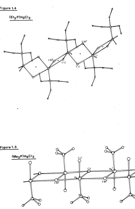
The complex ( Et^P^gCl^, Figure 1.4, shows even further extension via halogen-bridging and has been described as a chlorine-bridged polymer with mercury in a distorted trigonal bipyramidal
environment [ 5 ] . There are two short Hg - Cl bonds at o
2.42(1) and 2.56(1)A> Hg - Cl(2) and Hg - Cl(l) , and a short o
Hg - P bond at 2.35(1)A lying in the equatorial positions around mercury, and two longer apical H g - Cl contacts at 3.04(1) and
3.21(1)A , Hg - Cl(l’) and Hg - Cl(2’) . The angles within
the equatorial PHgCl^ unit range from 98.7(3)
[ci(l)
- Hg - 01(2)]to 145.4(4)° [ Cl(2) - Hg - P ], while the angle between the two longer apical contacts Cl(l’) — Hg - Cl(2f) is close to linearity at 170.8(3)°.
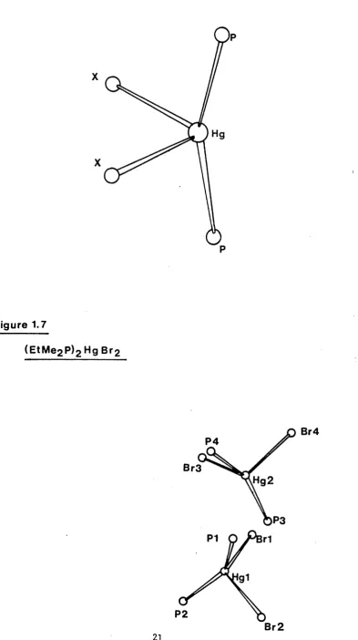
(Me^P^gC^ is found to have a polymeric arrangement [ 5] , which can be envisaged as being ’ionic’, containing
[ci
- Hg - PMe ] + and Cl" ions arranged alternatively in azig-zag chain [Figure l.s] . The [ Cl - Hg - PMe^ ] + cation is
almost linear
[cl
- Hg - P angle , 162.1(1)°] . The co-ordinationnumber of mercury is increased to five by three further Hg - Cl contacts: Hg-Cl(l), Hg - Cl(l') and Hg - Cl(l” ) at
o
2.782(4), 2.94(4) and 3.489(4)A respectively. The ’chain-like’ arrangement is different to that found in ( Et^P^gC^, as the bridging involves only one of the chlorine atoms of the
’C^HgPMe^’ unit. Preliminary photographs [ 5 ] indicate the bromo-analogue to be isostructural.
Figure 1.4
ci:
Figure 1.5
Cl 2
Cll
1.3.3 Crystallographic studies of (R^P)2HgX2 complexes
The (R2?)2HgX2 complexes fully characterised so far [6,7,8,14] are all tetraco-ordinate monomers with varying degrees of distortion from a regular tetrahedral geometry (Figure 1.6).
Of the structures reported, (Ph^P^Hg^tfc] has an almost
regular tetrahedral geometry around the mercury, as is evident from the I - Hg - I and P - Hg - P angles [110.43(9)° and
108.95(4)° respectively, Table 1.1 ] .
(Bu^P^HgC^ [ *-s more distorted, the P - Hg - P [ 139(2)°]
and Cl - Hg - Cl angles [ 105(2)°] being quite different.
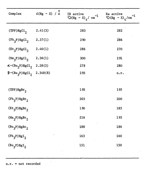
The structure of ( E tMe^P^HgB^ [l^] contains two independent monomer units in the unit cell (Figure 1.7), each having
mercury in a distorted tetrahedral environment, with P - Hg - P angles of 147(2) and 150(2)° for PI - Hgl - P2 and
P3 - Hg2 - P4 respectively (Table 1.1).
The remaining structure previously reported [7] is
(Et^P^HgC^, which is also a distorted tetrahedral species with a P - Hg - P angle of 158.5(5)° [Table l.l] .
Figure 1.6
X
X
Figure 1.7
(EtM e2 P)2 H g B r2
P4
Br3
,Hg2
P3
P1 r1
[image:24.564.87.481.93.796.2]Table 1.1
Crystal data for (R3P)2HgX2 complexes
Compound X - Hg - X /° P - Hg - P /° Ref
<Ph3P)2HgI2 110.43(9) 108.95(4) 6
(Bu3P)2HgCl2 105(2) 139(2) 8,14
(EtMe,P),HgBr, 102(1) 147(2) 14
107(1) 150(2)
(Et3P)2HgCl2 105.5(5) 158.5(5) 7
1.3.4 Spectroscopic studies of some (R^P)HgX2 complexes
The solid-state vibrational spectra of the complexes mentioned in Section l.'3.2 and those of their hromo- and iodo-
analogues have been previously examined [ 14 ] . Using the structure/spectra correlation technique (Section 1.2), various correlations were put forward between Q(Hg - Cl) spectral patterns and structural features.
Tables 1.2 and 1.3 show the ^O(Hg-Cl) assignments
made [ 14 . The following is a summary of detailed structure/spectra correlations given by Jones [ 14 ] , who was able to characterise spectroscopically (especially via infrared spectra) the five
separate structures described in Section 1.3.2: an asymmetric dimer, an almost symmetric dimer, a chain polymer, a tetramer and an ionic chain.
(a) In the structures of ( R ^ H g C ^ [ R^P = T P P , Ph^P, Bu^P, Et^P or Me^P ] , the common structural feature of one
short Hg - Cl bond is characterised by a band around
280 - 300 cm ^ in the infrared and Raman spectra, assigned to C(Hg - Cl)* (Table 1.2).
(b) The differentiation between each structural type was indicated by examination of the lower wavenumber regions
(c) The (Me^P^gCl^ 1 ionic1 chain structure was distinguished from the other structures by the lack of -Q(Hg - Cl)^ modes in the infrared between 140 - 300 cm
(d) Differentiation between the polymeric (Et^P^gC^ complex and the dimeric (Ph^P^gC^ and (TPP)HgCl2 complexes
is by observation of strong bands in the infrared spectrum
of the Et^P complex at 117 cm 1 and 90 cm 1 (Table 1.3) in addition to the bands found in all three spectra around
150 - 220 cm"1.
(e) The more complicated nature of the spectrum of
oc- (Bu^P^gC^ did not give rise to any information
which would enable a definite identification of that particular 'tetramer* structure.
Table 1.2
^Q(Hg - X) modes associated with 'short* Hg - X bonds f141 .
0
Complex d(Hg - X) / A IR active 1
Q(Hg - X)t/ cm” Ra active ^(Hg -X)t/cm"..
(TPP)HgCl2 2.41(3) 283 282
(Ph3P)HgCl2 2.37(1) 290 286
(Et3P)HgCl2 2.40(1) 286 270
(He3P)HgCl2 2.36(1) 300 291
«3C-(Bu3P)HgCl2 2.28(3) 278 280
p-(Bu3P)HgCl2 2.348(8) 295 n.r.
(TPP)HgBr2 195 195
(Ph3P)HgBr2 203 200
(Et3P)HgBr2 190 185
(Me3P)HgBr2 216 193
(Bu3P)HgBr2 188 186
(Ph3P)HgI2 163 160
Table 1.3
IR - active -Q (Hg - X), modes associated withb
Complex Structure d(Hg - X)/A O (Hg ~ X)b/cm_1
(TPP)HgCl2 asymmetric dimer 2.54(1),2.75(1) 219 156
(Ph3P)HgCl2 symmetric dimer 2.623(8),2.658(8) 188 183
(Et3P)HgCl2 polymeric chain 2.56(1),3.04(1),3.21(1) 203,198sh,117,105
(Me3P)HgCl2 * ionic1 chain 2.941(4),3.489(4),2.782(4) 141 and below
o(.-(Bu3P)HgCl2 tetramer 2.67(2),2.90(2),3.38(3) 252,218,179
P-(Bu3P)HgCl2 symmetric dimer 2.720(6),2.736(6) 181 173
(TPP)HgBr2 151 117
(Ph3P)HgBr2 137 117
(Et3P)HgBr2 144,91,84,72
(Me3P)HgBr2 166
(Ph3P)HgI2 117 89
H 8 _ bridge bonds [14].
3:.3.5 Spectroscopic studies of (R^P)2HgX2 complexes
These complexes have also been studied [ 14] with the use of infrared and Raman spectra, in terms of C^v symmetry, for which two
0(Hg - X) modes are predicted in the infrared and Raman spectra (point group analysis).
The vibrational assignments of 2:1 complexes are given in Table 1.4, together with the magnitudes of the Hg - X bond lengths and the P - Hg - P angles. The magnitude of the Hg - X bond lengths and the P - Hg - P angles were shown [ 14] to
influence the position of the 0(Hg - X) bonds. Thus the 0(Hg - X) modes were found to be in the ranges 160-240, 110-166 and 90-140 cm ^
for the chloro-, bromo- and iodo- complexes respectively, with the lower wavenumber bands in each series characteristic of compounds having the larger P - Hg - P angles and longer Hg - X bonds.
This correlation between O(Hg - X) and molecular parameters can thus be used in the study of other (R P) 0jgX2complexes to indicate the degree of distortion from a regular tetrahedral structure
Table 1.4
Relationship between the structures and spectra of some
^R3P^2HgX2 comPlexes •
Parameter (R3P)2HgCl2 (R3P)2HgBr2 (R3P)2HgI2
Ph^P Bu3P EtMe2P Et3P Ph3P Bu3P EtMe£P Et3P Ph3P Bu3P EtMe£P Et3P
P-Hg-P
angle/ 139 158.5 148.5 103-95
av.Hg-X
Length/A 2.60 2.68 2.79 2.75
^0 as(Hg-X) 223 205 181-169 176 155 127 112 132 129 107 94 109
1.4 Survey of stereochemistry of CdX^ complexes of neutral unidentate
ligands
1.4.1 Introduction
In the solid-state it is known that cadmium (II) halide addition complexes of the type L^CdX^ [ L = neutral unidentate ligand; n = 1 or 2 ] generally pack together in such a way that
extension from the monomer units is obtained by cadmium-halogen bridging interactions [18-34] . These interactions are
usually longer than the sum of the covalent radii (Cd - Cl = o
2.40; Cd - Br - 2.55; Cd - I = 2.74A), but within the sum of the van der Waals1 radii (Cd - Cl = 3.21; Cd - Br = 3.36; Cd - I = 3.56A).
The structures of the complexes, (R^P)nHgX2 (n = 1 or 2)
have been discussed in Section 1.3.2 and it was thought that similar structural variations might occur in the analogous cadmium complexes. However, despite the fairly close relationships found [l5-17] between some trihalogeno salts of the two metals, [MX^ ] , the mercury structures (Section 1.3.2) showed no relationship to the double-chained structures reported in the literature for LCdX^ type complexes (Section 1.4.2). Similarly,
the halogen-bridged chain structure of systems aPPaars
common for Cd [18-20] complexes but is rare for Hg.
1.4,2 Crystallographic studies of LCdX^ complexes
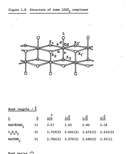
At the start of the present project the structures of a number of complexes had been reported, three of which are of a similar
structural type (Figure 1.8). They are (MeNHCONI^CdC^ [is] , (imidazole)CdCl^ [ 19] and (MeCSNH^CdC^ [ 2o] , where the donor atom of the ligand is oxygen, nitrogen and sulphur, respectively. The structure is that of a double-chain arrangement, in which two chains of cadmium atoms are linked by halogen bridges. The bridging units are not perfectly symmetrical. The two polymeric chains are linked together by further Cd - X interactions
( V in Figure 1.8), with the ligand occupying the sixth co-ordination position on the cadmium atoms.
While the structures mentioned above have their ligands acting as neutral unidentate ligands, in the following two 1:1
complexes the ligands are bidentate. The pyridine-N-oxide
complex (C^H^NO) Cdl^ [ 21] contains ’monomer' units which are linked together to form a polymeric chain, alternately bridged through pairs of iodine and oxygen atoms. The environment of the pentaco-ordinate Cd atom is distorted trigonal bipyramidal
(Figure 1.9).
The structure of dichloro(dicyandiamide) cadmium (II) (Figure 1.10) [22] contains cadmium atoms surrounded by four chlorine and two nitrogen atoms,giving rise to a slightly distorted octahedral environment about the metal atoms. The chlorine atoms form bridges between adjacent metal atoms to form polymeric chains running parallel to each other. These chains are linked together
by dicyandiamide bridges.
Figure 1.8 Structure of some LCdX^ complexes
Cd
Bond lengths / %
L
MeNKONH,
C3H4N2 MeCSNH.
X Cl Cl Cl
o a/A 2.67
b/A 2.62
c/%
2.60
,o d/A
2.18 2.729(3) 2.604(2) 2.676(3) 2.243(5) 2.760(2) 2.570(2) 2.630(2) 2.55(1)
Bond angles ( ) L
MeCSNH
C3H4N2
L-Cd-Xl L-Cd-X2 L-Cd-X3 X2“Cd-X3 X^d-0^
98.2(7) 81.7(6) 158.6(4) 79.5(4) 90.6(4)
87.5 90.8
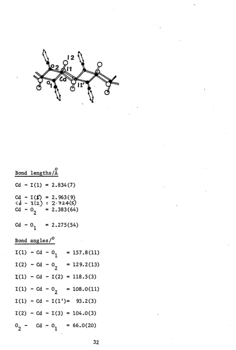
Figure 1.9 Structure of (C^H^N0)Cdl2
Bond lengths/A
Cd - 1(1) = 2.834(7) Cd - I(J') = 2.963(9) cd - 1(2) = 2-724CS) Cd - 02 = 2.383(64)
Cd - 01 = 2.275(54)
Bond angles/0
1(1) - Cd - 0 1 = 157.8(11)
1(2) - Cd - 02 = 129.2(13)
1(1) - Cd - 1(2) = 118.5(3)
1(1) - Cd - 02 - 108.0(11)
1(1) - Cd - 1(1*)= 93.2(3) 1(2) - Cd - 1(3) = 104.0(3) 02 - Cd - 01 = 66.0(20)
Figure 1,10
Structure of dichloro(dicyandiamide) Cd(II)
N3 6N4
CI2
CI1
N2
N1
CI2
C MN
-— N2
N
-Bond lengths/ A
Cd - Cl(l) = 2.607(3) Cd - Cl(2) = 2.647(3) Cd - C l d 1) = 2.572(3)
Bond angles/ 0 Cl(l) - Cd - N(l) Cl(l) - Cd - Cl(l!) Cl(l) - Cd - Cl(2f') Cl(l) - Cd - N(21T1) Cl(l$ - Cd - N(l)
Cd - Cl(211) = 2.630(3)
Cd - N(l) =2.358(4)
Cd - N(2Tf*) = 2.430(4)
87.0(1) Cl(2) - Cd - Cl(2f1)
85.03(4) Cl(2) - Cd - N(2,ff)
97.36(4) N(l) - Cd - Cl(2” )
97.6(1) N(l) - Cd - N(2f11)
89.1(1) Cl(l') - Cd - N(2,f)
= 84.68(3)
1.4.3 Crystallographic studies of L^CdX^ complexes
Two types of structure for these 2:1 complexes have been authenticated in the literature. The most common is that of a single chain polymeric structure having the cadmium atoms in an octahedral environment (Figure 1.11) formed by halide- bridges between cadmium atoms. The fifth and sixth co-ordination positions of the cadmium are taken up by the neutral monodentate ligand. This type of structure is observed in (Py)2CdX2
[ X = Cl, or Br] [23,24] , ( imidazole)2 CdC^ [25] and
^n*3^2 ^ X2 [ X = Cl or Br] [26] , which all have nitrogen-donor ligands, and by five complexes with oxygen as the donor atom,
viz. (urea^CdC^ [ 27] , (methylurea^CdC^ [ 28 ] , (biuret)2CdCl2 [ 29] , (acetamide^ CdC^ [30] and (HCONH2)2CdCl2 [3l] .
The second structural type found for these 2:1 complexes is that of a tetrahedral monomer (Figure 1.12). Three complexes of this type have been reported previously, one involving a sulphur-donor
ligand, (thiourea)2CdCl2 [32] r one with phosphorus as the donor
atom, (Ph^P^CdC^ [33] , and one of a nitrogen donor, (aniline^ Cdl2 [ 34 ] . In all cases the arrangement about cadmium is
close to that of a regular tetrahedron.
Figure 1.11
Structure of some L^CdX^ complexes
Bond lengths / A
L **X a/Ao b/Ao c/A,o
Py Cl ‘ 2.65 2.65 2.35
Im Cl 2.731 2.706 2.248
nh3 Cl 2.71 2.71 2.10
nh3 Br 2.86 2.86 2.10
biuret Cl 2.62 2.55 2.34
acetamide Cl 2.58 2.58 2.23
HCONH_2 Cl 2.383 2.912 2.34
Bond angles /°
L X Xx - Cd X2 L1 - Cd - X l Lx - Cd
Figure 1.12 Structure of (Ph^P^CdCl^ (tu^CdC^
X
Bond lengths /A
Complex £
2.635(5) (Ph3P)2CdCl2
(tu)2CdCl2
Bond angles / Complex
2.45
2.632(6) 2.504(6)
2.45 2.51
(Ph3P)2CdCl2
(tu)2CdCl2
L - Cd - L1 2
107.6(2)
L 1 - Cd - X
104.5(2)
d
2.440(6)
2.50
L - Cd - X1 2
105.7(2)
Complex (Ph3P)2CdCl2 (tu)2CdCl2
X x - Cd - X 2 113.9(2) 103
x-i-cd - l 2
112
.
2(
2)
106
x 2 - c d
- l 2112
.
1(
2)
105
1.4.4 Spectroscopic studies of L^CdX^ complexes
a) LCdX2
(i) Double-stranded halogen-bridged chain: One of the best examples of spectral characterisation in terms of this structure is a
Line group analysis was used to predict
fO(M-X) = 3 Ag(Ra) + 2 Bg(Ra) + 2 Au(I.R.) + 2 Bu(I.R.)
R)(M-*0 = A (Ra) + B (I.R.)
g u
The 3-4 0(M-X) and one Q(M-N) bands located in the infrared and Raman spectra (Table 1.5) were consistent with this line group analysis.
(ii) (R^P)CdX2 complexes: These have all been suggested to have halogen-bridged dimeric structures [36,37] since the observed bands (Table 1.5) correlate with predictions for a halogen- bridged dimer from group theory i.e.
study of (pyridine)MX^ complexes [m - Cd; X = Cl or Br] [35],
pQ(Cd-X) = A (Ra) + B (Ra) + A (I.R.) + B (I.R.) b g ^ u u
[O(Cd-P) « Ag(Ra) + B^d.R.)
For example, (Bu^P)CdX^ [ 36 ] complexes showQ(Cd-X)t at 285, 198 and 159 cm ^ for X ® Cl, Br or I respectively,
and ^(Cd-X) at 208, 148 cm ^ for X = Cl, Br respectively, b
b) L2CdX2
(i) Octahedral halogen-bridged chain structure^ The spectral characterisations of (Py)2CdX2 [ 38 ] and (aniline)^CdX2
[ 39] have been reported. The line group theory prediction
was (I>2h):
R(Cd-X) *= A (Ra) + B (Ra) + B. (I.R.) + B. (I.R.) g g lu ju
fl)(Cd-dT) = Ag (Ra) + B^d.R.)
However the observed number of bands (Table 1.6) was
substantially in excess of the predicted number, indicating the need for a different group theory approach.
(ii)Tetrahedral monomers: The (R^P)^CdX2 complexes i^iichhave been suggested to be tetrahedral monomers
[ 37, 40, 41 ] , show two ^(M-X^ modes in the infrared and Raman spectra (Table 1.6). This is consistent with a monomeric structure ( C ^ point group) . The (Cd-X2) modes are located in the ranges 260-270, 180-200, and
145-165 cm ^ for the chloride, bromide and iodide complexes respectively. The Q(Cd-P) modes were located around 130-140 cm ^ in all the infrared and
Raman spectra.
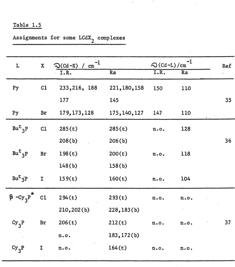
c) Summary of spectroscopic studies in LnCdX2 complexes
The spectra of LCdX2 complexes show differentiation between the halide-bridged dimeric structure and the double-stranded halogen-bridged structure. Thus the halogen-bridged dimers give a O(Cd-X)^ baid around 290, 200 and 165 cm * for Cl,
Br and I respectively and Q(Cd-X) bands correspondinglyb
around 210, 150 and 100 cm However the octahedral structure has 3-4 bands in the range of 170-240 cm ^
for chloride and 128-179 cm ^ for the bromide (all bands being in the infrared).
The 2!1 complexes are mainly tetrahedral monomers xdiich give rise to two (H-X^) bands. The octahedral polymeric structures give rise to a more complex
Table 1.5
Assignments for some LCdX^ complexes
L X ^(Cd-X) / cm”1 ^0 (Cd-L)/cm 1 Ref
I.R. Ra I.R. Ra
Py Cl 233,216, 188
177
221,180,158 145
150 110
35
Py Br 179,173,128 175,140,127 147 H O
Buc3P Cl 285(t)
208(b)
285(t) 206(b)
n.o. 128
36
But3P Br 198(t)
148(b)
200(t) 158(b)
n.o. 118
Bu^'P I 159(t) 160(t) n.o. 104
P -cy3** Cl 294(t)
210,202(b)
293 (t) 228,183(b)
n.o. n.o.
Cy3P Br 206(t)
n.o.
212(t) 183,172(b)
n.o. n.o. 37
Cy3P I n.o. 164(t) n.o. n.o.
n.o. = not observed, t = terminal, Cd-X, b = bridging Cd-X.
* Another crystalline form (cL-) is known [37] : i.r. bands 273,249, 210-200 and Ra at 277, 273, 252, 248, cm"’1.
Table 1.6
Assignments for some L^CdX^ complexes
L X Q (Cd-X)/cm"1 '0(Cd-L)/cm 1 Ref
I.R. Ra I.R. Ra
Ph3P Cl 268 136
261 40
Br 195 134
176
I 166 133
145
(m-CH3C6H4)3P Cl 270 263 130 . 132
Br 196 136 130 41
181 182
I 165 164 127 n.o.
133 133
(p^H3C6H4)3P Cl 270 262 138 n.o.
Br 198 199 136 n.o. 41
177 175
I 165 165 n.o. n.o.
140 140
(p-ch3oc6h4)3p Cl 270 262 n.o. n.o.
Table 1.6 ctn
L X *0(Cd-X)/
I.R.
-T— cm
Ra
«0(Cd-L)/cm ^ I.R. Ra
Ref
(p-(ch3)2nc6h4')3p Cl 266 250 n. o. n.o.
250
Br 187 190 n.o. n.o. 41
175 174
I 154 155 n.o. n.o.
139 137
Cy3P Cl 260 n.o. n.o. n.o.
Br n.o. n.o. n.o. n.o.
I n.o. n.o. n.o. n.o. 37
C6H5NH2 Cl 223 386,378 374
208 328,311 324
Br 140 131 373 374 33
111 106 324 324
Py - Cl 127-170 199
Br 105 167,180 38
n.o. - not observed
CHAPTER 2
CRYSTALLOGRAPHIC AND SPECTROSCOPIC STUDIES OF SOME (R_P) HgX„ COMPLEXES [ X = Cl, Br or I;
______ o n z u
Contents
Page 45
2.1 Introduction.
2.2. Crystallographic examination of (Ph^P)^HgCl^. 46
2.3 Crystallographic examination of (Cy^HgC^. 53
2.4 Spectroscopic studies of (Cy3P)nHgX2.
complexes (n = 1 or 2; X - Cl, Br or I). 60
2.4.1 (Cy3P)2HgX2. 60
2.4.2 (Cy3P)HgX2 . 64
2.1 Introduction
Chapter 1 outlined the structural variations within the two
series of complexes, (R-P) HgX„ f X = Cl, Br or I; n = 1 or 2 1 .3 n 2 *• J
A variety of structures was observed for the (R3P)HgX2 complexes ranging from discrete dimers, to a loosely held tetramer, to polymeric chain structures. Discrete tetraco-ordinate
monomers, with varying degrees of distortion from a regular tetrahedral geometry, were observed for the (R^P)2HgX2
complexes. It was suggested [ 14] that the degree of association
in the (R2P)HgX2 complexes and the extent of tetrahedral
distortion of the (R^P) 2*^X2 complexes could be related to the size and the donor strength of the phosphine ligand.
In order to investigate further the factors influencing the
solid-state structures of (R0P) HgX_ | n = 1 or 2 1 complexes,0 n 2 L J
the crystallographic and spectroscopic study reported in Chapter 1 has been extended. In particular the
Crystallographic examination of (Ph^P^HgC^
Crystal data
C36H30C12HsP2 Mr “ Monoclinic
a = 9.465(5), b = 17.525(6), c = 19.664(5)A,
U = 3261.60&3 , oL = 90.00, p = 90.10(6),
"Xs 90.00°. m D = 1.60g cm 3 (by flotation using CHC10/CHI<3) ,j j
D = 1.62. a cm”3 Z = 4, F(000) = 1560 1, c
/i(Mo - K«t) - 47.37 cm ^
Systematic absences:
OkO reflections are absent for k = 2n + 1
hO 1 " " 1 = 2n + 1
Space group: P21/c
Data collection and structure refinement
A colourless crystal, approximate dimensions, 0.40 x 0.25 xO. 25 m m. A,- <Wlb coiMCiAenl*
was mounted with thej^rotation (u>) - axis of a Stoe Stadi 2 two circle diffractometer. Data were collected using the background
-(*) scan - background technique. Lorentz and polarisation corrections were applied. Those reflections having I/<r(I) greater than 3.0 were considered to be observed. [ The net intensity I = T-B, where T = scan count, B = mean background count over the scan width,
<t(I) = (T-Bc/2t) *, where c = scan time, t = time for background
measurements at each end of the scan] .
Thus out of 3824 unique reflections collected, 2763 had I/<r(I) "7/3.0 and were used for subsequent refinement. The positions of the
function and the remaining non-hydrogen atoms were located from successive difference electron-density maps. Scattering factors were calculated [42] using an analytical approximation. The carbon atoms of the phenyl rings, after location from electron density maps, were fixed in ideal positions to give C-C bond
lengths = 1.395 & and C-C-C angles = 120.0°
The weighting scheme adopted was:
w * 1.3204/ [ qp2(Fo) + 0.0015(Fo)2 ] .
The structure was refined with full matrix refinement with anisotropic temperature factors of the form:
T « 2
.
2 * 2 , 2 *2 .2 *2 * *exp [ - 2-rr (Un h' a z + U^k^b + 0^1 c * + 2U12hka b +
2U13 h I a c + 2U23k 1 b c )J .
2
The function minimised in the refinement was Iw( I Fol - IFcl ) and the final R value is given by:
R _ ifcot - iFej I
I M
The Rf function is given by:
Rt = £>/w ||Fq1 - [Fell
I /w |Fo| [ w - weighting function]
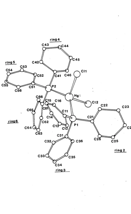
Description of structure:
(Pli^P)^HgCl^ crystallises with four monomeric units in the unit cell. The geometry of these monomers is shown in Figure 2.1. The mercury is tetraco-ordinate, being bonded to two phosphorus
o
atoms, at distances of 2.55€(9) and 2.476(13)A away. The co-ordination polyhedron about mercury is distorted with-
angles ranging from 135.2(4)° (Pl-Hg-P2) to 99.3(4)°
(Cl(2)-Hg-Pl). Figure 2.2 shows the packing of the four monomers in the unit cell. The geometry of the complexed Ph^P ligand is compatible with that observed for the free ligand [ 43 ] . The average P-C distance found (average = 1.79&) is close to that observed for the free Ph^P ligand (1.82&). The average OP-C angle in the present complex (106.5°) is less distorted from a regular tetrahedral geometry than that found in the free ligand (average - 103.0°). The C-P-Hg angles in this complex are slightly larger than that required for regular tetrahedral geometry (Table 2.1). Appendix A.3 contains relevant mean planes data for (Ph^P)^HgCl^. The angles between the phenyl rings are very similar to that found in the free ligand and in
(Ph3P)HgCl2 (Table 2.2)
While both (Ph^P^HgC^ and (Ph^P^Hgl^ [ 6 J adopt monomeric structures, there is a striking difference in the value of the P-Hg-P angled 135.2(4)° for the chloride complex, 108.95(4)° for the iodide complex . This difference can be attributed to both the nature of the phosphine ligand and also of the halogen atom and will be discussed in Chapter 5.
Figure 2.1 The molecular structure of ( Ph3 P)2
HgCl2-ring 4 C43 £ 4 4
C45
ring 5 C42
C53
C54 CM
£41 C46 C52
C55 C51 P 2
C56
Hgi C66
:15 C16 CI2
C22 C23
(C61
C6 £11
C21
C14
ring 6 C62
P1
C1 C24
C64
C63 C31
C2 C25
C36 C32
ring 2
C33 C35
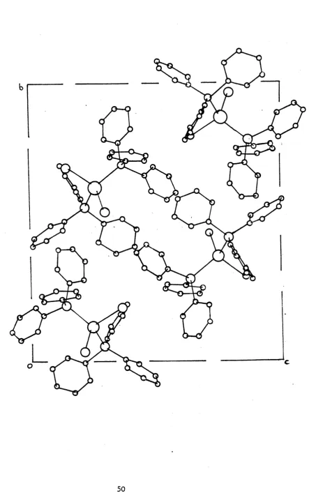
[image:52.556.97.535.68.785.2]Figure 2.2 The Crystal structure of ( P I ^ P ^ H g C ^
Table 2.1
Bond distances and angles for (Ph^P^HgC^ with estimated standard
deviations in parentheses*
o a) Bond distances/A
Hg - Cl(l) = 2.558(9) PI - C21 = 1.726(39)
Hg - Cl(2) « 2.476(13) PI - C31 = 1.790(24)
Hg - PI = 2.476(8) P2 - C41 = 1.814(24)
Hg - P2 = 2.378(13) P2 - C51 = 1.763(32)
PI - Cll = 1.792(29) P2 - C61 = 1.856(26)
All C-C bond lengths were fixed at 1.395&.
b) Bond angles/0
Cl(l) " Hg — Cl (2) = 113.3(4) Hg - P2 - C51 = 105.1(14)
Cl(l) - Hg - PI = 103.8(3) Hg - P2 - C61 110.8(11)
Cl(l) ‘ Hg - P2 = 104.0(4) Cll - PI - C2l = 104.2(15)
Cl(2) - Hg - PI = 99.3(4) Cll - PI -•C31 = 108.2(15)
Cl (2) - Hg - P2 = 100.9(4) C21 - PI - C31 = 106.0(13)
PI - Hg - P2 = 135.2(4) C41 - P2 - C51 = 105.1(14)
Hg - PI - Cll = 113.7(11) C41 - P2 - C61 = 106.8(11)
Hg - PI - C21 = 112.3(10) C51 - P2 - C61 = 108.9(17)
Hg - PI - C31 = 111.8(9)
Hg - P2 - C41 = 114.4(14)
Table 2.2
Mean planes3 Free ligand [431 (PhjDHgClj [ fc ] (Ph3P)HgCl2
A-B 84.0° 84.4°
PI 86.4°
P2 91.8'
B-C 78.1° 80.1° 60.7° 88.5'
C-A 76.5° 75.4° 77.8° 89.6'
2.3 Crystallographic examination of (Cy^P^gC^
Crystal data:
C18H33^2H^i>: Mr = 551.93, Triclinic,
a = 10.8431(5), b = 14.1183(4), c - 14.7919(3) A.
U « 2088.80S3 , o 94.7630(6) £ » 80.3793(5),
7f a 110.6^23(6)°, Dm = 1.70 g cm 3 (by flotation using CHCl^/CHBr^)
Dc= 1.75 g cm"*3, Z = 4 F(000) = 1030 ,
js(Ho - K«*) ■* 73.97 cm
Space group;
Preliminary photographs did not distinguish between PI and PI, but subsequent analysis showed the centrosymmetric space group, PI, to be correct.
Data collection and structure refinement:
The data set used for this structure was collected on an Enraf Nonius Cad-4 diffractometer . Details of the data collection are given in Appendix A.5.
A colourless crystal, approximate dimensions, 0.30 x 0.25 x 0.30 mm was used for intensity measurements. 5116 unique reflections were
collected of which 3563 had I/or(I) ^3.0 and were used for
refinement. Correction for absorption effects have been made to the data along with corrections for Lorentz and polarisation effects. Common isotropic temperature factors were applied to the hydrogen
The weighting scheme adopted was:
w = 1.5221 / [cr2(Fo) + 0.00031 (Fo)2] .
The final R values were R = 0.039 and R f = 0.040. Final atomic and thermal parameters are listed in Table A.1.2 (Appendix A.l) and calculated/observed structure factors are given in
Appendix A.2. Bond angles and distances are given in Table 2.3.
The relevant mean planes data are contained in Appendix A.3.
Description of structure
The structure consists of two independent dimers within the unit cell (Figure 2.3). Figure 2.4 shows the arrangement of the two dimer units in the unit cell and the two Hg^Cl^ bridging units are
found to be almost 90° to each other. Both independent mercury atoms lie in distorted tetrahedral environments with angles about mercury ranging from 95.9(1) to 132.0(1)°, Hgl, and 84.1(2) to
139.6(2)°, Hg2. The Cy^P ligands are arranged mutually trans and
the four-membered ring is planar and contains a centre of
symmetry.
One of the main differences between the two dimer units is shown by the Hg-Cl bridging distances. The Hg(2) dimer contains a very
symmetric bridging arrangement with Hg(2) - Cl(22) and Hg(2) - Cl(22f) o
equal to 2.642(4) and 2.665(5)A respectively. In contrast the dimer containing Hg(l) has very asymmetrical bridging with 2.559(3)
[ Hgl - Cl(12r) ] and 2.778(3)& Hgl - Cl(12) .
The P - Hg - Cl ° terminal. .. angles also differ between the two dimers,
with values of 132.0(1) [ PI - Hgl - Cl(ll) ] and 139.6(1)° '
[p2 - Hg2 - Cl(21)] being found.
The cyclohexyl rings adopt the preferred chair conformation with all
Figure 2 .4
Table 2.3
Bond distances and angles for (Cy^P^gC^ with estimated standard deviations
in parentheses
Symmetry code
none x, y, z.
1 1”X, -y, 1-z.
1-x, -1-y, Z. *
Bond lengths
ll
Hgl - Cl(ll) = 2.413(3) Hg2 - Cl(21) = 2.391(5)
Hgl - c i a f i = 2.778(3) Hg2 - Cl(22) = 2.642(4)
Hgl - Cl(12 ) = 2.599(3) Hg2 - C1(22T ') = 2.665(5)
Hgl - PI = 2.412(3) Hg2 - P2 = 2.416(3)
PI - c m a 1.853(14) P2 - C211 = 1.839(10)
PI - C121 = 1.849(11) P2 - C221 = 1.827(17)
PI - C131 = 1.833(10) P2 - C231 = 1.825(11)
c m - cii2
= 1.537(15) C211 - C212 - 1.559(16)C112- C113 = 1.552(23) C212 - C213 = 1.491(16)
C113- C114 = 1.508(22) C213 - C214 = 1.480(24)
C114- C115 = 1.490(20) C214 - C215 = 1.533(19)
C115- C116 . = 1.513(22) C215 - C216 = 1.530(16)
C116- Clll = 1.537(18) C216 - C211 = 1.536(20)
C121- C122 = 1.494(15) C221 - C222 = 1.481(20)
C122- C123 = 1.538(18) C222 - C223 = 1.481(27)
Cl23- C124 = 1.492(23) C223 - C224 = 1.412(23)
C133 - 0134 = 1.532(20) C233 - C234 1.522(21)
C134 - C135 = 1.531(22) C234 - C235 1.452(21)
C135 - C136 = 1.529(16) C235 - C236 1.463(21)
C136 - C131 = 1.556(17) C236 - C231 1.505(15)
Hgl -- - Hgl ' = 3.938(1) Hg2 -— Hg211 * 3.774(1)
Bond angles /°
Cl(ll) - Hgl - Cl(12) = 106.1(1) 01(21) - Hg2 - 01(22). * 95.2(2)
Cl(ll) “ Hgl - 01(12')= 101.5(1) 01(21) - Hg2 - 01(22” )= 92.6(3)
Cl(12) - Hgl - 01(12’)* 85.9(1) 01(22) - Hg2 - 01(22” )= 84.1(2)
Cl(ll) - Hgl - PI * 132.0(1) 01(21) - Hg2 - P2 • = 139.6(2)
Cl(12) - Hgl -PI * 99.8(1) 01(22) - Hg2 - P2 111.8(1)
01(12') - Hgl - PI * 120.3(1) 01(22")- Hg2 - P2 108.6(1)
Hgl’ - 01(12)- Hgl = 94.1(1) Hg2” - 01(22)- Hg2 = 95.9(1)
Hgl PI - c m = 112.2(4) Hg2 - P2 - 0211 ■110.2(4)
Hgl - ' PI - 0121 = 108.0(4) Hg2 - P2 - 0221 s 107.4(5)
Hgl - PI - 0131 = 111.0(5) Hg2 - P2 - 0231 = 112.1(4)
Clll - PI - 0121 = 105.5(6) 0211 - P2 - 0221 = 107.5(6)
Clll - PI - 0131 = 110.9(5) 0211 - P2 - 0231 = 107.8(5)
0121 - rPl - 0131 = 108.7(5) 0221 - P2 - 0231 = 111.8(6)
PI - Clll - 0112 = 112.9(10) P2 -.0211 - 0212 = 110.1(8)
PI - Clll - 0116 = 115.0(8) P2 - 0211 - 0216
,
= 112.7(8)Clll - 0112 - 0113 = 108.5(12) 0211 - 0212 r 0213 =5 111.8(10)
0112 - 0113 - 0114 = 110.5(11) 0212 - 0213 - 0214 = 113.6(12)
0113 - 0114 - 0115 = 111.7(12) 0213 - 0214 - 0215 = 111.1(12)
0114 - 0115 - 0116 = 113.0(14) 0214 - 0215 - 0216 = 111.7(11)
Cl 15 - 0116 - Clll = 110.6(22) 0215 - 0216 - 0211 = 110.5(11)
0116 — Clll - 0112 = 109.3(10) 0216 - 0211 - 0212 . a 109.6(9)
PI - C121 - C122 = 110.8(7)
PI - C121 - C126 = 111.7(8)
C121 - C122 - Cl23 = 111.6(9)
C122 - C123 - C124 = 111.5(11) C123 - C124 - C125 = 112.0(16)
C124 - Cl25 - C126 = 112.0(10)
C125 - C126 - C121 = 109.5(14)
C126 - C121 - C122 = 112.9(11)
PI - C131 - C132 = 110.9(7)
PI - C131 - C136 = 109.6(7)
C131 - C132 - C133 = 111.2(1)
C132 - C133 - C134 = 111.7(11)
C133 - C134 - C135 = 108.5(14) C134 - C135 - C136 = 113.3(11) C135 - C136 - C131 = 109.1(10)
C136 - C131 - C132 = 111.5(12)
P2 - C221 - C222 = 119.5(11)
P2 - C221 - C226 = 119.6(13)
C221 - C222 - C223 = 114.3(13) C222 - C223 - C224 = 117.9(15) C223 - C224 - C225 = 115.4(17) C224 - C225 - C226 = 113.5(15) C225 - C226 - C221 = 120.4(16) C226 - C221 - C222 = 116.4(14)
P2 - C231 - C232 = 114.5(9)
P2 - C231 - C236 = 113.4(9)
C231 - C232 - C233 = 109.4(13)
C232 - C233 - C234 111.6(14)
C233 - C234 - C235 = 111.4(13) C234 - C235 - C236 = 112.6(14)
C235 - C236 - C231 = 109.6(11)
2.4 Spectroscopic studies of (cy3P)nHgX2 complexes
(n = 1 or 2; X = Cl, Br or I). 2.4.1 (Cy3P)2HgX2
The structures of (R3P)2HgX2 complexes are all tetrahedral monomers (Section 1.3.5), and the same structure is reasonably assumed for the Cy^P complexes. On the basis of C2v point group symmetry, the following modes are predicted:
fTnt * 4A1(I.R.,Ra) + A2(Ra) + 2B1(I.R.,Ra) + 2B2(I.R.,Ra)
|0(Hg-X) = A 1(I.R.,Ra) + B (I.R.,Ra) .
pd(Hg-P) = A1(I.R.,Ra) + B (I.R.,Ra).
The infrared spectrum is shown in Figure 2.5 while Table 2.4 contains the vibrational assignments. The infrared assignments
for the chloride are in good agreement with previous suggestions [37],
with the ^(Hg-Cl) A^ and B^ modes located at 209 and 197 cm
These results are consistent with the Raman spectral assignments [ 37] . Raman spectra of the bromide and iodide complexes show
[37] Q(Hg-X) at 144, 132 cm ^ (bromide) and 103 cm ^ (iodide).
The average position of the ^0 (Hg-Cl) modes can be correlated to the magnitude of the Hg-Cl bond lengths and the P-Hg-P
angles as explained in Section 1.3.5. In the case of (Cy^P^HgC^ the average position of theO(Hg-Cl) infrared modes is close to that of (Bu° 3P)2HgCl2 (Table 2.5). This implies that (Cy3P)2HgCl2 has similar Hg-Cl bond lengths and P-Hg-P angle to (Bun'^P) 2HgCl2. This in turn suggests that the Cy^P-Hg and Bu^P-Hg interactions in these complexes are of similar magnitude.
Figure 2.5 Infrared spectrum of(CygP^ HgCl
2
ca.300K .
300
~
200 cm’ 1
Figure 2.6 Infrared spectrum of (Cy
3
P)HgCl
2
ca.300K .
1
Table 2.4
Vibrational assignments for (Cy^P^HgX^ (X = Cl, Br or I) (cm *) .
Cl Br I Assignments
I.R. Ra X .R • Ra I.R. Ra
209 208* 144* 103* ^(Hg-X)
197 187*
in*
•Q (Hg-X) ,*
134 132* ^.(Hg-P)
128* ^(Hg-P)cL
* Literature values
Table 2.5
Relationship between the Hg-Cl bond lengths and P-Hg-P angles
of (R^P^HgC^ complexes with Q(Hg-Cl) mode band positions.
Complex o) (Hg-Cl)/ cm”1 d(Hg-Cl)*
/ i
P-Hg-P angle/0
(BU" 3T)2HsC12 205 2.60 139
(Cy3P)2HgCl2 203 no structure obtained
(Et3P)2HgCl2 176 2.68 158.5
2.4.2 (Cy3P)HgX2
The structure of (Cy3P)HgCl2 consists of two independent
centrosymmetric dimers per unit cell with no significant interaction
between them (Section 2.3). A point group treatment can thus be
applied as for (Et3P)CdI (Appendix A.4), and for each dimer the following modes are predicted:
[""tot = 7 A + 5B + 4A + 8B
l g g u u
H.-C1) = A (Ra) + B (I.R.)J t g u
Rxm-P) = A (Ra) + B (I.R.)
1 * g u
R)(Ha“Cl). = A (Ra) + B (Ra) + A (I.R.) + B (I.R.)3 b g g u u
Thus 1^0(H<pCl) band and two ^(Hg-Cl)^ bands in each of the infrared and Raman spectra (mutually exclusive) are predicted for each dimer unit. The spectraorg shown in Figure 2.6 and Table 2.6 contains the vibrational assignments.
The two infrared bands in the spectrum of (Cy3P)HgCl2 at 293 and 276 cm ^ are unquestionably both (Hg-Cl) modes, but since only one such mode is allowed for a simple dimer, they must each be due to one of the different dimers present. An analogous argument obtains
for the two Raman bands observed at 282 and 272 cm Evidently, the
293 and 282 cm ^ bands are respectively the antisymmetric (B^) and symmetric (A^) o)(Hg-Cl)t modes of the dimer having the shorter Hg-Cl^ bonds (2.391&), while the 276 cm ^ (B^) and 272 cm ^ (A^) bands are 0(Hg-Cl)t of the other dimer (Hg-Cl^ bonds of 2.413&).
It is in many respects remarkable that the separate dimers can be
observed from the spectra, but there is no other plausible alternative.
The ^(Hcj-Cl)^ modes are more difficult to interpret in this way as they appear as very broad bands at ca. 183 cm ^ (I.R.) and 186 and
147 cm ^ (Ra). This failure to identify (Hg-Cl), bands characteristicb
of the individual dimers is somewhat surprising in view of the above, particularly because the bridging arrangements in the two dimers are distinctly different (see Section 2.3).
The previous interpretation of the spectra of (Cy^P^gC^ [37]
was in terms of an assumed non-centrosymmetric dimeric structure; the four bands observed (283, 273 cm ^ (I.R.) and 282, 270 cm ^ (Ra) ) were assigned as ^0(Hg-Cl) modes from the same dimer unit. However with the knowledge of the true structure,it is now clear that this
interpretation is erroneous. This demonstrates quite vividly the
dangers of making structural deductions without a suitable crystallographically determined base.
The infrared spectrum of (Cy^P^gB^ contains one ZO(Hg-Br)t band at
194 cm ^ and a 'O(Hg-Br)^ mode at 139 cm These are in the positions
expected for a centrosymmetric dimeric structure as exemplified, for
example, by (Ph^P^gB^ (corresponding bands at 190 and 137 cm \
[14] ). The Raman spectrum similarly is indicative of a simple
centrosymmetric dimeric structure, with (Hg-Br)^ at 180 and
(Hg-Br)b Ag at 141 cm _1 [ 37] .
In conclusion, the spectrum of (Cy^P^gC^ demonstrates the existence of two independent dimers in the unit cell whereas the spectra of the bromide and iodide complexes are readily interpreted in terms of a simple centrosymmetric dimeric structure.
Table 2.6
Vibrational assignments for (Cy3P)HgX2 complexes (X = Cl, Br or I)
(cm 1)
Cl Br I
Assignments *
I.R. Ra I.R. *Ra *Ra
293 j 194 -O(Hg-X) B t u
276 >
282} 180 154 -O(Hg-X) A
t g
272*
183 139*•* ^(Hg-X)bAu
and Bu
186 J 141** 104 -0(Hg-X).A
b g 147 >
wjc
139
and B g
139 3(Hg-P)Bu
146 141** 137 -0(Hg-P)A
* Literature values [37],reassigned for the chloride in the present work
CHAPTER 3
CRYSTALLOGRAPHIC STUDIES OF SELECTED CADMIUM (II) HALIDE COMPLEXES (R3P)CdX2 [ R3P = Et^P, Cy3P, Me2PhP;
X = Cl, Br or 11
Contents
Page
3.1 Introduction 70
3.2 Crystallographic examination of (Me2PhP)CdX2
complexes (X = Cl, Br and I). 71
3.3 Crystallographic examination of (Et^P^dl . 81
3.1 Introduction
Crystallographic studies reported for . (LOCdX^fL = neutral unidentate ligand and X ** Cl, Br or ]], indicate that these complexes attain some degree of extension from the monomer unit by halogen-bridging. The ligands containing nitrogen-, oxygen- and sulphur- donor atoms all tend to give a double-chain
arrangement (section 1.4.2), with the exception of
(pyridine-N-oxide)Cdl2, which forms a polymeric pentaco-ordinate cadmium structure (section 1.4.2, Figure 1.9). However the 1:1 stoichiometric complexes of the type (R^P^dX^ have not been structurally studied in a systematic manner.
The structural work reported has been mostly spectroscopic and suggests that the complexes are halogen-bridged dimers. It was decided systematically to study the crystal and molecular
structures of selected (R^P)CdX2 complexes in order .to examine the factors influencing the types of structure adopted in the solid-state. In addition, it was hoped that these crystallographic studies
would provide a sound basis for interpretation of spectroscopic data for such complexes.
3.2 Crystallographic examination of (l^PhP^c^ complexes
fx-Cl, Br and 1 1 Crystal data:
The three complexes are isostructural and the crystal data are summarised in Table 3.1.a.
Data collection and structure refinement:
The relevant parameters for data collection are listed in Table 3.1.b. The crystals for all three compounds were
mounted with their a-axes coincident with the rotation (CO)-axis of a Stoe Stadi 2 two-circle diffractometer. Data were
collected using the background- coscan- background technique, Lorentz and polarisation corrections were applied and corrections were also made for absorption effects. Those reflections
having 1/ <jr(I) greater , than the indicated value were considered to be observed. The relevant parameters concerned with structure refinement are listed in Table 3.I.e. The positions of the
cadmium atoms were determined from the three-dimensional Patterson function for all three compounds. The remaining non-hydrogen atoms were located from successive difference electron-density maps. Scattering factors were calculated [ 42] using an analytical approximation. Hydrogen atoms were included in ideal positions calculated to give C-H = 1.08&. Common
isotropic temperature factors were applied to methyl and
Table 3.1
Crystal data and details of data collection for (Me2PhP)CdX2
complexes
[x
= Cl, Br and ij.a) Crystal data (Me2PhP)CdCl2
Mr 321. S
Crystal system Monoclinic
7.057(5) a/A
b/X c/A
* / °
P / °
K/°
u/£3
D/g cm m
D /g cm c
-3 -3
Z
F(000)
jt(Mo-K ) /cm Space group -1 12.471(7) 12.905(8) 90.00 93.08(5) 90.00 1134.17 1.90 1.88 4 624 23.0 P21/n (non standard (Me2PhP)CdBr2
410.26
Monoclinic 7.361(8) 12.599(7) 13.012(6) 90.00 93.13(5) 90.00 1204.84 2.39 2.26 4 7 AS 82.7 P2x/nsettings of P2^/c)
(Me2PhP)CdI2 504.07 Monoclinic 7.839(8) 12.868(7) 13.526(8) 90.00 94.17(6) 90.00 1360.83 2.46 2.46 4 912 57.0 P21/n
b) Collection of intensity data
size of crystal/mm 0.10x0.10x0.10 0.40x0.14x0.18 0.41x0.23x0.2
3107 3651
number of 3314
reflections collected
number of 2820
reflections observed 1783 2262
number of layers 10(0kl-*9kl) ll(0kl-*10kl) ll(0kl-»10kl)
collected
1/ a (I) 3.0 3.0 4.0
c) Refinement data
o2 U (methyl) /A
U(phenyl)
weighting scheme values
£
b R R*
(Me2PhP)CdCl2
0.170(30)
0.157(10)
1.0000 0.0086 0.037 0.047
(Me2PhP)CdBr2
0. 127(28)
0.111(28)
1.5319 0.0034 0.067 0.074
(Me2PhP)CdI2
0.154(60)
0.125(50)
1.0000
0.0180 0.098
The weighting scheme adopted was:
w = a / [a^(Fo) + MFo)2 ]
The relevant £ and Id values are contained in Table 3.I.e.
The three structures were refined with full matrix refinement with anisotropic temperature factors for all non- hydrogen atoms.
The final R and R r values are listed in Table 3.I.e. The final atomic and thermal parameters are listed in Tables A.1.3 to A.T.5
(Appendix A.l) and calculated and observed structure factors are contained in Appendix A.2. Bond angles and distances are given in Table 3.2.
Description of the structures:
The three complexes have similar structures (Figure 3.1), consisting
*
of a halogen-bridged polymeric arrangement, in which the cadmium atom is pentaco-ordinate. The cadmium atoms lie in distorted trigonal bipyramidal environments, with two relatively short Cd-X
bonds
[ x = C l
2.481(1), 2.497(1); X = Br 2.569(2), 2.603(2);X = I 2.786(2), 2.759(2) X ] and a short Cd-P bond [ 2.560(1)X
for X = Cl, 2.531(4)
X
for X = Br and 2.553(4)X
for X = I ] ,lying in the equatorial positions. The two apical positions are
taken up by two longer Cd-X contacts [ 2.745(1), 2.734(1)
X
for X = Cl2.914(2), 2.918(2) A for X = Br; 3.242(2), 3.201
X
for X = i] .The cadmium atoms are essentially coplanar with P, XI and X2; the deviations from the mean plane are listed in Appendix A.3,
with the largest derivation being 0.019(10)
X.
The trigonalarrangement around the central cadmium atom however, is distorted with angles ranging from 110.50(2), 111.3(1), 112.3(1)° [ XI - Cd - X2 for X = Cl, Br and I respectively ] to 130.00(4), 126.9(1),
125.3(1)° [ XI - Cd - P for X = Cl, Br and I respectively]
The angle between the two apical contacts XI* - Cd - X2M is close to linearity at 178.60(4), 178.8(1) and 177.2(1)° for the chloride,
bromide and iodide complexes respectively.
The phenyl rings are twisted out of the Cd, P, XI, X2 mean plane by 51.9°, 49.4° and 49.8° for the chloride, bromide and iodide complexes respectively, presumably to minimise steric interactions within the chain.
There are two polymeric chains per unit cell running parallel to the ji-axis, (Figure 3.2). Similar packing arrangements are found in the bromide and iodide complexes.
The resulting crystal structures are quite different from the arrangements found in other (L)CdX^ systems [Figure 1.7, Section
76
[image:79.549.29.788.70.510.2]Table 3.2
Bond distances and angles for (M^PhP^CdX^ structures
fx
- Cl, Br andI ]
with standard deviations in parentheses.a) Symmetry code:
None
Cl
x, y, z -x, -y, -z 1.0-x, -y, -z
Br x, y, z
2:0 -x, -y, 1.0-z
1.0-x, -y, 1.0-z
x, y, z
-x, -y, 1.0-z
1.0-x, ;-y, 1.0-z
b) Metal co-ordination: o
Bond lengths/A :
X Cl Br I
Cd - XI 2.481(1) 2.569(2) 2.768(2)
Cd - X2 2.497(1) 2.603(2) 2.759(2)
Cd-- XI* 2.745(1) 2.914(2) 3.242(2)
Cd - X2" 2.734(1) 2.918(2) 3.201(2)
Cd - P 2.560(1) 2.531(4) 2.553(4)
Cd - Cd' 3.826(1) 3.952(1) 4.138(1)
Cd - Cd" 3.826(1) 4.868(1) 4.138(1)
Bond angles/0
X Cl Br I
XI - Cd - X2 110.50(2) 111.3(1) 112.3(1)
XI - Cd - XI1 86.00(2) 88.0(1) 90.9(1)
XI - Cd - X2 ’' 92.60(2) 91.7(1) 114.4(1)
XI - Cd - P 130.00(4) 126.9(1) 125.3(1)
X2 - Cd - XI1 93.40(2) 92.4(1) 89.6(1)
X2 - Cd - X2" 87.10(2) 88.8'(1) 92.4(1)
X2 - Cd - P 119.50(3) 121.8(1) 122.5(1)
XI' -- Cd - X2" 178.60(4) 178.8(1) 177.2(1)
XI’ - Cd - P 88.70(2) 88.1(1) 89.2(1)
X2n -- Cd - P 92.20(1) 91.2(1) 77.9(1)
Cd - P - Cll 108.90(10) 109.9(5) 110.4(6)
Cd - P - C2 115.50(20) 115.4(7) 116.4(6)
Cd - P - C3 115.70(30) 114.4(7) 116.2(10)
Cd - XI - Cdf 94.0(1) 92.0(1) 89.1(1)
Cd - X2 - Cd" 92.9(1) 91.2(1) 87.6(1)
c) Ligand co-ordination: Bond lengths/^
X Cl Br I
P - Cll 1.825(5) 1.811(15) 1.784(20)
P - C2 1.815(6) 1.787(20) 1.843(22)
P - C3 1.803(7) 1.808(16) 1.764(27)
Cll - C12 1.384(7) 1.390(21) 1.412(27)
Bond angles/0
X Cl Br I
Cll - P - C2 105.8(3) 108.2(8) 104.1(10)
Cll - P - C3 106.8(3) 105.0(8) 106.4(12)
C2 - P - C3 103.7(4) 103.3(10) 102.0(14)
P - Cll - C12 123.3(5) 123.6(13) 128.2(21)
P - Cll - C16 117.4(4) 117.2(11) 119.2(13)
C12 - Cll - C16 119.2(5) 119.1(15) 112i 7(22)
Cll - C12 - C13 119.1(6) 118.0(19) 124.2(30)
C12 - C13 - C14 122.4(6) 123.7(20) 119.3(22)
C13 - C14 - C15 119.8(6) 118.9(19) 120;8(27)
C14 - C15 - C16 119.1(6) 118.6(18) 119.4(32)
C15 - C16 - Cll 121.1(5) 121.6(15) 123.6(23)