R E S E A R C H A R T I C L E
Open Access
Is the relationship between increased knee
muscle strength and improved physical
function following exercise dependent on
baseline physical function status?
Michelle Hall
1, Rana S. Hinman
1, Martin van der Esch
2, Marike van der Leeden
2, Jessica Kasza
3, Tim V. Wrigley
1,
Ben R. Metcalf
1, Fiona Dobson
4and Kim L. Bennell
1*Abstract
Background:Clinical guidelines recommend knee muscle strengthening exercises to improve physical function. However, the amount of knee muscle strength increase needed for clinically relevant improvements in physical function is unclear. Understanding how much increase in knee muscle strength is associated with improved physical function could assist clinicians in providing appropriate strength gain targets for their patients in order to optimise outcomes from exercise. The aim of this study was to investigate whether an increase in knee muscle strength is associated with improved self-reported physical function following exercise; and whether the relationship differs according to physical function status at baseline.
Methods:Data from 100 participants with medial knee osteoarthritis enrolled in a 12-week randomised controlled trial comparing neuromuscular exercise to quadriceps strengthening exercise were pooled. Participants were categorised as having mild, moderate or severe physical dysfunction at baseline using the Western Ontario and McMaster Universities Osteoarthritis Index (WOMAC). Associations between 12-week changes in physical function (dependent variable) and peak isometric knee extensor and flexor strength (independent variables) were evaluated with and without accounting for baseline physical function status and covariates using linear regression models.
Results:In covariate-adjusted models without accounting for baseline physical function, every 1-unit (Nm/kg) increase in knee extensor strength was associated with physical function improvement of 17 WOMAC units (95% confidence interval (CI)−29 to−5). When accounting for baseline severity of physical function, every 1-unit increase in knee extensor strength was associated with physical function improvement of 24 WOMAC units (95% CI−42 to−7) in participants with severe physical dysfunction. There were no associations between change in strength and change in physical function in participants with mild or moderate physical dysfunction at baseline. The association between change in knee flexor strength and change in physical function was not significant, irrespective of baseline function status.
Conclusions:In patients with severe physical dysfunction, an increase in knee extensor strength and improved physical function were associated.
Trial registration:ANZCTR 12610000660088. Registered 12 August 2010.
Keywords:Exercise, Knee muscle, Physical function, Knee osteoarthritis
* Correspondence:[email protected]
1Centre for Health, Exercise and Sports Medicine, Department of
Physiotherapy, School of Health Sciences, The University of Melbourne, Melbourne, VIC 3010, Australia
Full list of author information is available at the end of the article
Background
People with knee osteoarthritis (OA) often have diffi-culty performing activities of daily living and physical dysfunction is a key driver for total knee arthroplasty eligibility [1]. Knee muscle weakness is a typical feature of knee OA [2] and is associated with physical dysfunc-tion in people with the disease [3]. Clinical guidelines recommend knee muscle strengthening exercises to im-prove physical function [4, 5]. However, the amount of knee muscle strength increase needed for clinically relevant improvements in physical function is unclear. Understanding how much increase in knee muscle strength is associated with improved physical function could assist clinicians in providing appropriate strength gain targets for their patients in order to optimise out-comes from exercise.
In people with knee OA, deficits in knee extensor and flexor strength, relative to body mass, range be-tween 20 and 40% compared to individuals without knee OA [6–10]. Evidence from observational [11, 12] and pre–post exercise [13, 14] studies supports an as-sociation between change in knee muscle strength and change in self-reported physical function in people with [11–14] or at risk for [11] knee OA. Although these study designs preclude causal infer-ences, these studies provide some insight into the magnitude of increase in knee muscle strength poten-tially associated with physical function improvement. However, interpretation is limited by the assumption that the relationship between change in strength and function is consistent across all patients, irrespective of baseline dysfunction. Previous research has deter-mined that the magnitude of the minimal clinically important improvement (MCII) in physical function in people with knee OA depends on baseline physical function status [15]. Specifically, patients with less difficulty with physical function require less improvement in physical function to have a clinical meaningful improve-ment compared to patients with more severe physical dys-function [15]. Therefore, it is possible that relationships between changes in knee muscle strength and physical function may be influenced by baseline physical function status. However, this has not been evaluated to date.
In a randomised controlled trial (RCT), we compared outcomes at 12 weeks from two exercise programmes (weight-bearing neuromuscular exercise versus non-weight-bearing quadriceps strengthening) in people with medial tibiofemoral knee OA and varus alignment [16]. Comparable between-group improvements in self-reported physical function and knee muscle strength over the 12-week study period were found with both exercise programmes [16]. Using pooled data from this RCT, the purpose of this exploratory study was to evalu-ate the association between change in knee muscle
strength and change in self-reported physical function in patients following 12 weeks of exercise and to evaluate magnitudes of association according to baseline severity in physical dysfunction.
Methods Participants
Data from people who participated in a RCT were used [16]. Between July 2010 and July 2011, 100 people aged > 50 years with medial tibiofemoral compartment knee OA and varus malalignment were recruited from the community. Individuals were eligible if they had: radiographic medial tibiofemoral knee OA (defined as Kellgren and Lawrence grade 2 or greater [17]) with greater medial tibiofemoral joint space narrowing com-pared to lateral tibiofemoral joint narrowing [18], and medial compartment osteophyte severity greater than or equal to lateral compartment osteophyte severity [18]; static varus alignment on radiograph (defined as a mech-anical axis angle of < 181° for females and < 183° for males) [19]; and average knee pain over the past week of≥25 on a 100-mm visual analogue scale (VAS). Exclu-sion criteria included: knee surgery or an intraarticular corticosteroid injection within the last 6 months; oral corticosteroid use current or within the past 4 weeks; systemic arthritic conditions; prior joint replacement (hip or knee) or tibial osteotomy surgery; other non-pharmacologic treatment within the past 6 months; or body mass index > 36 kg/m2. The most symptomatic knee was deemed the study knee in cases of bilaterally eligible cases. Ethical approval was obtained from the University of Melbourne Human Ethics Committee and all participants provided written informed consent.
Interventions
Participants were randomised to one of two 12-week home-based exercise programmes (either neuromuscular exercise or traditional quadriceps strengthening exer-cises) [16]. Participants were supervised by a physiother-apist for 14 sessions and were instructed to exercise a minimum of 4 days per week [16]. Further detail on the exercise programmes and the trial protocol has been published previously [20].
Dependent variable (outcome)
knee OA with mild, moderate and severe physical dys-function respectively [15].
Independent variables
A KinCom 125-AP isokinetic dynamometer (Chattecx, Chattanooga, TN, USA) was used to assess maximal iso-metric knee extensor and knee flexor strength at 60° knee flexion. Following submaximal efforts, participants performed three maximal trials while receiving strong verbal encouragement to“push/pull as hard as you can” for the knee extensor and knee flexor muscles respect-ively. The distance from the ankle cuff to the rotation axis of the dynamometer was used as the lever arm length. The peak force (Newtons) from three maximal contractions (gravity compensated) was multiplied by the lever arm length (m) and divided by body mass (kg). Muscle strength was normalised to body mass (kg) as a large proportion of the items on the WOMAC physical function subscale involve weight-bearing activities [21].
Other measures
Average overall knee pain during the past week was assessed using a 100-mm VAS with endpoints of “no pain” and “worst pain possible” [22]. Disease severity was assessed using the Kellgren and Lawrence grading scale [17]. Participants were graded as either grade 2 (definitive osteophytes with possible narrowing of joint space), grade 3 (moderate multiple osteophytes, definite narrowing of joint space and some sclerosis and possible deformity of bone ends) or grade 4 (large osteophytes, marked narrowing of joint space, severe sclerosis and definite deformity of bone ends). Anatomic knee alignment was assessed from radiographs according to previously described methods [23].
Statistical analysis
Analyses were performed using Stata software, version 13.1 (Statacorp, College Station, TX, USA) and signifi-cance was set at p< 0.05. Data from both exercise groups were pooled because no significant interactions between exercise group (i.e. neuromuscular exercise and quadriceps strengthening groups) and strength were observed (Additional file 1: Table S1). One-way analyses of variance and Pearson chi-squared tests were used to compare baseline participant characteristics across the three levels of physical dysfunction severity for continu-ous and categorical data respectively. For descriptive purposes, paired t tests were used to determine change in knee muscle strength and symptoms for the cohort and each category of each physical dysfunction.
Missing follow-up data were imputed (n= 8 self-reported physical function; n= 20 knee muscle strength) using chained equations with predictive mean matching and a neighbourhood size of three. The multiple
imputation model included change in knee extensor strength, change in knee flexor strength, change in VAS pain, change in WOMAC pain, change in WOMAC phys-ical function, sex, age, BMI and the baseline score of each measures of knee strength (extensor and flexor) and symptoms (pain and function). Estimates from 20 imputed data sets were combined using Rubin’s rules. Sensitivity analyses were performed using complete case analyses (n = 80) (Additional file 2: Table S2). Using previously described cut-off points in WOMAC physical function (0–100) [15], scores≤35.5 were classified as mild physical dysfunction, scores ranging between 35.4 and 51.5 were classified as moderate physical dysfunction and scores > 51.5 were classified as severe physical dysfunction.
Separate linear regression models for knee extensor strength and knee flexor strength were used to estimate the association between change in strength (independent variable) and change in physical function (dependent vari-able), initially without considering baseline physical dys-function. In models that considered changes in strength by baseline physical dysfunction severity, baseline physical dysfunction severity and an interaction term between the change in muscle strength and baseline physical function severity (e.g. 12-week change in knee extensor strength × baseline physical function severity) were also included. Re-gression models were unadjusted, as well as adjusted for age, sex, exercise group, baseline strength and change in pain (VAS). For each measure of strength, the interaction term (change in knee extensor strength × baseline physical dysfunction severity) was interrogated to yield a coefficient for each level of physical dysfunction. Residuals of each linear model were inspected using plots of for normality (normal quantile–quantile plots) and constant variance (scatter plots).
Results
Knee extensor strength
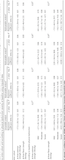
Without accounting for baseline physical function status, change in knee extensor muscle strength along with all co-variates considered in this study explained 10% of the vari-ation in the change in physical function. A 1-unit increase (Nm/kg) in peak knee extensor strength was associated with a 17-unit improvement (95% CI −29 to −5) in WOMAC physical function. In another model, accounting for baseline physical function status and an interaction with change in knee extensor strength along with all covariates, 33% of the variation in the change in physical function was
[image:4.595.58.542.110.380.2]explained. In this model, a 1-unit increase (Nm/kg) in peak isometric knee extensor strength corresponded to a 24-unit improvement (95% CI −42 to −7) in WOMAC physical function score in participants with severe baseline dysfunc-tion. Notably, the association between change in knee ex-tensor strength and change in physical function was not significant for participants with mild and moderate baseline physical dysfunction (Table 3). Sensitivity analyses using only complete cases (n= 80) demonstrated similar results for change in knee extensor strength as the independent variable (Additional file 2: Table S2).
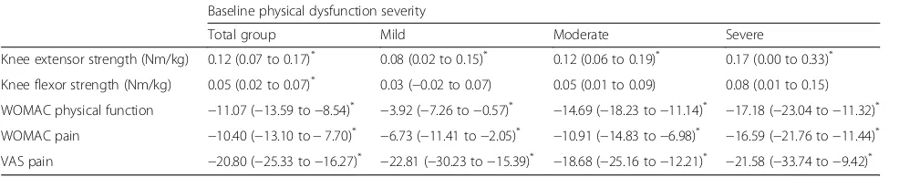
Table 2Change (follow-up minus baseline) in knee muscle strength
Baseline physical dysfunction severity
Total group Mild Moderate Severe
Knee extensor strength (Nm/kg) 0.12 (0.07 to 0.17)* 0.08 (0.02 to 0.15)* 0.12 (0.06 to 0.19)* 0.17 (0.00 to 0.33)*
Knee flexor strength (Nm/kg) 0.05 (0.02 to 0.07)* 0.03 (−0.02 to 0.07) 0.05 (0.01 to 0.09) 0.08 (0.01 to 0.15)
WOMAC physical function −11.07 (−13.59 to−8.54)* −3.92 (−7.26 to−0.57)* −14.69 (−18.23 to−11.14)* −17.18 (−23.04 to−11.32)*
WOMAC pain −10.40 (−13.10 to−7.70)* −6.73 (−11.41 to−2.05)* −10.91 (−14.83 to−6.98)* −16.59 (−21.76 to−11.44)*
VAS pain −20.80 (−25.33 to−16.27)* −22.81 (−30.23 to−15.39)* −18.68 (−25.16 to−12.21)* −21.58 (−33.74 to−9.42)* Data presented as mean (95% confidence interval)
[image:4.595.56.549.610.707.2]WOMACWestern Ontario and McMaster Universities Osteoarthritis Index,VASvisual analogue scale *p< 0.05
Table 1Baseline participant characteristics in the entire cohort and when categorised based on baseline physical dysfunction
severity
Total cohort (n= 100) Baseline physical dysfunction severity
Mild (n= 38) Moderate (n= 43) Severe (n= 19)
Age (years) 62.4 ± 7.3 62.4 ± 6.8 62.2 ± 7.8 63.0 ± 7.6
Women,n(%) 52 (52%) 20 (53%) 22 (51%) 10 (53%)
Height (m) 1.67 ± 0.10 1.67 ± 0.10 1.68 ± 0.09 1.65 ± 0.11
Mass (kg) 82.7 ± 14.3 82.6 ± 15.2 84.0 ± 14.7 79.8 ± 11.5
Body mass index (kg/m2) 29.64 ± 4.08 29.67 ± 4.29 29.69 ± 4.08 29.50 ± 3.85
Knee alignmenta(degrees) 176.8 ± 3.5 176.6 ± 3.7 176.5 ± 3.4 177.9 ± 3.0
Neuromuscular exercise: quadriceps strengthening group
50:50 20:18 24:19 6:13
Bilateral osteoarthritis, yes:no 47:53 15:23 23:20 9:10
Radiographic disease severityb
Grade 2 22 (22%) 7 (18%) 11 (26%) 4 (21%)
Grade 3 43 (43%) 16 (42%) 17 (40%) 10 (53%)
Grade 4 35 (35%) 15 (39%) 15 (35%) 5 (26%)
VAS average knee pain in the past week, 0–100 mmc
54.1 ± 15.0 52.8 ± 15.1 51.3 ± 12.5 62.8 ± 17.7d,e
WOMAC physical function, 0–100c 39.8 ± 14.1 26.2 ± 8.5 43.2 ± 4.5d 59.3 ± 8.2d,e
Knee extensor strength (Nm/kg) 1.45 ± 0.45 1.49 ± 0.48 1.50 ± 0.43 1.27 ± 0.39
Knee flexor strength (Nm/kg) 0.69 ± 0.22 0.69 ± 0.19 0.70 ± 0.24 0.64 ± 0.23
WOMACWestern Ontario and McMaster Universities Osteoarthritis Index,VASvisual analogue scale
a
Anatomic alignment, where neutral alignment is 181° for females and 183° for males. Varus is < 181° for females and < 183 ° for males [19]
b
Kellgren and Lawrence grade
c
Higher scores indicates greater pain/dysfunction
d
Significantly different to mild physical dysfunction (p< 0.05)
e
Knee flexor strength
Without accounting for baseline physical function status, increased knee flexor muscle strength along with all covariates considered in this study explained 6% of the vari-ation in the change in physical function. However, the asso-ciation between change in knee flexor strength and change in physical function was not statistically significant in the model unadjusted for covariates or in the models adjusted for any or all covariates (Table 3). When accounting for baseline physical function status and an interaction with change in knee flexor strength along with all covariates, 27% of the variation in the change in physical function was explained. In this model, the association between change in knee flexor strength and change in physical function was not significant, irrespective of baseline physical function status. Sensitivity analyses using only complete cases (n= 80) demonstrated a statistically significant association be-tween increased knee flexor strength and improvement in physical function without accounting for baseline physical function status. In the sensitivity analyses accounting for baseline physical function status and an interaction with change in knee flexor strength, there was a significant asso-ciation between increased knee flexor strength and im-proved physical function in those with severe physical dysfunction only (Additional file 2: Table S2).
Discussion
We observed a statistically significant association between increased knee extensor strength and improved self-reported physical function in people with knee OA who underwent exercise therapy. However, when investigating the cohort according to baseline severity of physical dys-function, there was limited evidence of associations be-tween change in knee muscle strength and change in physical function for participants with mild and moderate physical dysfunction at baseline. Conversely, for partici-pants with severe physical dysfunction at baseline, there was a significant association between increased knee ex-tensor strength and improved self-reported physical func-tion. Taken together, the findings of this study provide preliminary evidence to suggest that the relationship be-tween increased knee extensor strength and self-reported physical function improvement may depend on baseline levels of physical dysfunction.
Understanding the association between change in muscle strength and improved physical function following exercise is important. Exercise is a cornerstone treatment for knee OA [4, 5] and self-reported physical function using the WOMAC is recommended as an end-point in OA clinical trials [22]. Similar to previous research [13, 14, 24] we ob-served a statistically significant association between in-creased knee extensor strength and improvement in physical function without accounting for physical dysfunc-tion severity at baseline. Based on the regression equadysfunc-tion,
the 0.12 Nm/kg (8%) average increase in knee extensor strength was associated with a 2.05-unit improvement (95% CI−3.5 to−0.6) in WOMAC physical function. In the con-text of strength gains in people with knee OA, the strength gains in the current study are lower than mean increases of 17% (range 10.5% decrease to 49.5% increase) in response to resistance training in people with knee OA reported in a systematic review [25]. Notably, the relationship between self-reported physical function and variables of knee muscle strength, age, sex, exercise group, baseline strength and change in pain (VAS) explained 6–10% of the variation in self-reported physical function. In contrast to a 38-week longitudinal study where participants performed exercise [14], we did not observe an association between increases in knee flexor strength and improved self-reported function on the WOMAC despite the significant improvement in knee flexor strength we reported previously [16]. Several between-study differences such as strength measurement protocols, study duration and participant characteristics may account for the inconsistent findings. Further to this, the exercise interventions used in the current study did not specifically target knee flexor strength. Thus, changes in knee flexor strength were relatively small (Table 2), which may have contributed to a lack of statistical power to detect a statically significant association. Nonetheless, we observed potential evidence to support an association between change in knee flexor strength and improvement in phys-ical function (48.6-unit improvement (95% CI −100.7 to 5.6) in WOMAC physical function), and hence we are hesi-tant to disregard the potential for a relationship. There was a statistically significant association between increased knee flexor strength and physical function improvement when analysing complete cases (Additional file 2: Table S2).
severe physical dysfunction at baseline. Interestingly, only one of the 19 participants with severe physical dysfunction at baseline increased knee extensor strength≥0.85 Nm/kg over the 12 weeks. Overall, it appears that an increase in knee extensor strength associated with a clinically relevant improvement in physical function is potentially achievable for few patients. Our findings suggest that resistance pro-grammes should aim for greater increases in knee extensor strength, as this may yield greater self-reported physical function improvement for people with severe physical dys-function at baseline. It is important to acknowledge that a large proportion of the variation in physical function re-mains unexplained (67%) by a linear relationship between physical function and knee muscle strength together with covariates. Future research is required to validate whether these estimates of knee extensor strength increases yield a MCII in physical function for knee OA patients with se-vere physical dysfunction in response to exercise.
In contrast to participants with severe physical dysfunc-tion, changes in knee muscle strength were not associated with changes in physical function for participants with mild or moderate physical dysfunction at baseline. Rea-sons for differences in the associations depending on se-verity of baseline physical dysfunction are unclear. Increases in knee muscle strength were not statistically different across the levels of physical dysfunction, when accounting for baseline level of knee muscle strength (data not shown). Thus, factors other than increased maximal isometric knee extension/flexion strength appear to con-tribute to a clinically relevant improvement in physical function [15] following exercise for many participants. Various clinical and psychological factors predict deterior-ation in self-reported physical function [26] in people with knee OA. However, little is known regarding the associ-ation of these factors to improved physical function fol-lowing exercise. In people with OA, treatment expectation has been shown to moderate the effectiveness of cognitive behavioural therapy [27] and there is some evidence to suggest that treatment expectations are related to clinical outcomes from exercise and acupuncture [28]. A recent study suggests that knee OA patients with higher treat-ment expectancy for exercise have greater self-efficacy and fewer depressive symptoms compared to patients with lower treatment expectancy [29]. The unexplained vari-ation in self-reported physical function change observed in the current study may in part be accounted for by treat-ment expectation, self-efficacy and depressive symptoms. Future research should consider whether improvement in these factors, among others [30], differs according to physical dysfunction.
From a clinical perspective, our data suggest that ei-ther exercise programme used in this study is beneficial to improve physical function, irrespective of baseline physical function. Post-hoc analyses confirmed that
improvement in physical function was no different be-tween exercise groups according to the baseline level of physical dysfunction (interaction p= 0.39). Hence, this study does not question the efficacy of knee strengthening interventions to improve physical function. Instead, this study generates the hypotheses that physical function im-provement is associated with factors other than increased knee extensor strength for patients with mild or moderate physical dysfunction at baseline, and that gains in knee ex-tensor strength only partially account for improved phys-ical function in patients with severe physphys-ical dysfunction.
Evidently, a greater understanding of how physical function improves in patients with knee OA following strengthening exercise is needed so that exercise pre-scription can be improved to optimise treatments.
Conclusions
Overall, we found preliminary evidence to suggest that the association between change in knee muscle strength and improvement in self-reported physical function following 12 weeks of exercise therapy differs according to baseline difficulty with physical function. This may facilitate future research to optimise treatment effects of exercise. Further research is required to investigate factors that associate with improvement in physical function for knee OA patients, particularly those with mild to moderate physical dysfunction.
Additional files
Additional file 1: Table S1.Linear relationships between measure of
peak isokinetic knee muscle strength (independent variable) and WOMAC physical function 100 scale (dependent variable), including interaction
between‘exercise program’and‘measure of strength’. (DOCX 20 kb)
Additional file 2: Table S2.Linear relationships between change in
strength-related measures (independent variable) and change on WOMAC function (dependent variable) according to physical dysfunction severity at baseline (complete cases n =80). (DOCX 21 kb)
Additional file 3: Table S3.Linear relationships between 12-week
change in strength-related measures (independent variable) and 12-week change on WOMAC physical function (dependent variable) according to tertiles of physical dysfunction severity at baseline. (DOCX 26 kb)
Abbreviations
MCII:Minimal clinical important improvement; OA: Osteoarthritis;
RCT: Randomised controlled trial; VAS: Visual analogue scale; WOMAC: Western Ontario and McMaster Universities Osteoarthritis Index
Acknowledgements Not applicable.
Funding
The study was funded by the Australian National Health and Medical Research Council (Project # 628644). MH is supported by a Sir Randal Heymanson Research Fellowship from The University of Melbourne. RSH is supported by a Future Fellowship from the Australian Research Council (FT130100175). KLB is supported by a NHMRC Principal Research Fellowship (APP1058440).
Availability of data and materials
The datasets used and/or analysed during the current study are available from the corresponding author on reasonable request.
Authors’contributions
MH, RSH, MvdE, MvdL, TVW, JK, FD and KLB conceived the design of this study. BRM and MH acquired the data. MH and JK performed statistical analyses. MH, RSH, MvdE, MvdL, JK, TVW, FD and KLB interpreted the data. MH drafted the manuscript. All authors revised the manuscript for intellectual content and approved the final version.
Ethics approval and consent to participate
Ethical approval was obtained from the University of Melbourne Human Ethics Committee and all participants provided written informed consent.
Consent for publication
The authors obtained consent from each participant.
Competing interests
The authors declare that they have no competing interests.
Publisher’s Note
Springer Nature remains neutral with regard to jurisdictional claims in published maps and institutional affiliations.
Author details
1Centre for Health, Exercise and Sports Medicine, Department of
Physiotherapy, School of Health Sciences, The University of Melbourne, Melbourne, VIC 3010, Australia.2Amsterdam Rehabilitation Research Center Reade, Amsterdam, The Netherlands.3Department of Epidemiology and
Preventive Medicine, Monash University, Melbourne, VIC, Australia.
4Department of Physiotherapy, School of Health Sciences, The University of
Melbourne, Melbourne, VIC, Australia.
Received: 10 July 2017 Accepted: 16 November 2017
References
1. Skou ST, Roos EM, Laursen MB, et al. Criteria used when deciding on
eligibility for total knee arthroplasty—between thinking and doing. Knee.
2016;23:300–5.
2. Roos EM, Herzog W, Block JA, et al. Muscle weakness, afferent sensory
dysfunction and exercise in knee osteoarthritis. Nat Rev Rheumatol. 2011;7:57–63.
3. Berger MJ, Kean CO, Goela A, et al. Disease severity and knee extensor force
in knee osteoarthritis: data from the Osteoarthritis Initiative. Arthritis Care
Res. 2012;64:729–34.
4. Fernandes L, Hagen KB, Bijlsma JW, et al. EULAR recommendations for the
non-pharmacological core management of hip and knee osteoarthritis. Ann
Rheum Dis. 2013;72:1125–35.
5. McAlindon TE, Bannuru RR, Sullivan MC, et al. OARSI guidelines for the
non-surgical management of knee osteoarthritis. Osteoarthritis Cartilage.
2014;22:363–88.
6. Cheing GL, Hui-Chan CW. The motor dysfunction of patients with knee
osteoarthritis in a Chinese population. Arthritis Rheum. 2001;45:62–8.
7. Jan MH, Lai JS, Tsauo JY, et al. Isokinetic study of muscle strength in
osteoarthritic knees of females. J Formos Med Assoc. 1990;89:873–9.
8. Liikavainio T, Lyytinen T, Tyrvainen E, et al. Physical function and properties
of quadriceps femoris muscle in men with knee osteoarthritis. Arch Phys
Med Rehabil. 2008;89:2185–94.
9. Messier SP, Loeser RF, Hoover JL, et al. Osteoarthritis of the knee: effects on
gait, strength, and flexibility. Arch Phys Med Rehabil. 1992;73:29–36.
10. Palmieri-Smith RM, Thomas AC, Karvonen-Gutierrez C, et al. Isometric
quadriceps strength in women with mild, moderate, and severe knee
osteoarthritis. Am J Phys Med Rehabil. 2010;89:541–8.
11. Ruhdorfer A, Wirth W, Eckstein F. Longitudinal change in thigh muscle
strength prior to and concurrent with minimum clinically important worsening or improvement in knee function: data from the Osteoarthritis
Initiative. Arthritis Rheumatol. 2016;68:826–36.
12. Sanchez-Ramirez DC, van der Leeden M, van der Esch M, et al. Increased
knee muscle strength is associated with decreased activity limitations in established knee osteoarthritis: two-year follow-up study in the Amsterdam
osteoarthritis cohort. J Rehabil Med. 2015;47:647–54.
13. Baker KR, Nelson ME, Felson DT, et al. The efficacy of home based
progressive strength training in older adults with knee osteoarthritis: a
randomized controlled trial. J Rheumatol. 2001;28:1655–65.
14. Knoop J, Steultjens MP, Roorda LD, et al. Improvement in upper leg muscle
strength underlies beneficial effects of exercise therapy in knee osteoarthritis: secondary analysis from a randomised controlled trial.
Physiotherapy. 2015;101:171–7.
15. Tubach F, Ravaud P, Baron G, et al. Evaluation of clinically relevant changes
in patient reported outcomes in knee and hip osteoarthritis: the minimal
clinically important improvement. Ann Rheum Dis. 2005;64:29–33.
16. Bennell KL, Kyriakides M, Metcalf B, et al. Neuromuscular versus quadriceps
strengthening exercise in patients with medial knee osteoarthritis and varus
malalignment: a randomized controlled trial. Arithis Rhematol. 2014;66:950–9.
17. Kellgren JH, Lawrence JS. Radiological assessment of osteo-arthrosis. Ann
Rheum Dis. 1957;16:494–502.
18. Altman RD, Gold GE. Atlas of individual radiographic features in
osteoarthritis, revised. Osteoarthritis Cartilage. 2007;15(Suppl A):1–56.
19. Kraus VB, Vail TP, Worrell T, et al. A comparative assessment of alignment
angle of the knee by radiographic and physical examination methods.
20. Bennell KL, Egerton T, Wrigley TV, et al. Comparison of neuromuscular and quadriceps strengthening exercise in the treatment of varus malaligned knees with medial knee osteoarthritis: a randomised controlled trial protocol. BMC Musculoskelet Disord. 2011;12:276.
21. Bellamy N, Buchanan WW, Goldsmith CH, et al. Validation study of WOMAC:
a health status instrument for measuring clinically important patient relevant outcomes to antirheumatic drug therapy in patients with
osteoarthritis of the hip or knee. J Rheumatol. 1988;15:1833–40.
22. Bellamy N. Osteoarthritis clinical trials: candidate variables and clinimetric
properties. J Rheumatol. 1997;24:768–78.
23. Moreland JR, Bassett LW, Hanker GJ. Radiographic analysis of the axial
alignment of the lower extremity. J Bone Joint Surg Am. 1987;69:745–9.
24. Maurer BT, Stern AG, Kinossian B, et al. Osteoarthritis of the knee: isokinetic
quadriceps exercise versus an educational intervention. Arch Phys Med
Rehabil. 1999;80:1293–9.
25. Lange AK, Vanwanseele B, Fiatarone Singh MA. Strength training for
treatment of osteoarthritis of the knee: a systematic review. Arthritis Rheum.
2008;59:1488–94.
26. de Rooij M, van der Leeden M, Heymans MW, et al. Prognosis of pain and
physical functioning in patients with knee osteoarthritis: a systematic review
and meta-analysis. Arthritis Care Res. 2016;68:481–92.
27. Broderick JE, Keefe FJ, Schneider S, et al. Cognitive behaviroal therapy for
chronic pain is effective, but for whom? Pain. 2016;157:2115–23.
28. Foster NE, Thomas E, Hill JC, Hay EM. The relationship between patients and
practitioner expectations and preferences and clinical outcomes in a trial of
exercise and acupuncture for knee osteoarthritis. Eur J Pain. 2010;14:402–9.
29. Marszalet J, Price LL, Harvey WF, Driban JB, Wang C. Outcome expectations
and osteoarthritis: association of percieved benefits of exercise with
self-efficacy and depression. Arthritis Care Res. 2017;69:491–8.
30. Runhaar J, Luijsterburg P, Dekker J, Bierma-Zeinstra SM. Identifying potential
working mechanisms behind the positive effects of exercise therapy on pain and function in osteoarthritis; a systematic review. Osteoarthritis
Cartilage. 2015;23:1071–82.
• We accept pre-submission inquiries
• Our selector tool helps you to find the most relevant journal
• We provide round the clock customer support
• Convenient online submission
• Thorough peer review
• Inclusion in PubMed and all major indexing services
• Maximum visibility for your research
Submit your manuscript at www.biomedcentral.com/submit