D
E
V
E
LO
P
M
E
N
T
INTRODUCTION
Epithelia that line the animal body and internal organs synthesise and secrete proteins and polysaccharides at their apical surface to build specialised apical extracellular matrices (aECMs). Such aECMs may serve as protective barriers against environmental influences, provide apical attachment sites for the underlying epithelium, or temporarily assist in epithelial remodelling (Sebastiano et al., 1991; Jovine et al., 2002; Roch et al., 2003; Bökel et al., 2005; Moussian and Uv, 2005). Apical ECMs are rich in carbohydrates such as glucosaminoglycans and glycosyl-groups on proteins. These sugars are important both for the polarised deposition of aECM components and their assembly into functional aECMs. The glycosyl-groups bound up in aECMs originate from cytosolic residues that are imported into the secretory compartments or used directly by transmembrane enzymes to generate extracellular polysaccharides (Wilson, 2002; Huet et al., 2003; Moore, 2003; Mayor and Riezman, 2004). Many enzymes acting in protein maturation are identified, but their contribution to and requirements for aECM differentiation and organ morphogenesis in complex multicellular organisms has been little investigated.
Differentiation of the Drosophilaexoskeleton (cuticle), a stratified highly ordered aECM, relies on a carefully orchestrated apparatus for biosynthesis and modifications of proteins and the polysaccharide chitin. The Drosophilacuticle is deposited during late embryogenesis to cover the apical surface of the epidermis, as well as of the respiratory organ (tracheae) and the fore- and hindgut
(Neville, 1975). The first step in cuticle deposition is the formation of an outer impermeable envelope. Subsequently, the middle protein-rich epicuticle responsible for cuticle stiffness is formed and, finally, an innermost procuticle loaded with lamellar linear chitin is assembled to confer stability and elasticity to the cuticle (Locke, 2001).
Several Drosophilamutants have been identified that show defects in cuticle formation without affecting the overall embryonic patterning (Jürgens et al., 1984; Nüsslein-Volhard et al., 1984; Wieschaus et al., 1984; Ostrowski et al., 2002). One of these is krotzkopf verkehrt(kkv), which encodes the epidermal chitin synthase CS-1. CS-1 is a transmembrane enzyme that links cytosolic UDP-N-acetylglucosamine (UDP-GlcNAc) into long GlcNAc polymers (chitin) that extrude from the apical surface. Loss of chitin destabilises not only the procuticle, but also affects the integrity of the epicuticle during cuticle differentiation suggesting that a proper procuticle is required for epicuticle formation (Moussian et al., 2005a). Loss of function of two other factors, the predicted extracellular molecules encoded by retroactive(rtv) and knickkopf(knk), causes similar cuticle defects as mutations in kkvand is required for chitin filament assembly (Moussian et al., 2005b; Moussian et al., 2006). Another group of mutations that affects cuticle differentiation disrupt genes that encode components of the septate junctions (SJs). SJs share functions with vertebrate tight junctions, confer a paracellular diffusion barrier in Drosophilaepithelia (Lamb et al., 1998; Wu and Beitel, 2004) and are necessary for polarised deposition of components needed for cuticle assembly such as Knk (Moussian et al., 2006). Both groups of mutants additionally have impaired tracheal tube size regulation, which is associated with a defective temporary chitin-containing luminal matrix that is essential for uniform diameter growth (Tonning et al., 2005).
We report on the phenotypic and molecular characterisation of mummy(mmy), a mutation that is described to cause severe cuticle defects (Nüsslein-Volhard et al., 1984). The mmy mutant
Hormonal regulation of
mummy
is needed for apical
extracellular matrix formation and epithelial morphogenesis
in
Drosophila
Anna Tonning1, Sigrun Helms2, Heinz Schwarz3, Anne E. Uv1,* and Bernard Moussian2,*
Many epithelia produce apical extracellular matrices (aECM) that are crucial for organ morphogenesis or physiology. Apical ECM formation relies on coordinated synthesis and modification of constituting components, to enable their subcellular targeting and extracellular assembly into functional matrices. The exoskeleton of Drosophila, the cuticle, is a stratified aECM containing ordered chitin polysaccharide lamellae and proteinaceous layers, and is suited for studies of molecular functions needed for aECM assembly. Here, we show that Drosophila mummy(mmy) mutants display defects in epithelial organisation in conjunction with aberrant deposition of the cuticle and an apical matrix needed for tracheal tubulogenesis. We find that mmyencodes the
UDP-N-acetylglucosamine pyrophosphorylase, which catalyses the production of UDP-N-UDP-N-acetylglucosamine, an obligate substrate for chitin synthases as well as for protein glycosylation and GPI-anchor formation. Consequently, in mmymutants GlcNAc-groups including chitin are severely reduced and modification and subcellular localisation of proteins designated for extracellular space is defective. Moreover, mmyexpression is selectively upregulated in epithelia at the time they actively deposit aECM, and is altered by the moulting hormone 20-Hydroxyecdysone, suggesting that mmyis part of a developmental genetic programme to promote aECM formation.
KEY WORDS: Chitin, Knickkopf, Apical ECM, Cuticle, Epidermis, Trachea, Mummy, Pyrophosphorylase, Udp-Glcnac, Ecdysone, Drosophila Development 133, 331-341 doi:10.1242/dev.02206
1Department of Medical Biochemistry, Göteborg University, Sweden. 2Department of
Genetics, Max-Planck-Institute for Developmental Biology, Tübingen, Germany.
3Microscopy Section, Max-Planck-Institute for Developmental Biology, Tübingen,
Germany.
*Author for correspondence (e-mail: [email protected]) and [email protected]
D
E
V
E
LO
P
M
E
N
T
phenotype is similar to that of the so-called ‘Halloween’ mutants, which fail to produce the differentiation hormone 20-Hydroxyecdysone (Gilbert, 2004), and whose role is still an enigma during insect embryogenesis. We find that mmycodes for the DrosophilaUDP-GlcNAc pyrophosphorylase that functions in aECM formation by producing GlcNAc residues needed for chitin synthesis and protein glycosylation, and that dynamic mmy expression is hormonally regulated in aECM differentiating tissues.
MATERIALS AND METHODS Fly strains and chemical treatments
Unless otherwise noted, fly strains were obtained form Bloomington Stock Centre (Indiana, USA) and Tübingen Stock Centre (Germany). The mutant alleles of mmyused in this study weremmyIK63,mmyIL07, mmyKG04349and
mmyKG08617 (Nüsslein-Volhard et al., 1984; Bellen et al., 2004). The
deficiency uncovering the mmylocus and used in this work is Df(2L)BSC6. Other mutants used were shade7C(shd), shadow10D (sad), kkv14C73and kkvDZ8
(Jürgens et al., 1984). Germline clones were generated using the Flp/FRT technique (Chou et al., 1993). Tracheal expression of UAS-Nod-lacZ (a
chimeric construct of lacZand the microtubule minus-end motor Nod;
provided by Yuh-Nung Jan) was driven by Btl-Gal4, which expresses GAL4 in all tracheal cells from stage 12-13. The enhancer-trap marker 1-eve-1
(Tracheal-1) expresses lacZ in all tracheal cells from tracheal cell invagination at stage 11 and has been described previously (Samakovlis et al., 1996).
Transmission electron microscopy
For transmission electron microscopy (TEM), embryos were cryoimmobilised, sectioned and contrasted as previously described (Moussian et al., 2005a). For gold-labelling of chitin, thin-sections of embryos were incubated with biotinylated Wheat Germ Agglutinin (WGA; 1:500, Vector Laboratories) that was recognised by an anti-biotin antibody (1:300, Enzo Diagnostics) that in turn was detected by protein A conjugated to 10 nm gold particles (1:100, York Stierhof). Gold-labelled specimens were contrasted for only 3 minutes instead of 10 minutes.
Immunohistochemistry
Embryos were fixed and stained according to standard procedures. The following primary antibodies were used: tracheal lumen-specific antibody, mouse monoclonal IgM 2A12 (1:10; Developmental Studies Hybridoma Bank, DSHB), rabbit anti--gal (1:500; Jackson), rabbit anti-GFP (1:500; Molecular Probes, MP), mouse IgG1 monoclonal anti-Crumbs (1:10; DSHB), mouse IgG monoclonal anti-Discs large 1 (1:10; DSHB), mouse monoclonal IgG2a anti-Fasciclin 3 (1:10; DSHB) and rabbit IgG polyclonal anti-Piopio (1:20; M. Affolter). For fluorescent visualisation, the following secondary antibodies were used: Alexa488 goat anti-rabbit IgG (1:500, MP), Alexa488 goat anti-mouse IgM (1:500, MP), Alexa594 goat anti-mouse IgM (1:500, MP) and Alexa546 goat anti-mouse IgG (1:500, MP). Labelling with fluorescein-conjugated Chitin-binding probe (1:500, New England Biolabs, NEB) was performed according to manufacturers recommendations. A BioRad Radiance 2000 was used to obtain confocal images, and Nikon eclipse E1000 was used for obtaining fluorescent images. Images were processed using Adobe Photoshop 7.0.
Immunoblot analysis
Protein extracts were prepared form manually selected larvae in PLC-buffer or in the EndoH-buffer provided by the manufacturer (NEB), and correct genotypes were identified based on their absence of a GFP-marked balancer chromosome. For protein extraction, around 500 embryos were homogenised in PLC buffer on ice, the extract was centrifuged to remove cellular debris, and 15 g protein was loaded in each lane on SDS-gel electrophoresis. Separated proteins were blotted onto a nitrocellulose membrane, and probed with the primary antibodies against Knk (1:2000), Ttv (1:1000; I. The), Syx1A (1:5, DSHB) and ␣-tubulin (1:2000, Sigma). The primary antibodies were detected with HRP-conjugated secondary antibodies. EndoH treatment was performed according to the manufacturer (NEB).
In situ hybridisation
Whole-mount in situ hybridisation was performed with digoxigenin-labelled RNA sense and antisense probes as described previously (Tonning et al.,
2005). RNA probes for mmywere generated from the mmycDNA LD24639
with SP6 and T7 polymerases.
RESULTS
Cuticle composition and epidermal integrity are disrupted in mmymutant larvae
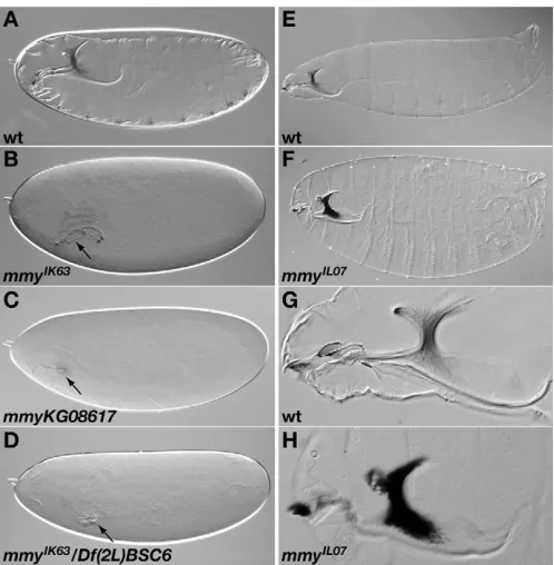
Two alleles of mmywere isolated in a screen for EMS-induced lethal zygotic mutations due to their effects on cuticle differentiation (Nüsslein-Volhard et al., 1984). Compared with the wild-type larval cuticle (Fig. 1A), the cuticle of larvae harbouring the strongmmyIK63 allele is hardly visible (Fig. 1B), whereas larvae mutant for the weak mmyIL07 allele develop a bloated cuticle and a deformed and strongly melanised head skeleton (Fig. 1F,H).
[image:2.612.314.563.57.311.2]In order to better understand the role of mmy in cuticle differentiation, we compared the mmymutant and wild-type larval epidermis by transmission electron microscopy (TEM). Wild-type cuticle is composed of three layers (Locke, 2001): the outermost envelope characterised by five alternating electron-dense and electron-lucid sheets, the underlying epicuticle built up by an upper electron-lucid and a lower electron-dense sublayer, and the innermost procuticle structured by lamellar chitin microfibrils and contacting the apical plasma membrane of the epidermal cells (Fig. Fig. 1. Defective differentiation of mmymutant cuticles.
D
E
V
E
LO
P
M
E
N
T
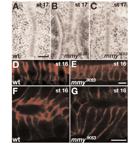
2A). All three cuticle layers are affected in mmyIK63 larvae (Fig. 2B). The outer envelope is thinner than in the wild type with only three sheets, and the electron-dense sub-layer of the epicuticle disintegrates and spreads into the upper electron-lucid sub-layer and the procuticle. The procuticle is also reduced in thickness and seems to be devoid of chitin microfibrils; occasionally, the cuticle detaches from the epidermal surface. The cuticle of larvae mutant for the weak mmyIL07 allele is stratified as in the wild type, and the procuticular chitin microfibrils appear correctly oriented (Fig. 2C). However, the mmyIL07 mutant procuticle contains abnormal inclusions of electron-dense material that are scattered below the epicuticle, presumably orphan proteins, suggesting that the coordinated assembly of the epi- and procuticle is impaired. Taken together, cuticle assembly requires mmyactivity.
As the chitin containing procuticle is affected in both mmymutant larvae, we tested for the presence of chitin using gold labelling with the lectin wheat germ agglutinin (WGA). WGA binds GlcNAc-groups on glycosylated proteins, glycosaminoglycans as well as chitin (D’Amico and Jacobs, 1995), but on a specimen embedded in Epon and sectioned for TEM, WGA recognises only chitin (Peters and Latka, 1986). Although chitin is present in wild-type and in the mmyIL07 mutant procuticle (Fig. 2D,F), no chitin is detected in
mutant larvae homozygous for the strong mmyIK63 allele (Fig. 2E), arguing that mmyis essential for chitin synthesis. However, mmy appears to have additional roles in cuticle differentiation, as loss of chitin causes a compensatory increase in cuticular protein deposition (Moussian et al., 2005a), which is not detected in mmyIK63mutants.
mmyis required for epithelial organisation
[image:3.612.51.520.58.357.2]D
E
V
E
LO
P
M
E
N
T
Fasciclin 3 (Fas3) and Discs large 1 (Dlg1). Both SJ proteins are normally distributed in the weak mmyIL07 epithelia, and the intracellular Dlg1 protein is also normally localised in the more severe mmyIK63mutants (not shown). By contrast, the transmembrane Fas3 is mislocalised along the lateral membrane in the columnar epithelia of the hindgut and salivary gland in some mmyIK63embryos (Fig. 3D-G). Thus, the disrupted SJ ladder in mmyIK63mutants may reflect a requirement for Mmy in the correct localisation of membrane-bound SJ components. As chitin-deficient embryos have no detectable defects in SJ assembly or function (Moussian et al., 2005a), and chitin-deposition appears unaffected in embryos with disrupted SJ components (Tonning et al., 2005), these results point to two parallel requirements for mmy in chitin synthesis and maturation of cell-cell contacts (SJs).
Mmy is required for generation of GlcNAc-groups, including tracheal luminal chitin
The tracheal (respiratory) tubular network is an epithelium that undergoes extensive cell rearrangements during embryonic development, and relies on a luminal (apical) matrix for uniform tube growth (Beitel and Krasnow, 2000; Ghabrial et al., 2003; Uv et al., 2003; Tonning et al., 2005). As chitin is an essential component of this luminal matrix, we initially tested whether mmymutant embryos, which lack cuticular chitin, also display tracheal tube size
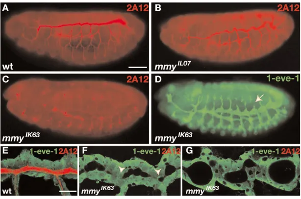
defects. The wild-type developing tracheal system can be visualised with the lumen specific antibody 2A12 (Fig. 4A). The same staining reveals a perfectly patterned tracheal system in embryos homozygous for the weak mmyIL07 allele, although the dorsal trunk (DT) lumen diameter is slightly irregular at stage 15 (Fig. 4B) and becomes excessively elongated at stage 16 (not shown). The tracheal mmyIK63 mutant lumen, however, fails to label with the 2A12-antibody, although the typical intracellular vesicular 2A12-staining is present (Fig. 4C). Tracheal tube shapes in mmyIK63mutants were therefore visualised with the pan-tracheal LacZ-enhancer-trap 1-eve-1. Detection of the 1-eve-1product shows that also mmyIK63mutants display normal tracheal branch patterning, but tube diameter is severely defective (Fig. 4D). The mmyIK63DT fusion branch lumens fail to expand and remain constricted, whereas other parts of the DT trunk become excessively dilated (Fig. 4E,F); during stage 16, these DTs develop huge cyst-like structures (Fig. 4G). Thus, mmyis indeed required for uniform tube expansion.
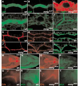
The luminal tracheal chitin matrix can be detected both with a FITC-conjugated chitin-binding probe (CBP) and WGA (Tonning et al., 2005). CBP appears specific for chitin and labels a broad filamentous chitin cable within the wild-type tracheal lumen (Fig. 5A) that is absent in kkvmutant embryos (Tonning et al., 2005). In mutants for the weak mmyIL07allele, CBP labels a luminal cable with similar intensity to the wild type (Fig. 5B), but its filamentous appearance is slightly perturbed, implying that the chitin matrix is not properly assembled. By contrast, embryos homozygous for the strong mmyIK63 allele completely fail to label with CBP (Fig. 5C), indicating a need for mmyalso in tracheal chitin synthesis. We also compared WGA labelling of wild-type and mmyIK63 mutant embryos. In the wild-type tracheae, WGA labels not only the broad luminal chitin cable, but also the luminal surface that probably represents glycosylated proteins (Fig. 5D). In the mmyIK63mutant tracheae, both the luminal and apical WGA-staining is severely reduced (Fig. 5E). This is different from chitin-deficient embryos, where WGA detects GlcNAc-groups along the apical cell surface and within the lumen, although the broad luminal cable is absent (Fig. 5F), arguing that mmyfunction is required not only for chitin synthesis, but also for other GlcNAc-containing components.
Despite the severe tracheal tube defects in mmyIK63mutants, we find that apicobasal polarity appears normal. The transmembrane protein Crumbs (Crb), which is required for cell polarisation and assembly of the adherens junctions (AJs) (Tepass et al., 1990), localises to the apical tracheal membrane in both wild-type and mmyIK63mutant embryos (Fig. 5H,K), and the microtubule minus end reporter, Nod:-gal (Clark et al., 1997), appears to accumulate correctly in the apical tracheal cell domain in the mutant (Fig. 5I,L). Furthermore, no significant defects in Fas3 localisation was observed in the mmyIK63 mutant tracheal epithelium (not shown) and apical secretion of the luminal protein Piopio (Pio), which is required to form narrow tubes with autocellular junctions (Jazwinska et al., 2003), was normal in mmyIK63mutants (Fig. 5G,J). This indicates that zygotic mmyis not generally required for apical protein secretion.
[image:4.612.52.281.58.312.2]We also observed that mmyIK63mutants display severely reduced WGA-labelling in embryonic tissues not known to produce chitin, including the early epidermis and the salivary glands (Fig. 5M-T). Moreover, labelling for Crb revealed local tube dilations in the single-cell-layered Malphigian tubules (both in the common ureter and in the individual tubules), a failure of the salivary gland lumens to enlarge in late embryogenesis, and a dorsal open phenotype in ~10 % of the mmymutant embryos (not shown), which may reflect a need for high levels of GlcNAc-containing substances for these processes.
Fig. 3. Septate junctions (SJs) are defective in mmymutant epithelia. (A-C) TEM-analysis of septate junctions (SJs) of wild-type,
D
E
V
E
LO
P
M
E
N
T
mmyencodes the Drosophila UDP-N-acetylglucosamine-pyrophosphorylase
In order to identify the gene disrupted in mmymutants, we first mapped the mutation to deficiency Df(2L)BSC6 on chromosome 2 (Fig. 1D). P-element induced mutant fly strains known to segregate lethal mutations in this region were subsequently tested in complementation crosses to the EMS-induced mmyIK63and mmyIL07 heterozygous flies. Two P-element insertions isolated in the BDGP Gene Disruption project (Bellen et al., 2004), KG08617 and KG04349, failed to complement the mmyEMS-induced mutations. KG08617 fails to complement the lethality of both mmy EMS-alleles, and embryos homozygous for this P-element insertion develop a cuticle phenotype similar to homozygotes for mmyIK63 (Fig. 1C and not shown), whereas KG04349 complements lethality, but the transhetereozygous flies are sterile.
KG08617 is located within the first intron of the predicted gene CG9535, and KG04349 resides in the 5⬘UTR of CG9535 (Fig. 6A). To verify that the P-element insertion in KG08617 was indeed responsible for the lethality and the mmyphenotype, we excised this P-element and established 13 independent excision lines. Three of these are homozygous viable and naturally complement mmyIK63, whereas 10 new lethal alleles were generated that fail to complement mmyIK63. To further prove that mmy is equivalent to CG9535, we sequenced the CG9535 open reading frame in both mmyIL07and mmyIK63EMS-induced mutant alleles (Fig. 6B). The mmyIK63allele harbours a base-pair change that causes the exchange of an invariant glycine261to valine, and in the mmyIL07allele a singe base-pair changes alters the isoleucine197codon to that of asparagine. Taken together, these experiments prove that CG9535 is indeed mmy.
CG9535 is predicted to code for the Drosophila UDP-N-acetylglucosamine-pyrophosphorylase (UDP-GlcNAc-Pyp), a cytosolic enzyme catalyzing the formation of UDP-N-acetylglucosamine (UDP-GlcNAc) from UTP and GlcNAc-1-Phosphate (Fig. 6C) (Merzendorfer and Zimoch, 2003). CG9535 is the only predicted UDP-GlcNAc-Pyp in Drosophila, and displays high amino acid homology to the UDP-GlcNAc-Pyp in both yeast and humans (39% and 51% identity, respectively) (Fig. 6B). As UDP-GlcNAc is the substrate for chitin synthases (Merzendorfer and Zimoch, 2003), a reduced level of UDP-GlcNAc in zygotic mmyIK63mutants would explain their chitin-deficiency in the luminal tracheal matrix and in the cuticle, as well as a general loss of GlcNAc-group in aECMs.
Protein modification is impaired in mmyIK63
embryos
Yeast and human UDP-GlcNAc-Pyp have been shown to exhibit also a UDP-N-acetylgalactosamine (UDP-GalNAc)-Pyp activity (Wang-Gillam et al., 2000; Peneff et al., 2001). Moreover, UDP-GlcNAc can be converted to UDP-GalNAc by UDP-N-acetylglucosamine 4-epimerase (Winans and Bertozzi, 2002; Mok et al., 2005). Both UDP-GlcNAc and UDP-GalNAc are essential components of the polysaccharide moieties that are added to secreted proteins upon glycosylation and GPI-anchor synthesis in the ER and the Golgi apparatus.
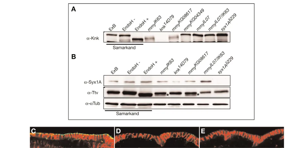
In order to test whether mmymutants display dysfunctional protein maturation along the secretory pathway, we compared the size of the extracellular cuticle protein Knickkopf (Knk) (Jürgens et al., 1984; Ostrowski et al., 2002) in wild-type and mmymutant larvae on Western blots (Fig. 7A). The 689 amino acids Knk protein is predicted to be N-glycosylated at three positions (N170, N354and N636), O-glycosylated at S209and T468, and to possess a GPI-anchor (Mayor and Riezman, 2004; Moussian et al., 2006). In extracts of mmyIK63 and mmyKG08617larvae, no wild-type sized Knk can be detected. Instead several proteins of smaller molecular weight are recognised by the Knk antiserum, indicating that post-translational modifications of Knk indeed are affected in the mutant larvae. In addition, smaller molecular weight species of the plasma-membrane protein Tout-velu (Ttv) (Bellaiche et al., 1998), which is predicted to be N-glycosylated at N71, N327 and N476, are present in mmy mutant larvae when compared with the wild type, whereas no size changes are observed for the intracellular membrane-bound Syntaxin1A (Schulze and Bellen, 1996) (Fig. 7B). Treatment of protein extracts from wild-type larvae with EndoH, an enzyme that removes N-glycosylated modification from proteins, produce Knk and Ttv proteins of even higher mobility than those seen in mmymutant larval extracts (Fig. 7A,B), indicating that protein glycosylation is not completely abolished in mmymutant larvae. By contrast, in protein extracts from mmyIL07 mutant larvae, which display strikingly similar cuticle phenotypes as knkand rtvmutants (Ostrowski et al., 2002; Moussian et al., 2005b), and mmyKG04349mutant larvae, Knk and Ttv migration is not detectably altered, indicating that protein modification in these mutant backgrounds is not dramatically impaired.
[image:5.612.50.357.59.262.2]To test whether impaired modification of Knk may affect its subcellular localisation, we analysed the distribution of Knk in the epidermis of stage 16 wild-type and mmyIK63embryos labelled with
Fig. 4. Mmy is required for uniform tracheal tube growth. (A-C) Lateral view of wild-type and mmymutant embryos labelled with the tracheal lumen-specific antibody 2A12. The 2A12 antibody marks the lumen of wild-type (A) and mmyIL07mutant (B) tracheae. The dorsal trunks (DTs) of mmyIL07embryos are slightly convoluted compared with wild type. The tracheal lumen of mmyIK63 mutant embryos fails to label with 2A12 (C).
(D) Expression of the pan-tracheal lacZmarker 1-eve-1in
mmyIK63mutants and labelling with
D
E
V
E
LO
P
M
E
N
T
the Knk antiserum (Fig. 7C,D). In the wild-type embryo, Knk is detected along the apical surface of the epidermal cell (Fig. 7C), whereas in mmyIK63mutant embryos, only low levels of Knk signal are detected at the apical epidermal surface in addition to a faint cytoplasmic signal (Fig. 7D). This indicates that modification of Knk is required for its stability and localisation to the apical plasma membrane.
mmyexpression is temporally up-regulated in developing epithelia
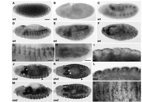
Although Mmy is a metabolic enzyme presumably required for housekeeping functions in most cells, the specific requirements for zygotic mmyin epithelial organisation and aECM formation lead us to investigate its developmental expression pattern. In situ hybridisations to detect mmymRNA produced a strong signal in early embryos before zygotic mid-blastula transcription starts (Fig. 8A), indicating a maternal supply of mmyRNA. Indeed, embryos derived from mmy mutant germline clones arrest development before cellularisation (not shown), suggesting that maternal mmyprovides UDP-GlcNAc for basic cellular needs during early embryogenesis. The zygotic mmyRNA is detected from stage 11, displaying a strong and temporary upregulation in different epithelial tissues. The mmy gene is first transcribed in tracheal cells just after they have invaginated as epithelial sacks from the ectoderm (Fig. 8C) (Manning and Krasnow, 1993), and continues to be expressed at high levels in
the tracheae until stage 15 (Fig. 8D,E,G). This mid-embryonic mmy expression correlates with luminal chitin deposition during tube expansion (Tonning et al., 2005). At stage 16, mmyis strongly expressed in the epithelium of the developing salivary glands (Fig. 8F,H), which are not known to synthesise chitin, but may require high levels of mmyto accommodate extensive protein secretion. First, at late stage 16, and concurrent with chitin deposition, the mmy transcript is detected in the epidermis (Fig. 8F,I).
mmyexpression is altered in mutants that disrupt 20-Hydroxyecdysone biosynthesis
The dynamic and tissue-specific zygotic expression of mmymay be subjected to feedback regulation triggered by UDP-GlcNAc consumption during aECM formation. However, a hypothetical accumulation or lack of UDP-GlcNAc in kkvor mmyIK63mutant embryos, respectively, did not detectably alter the levels of mmy transcript, as mmywas normally expressed in these mutants (not shown).
[image:6.612.251.564.60.403.2]As mmybelongs to the group of Halloween mutants, including shadow (sad) (Warren et al., 2002) and shade(shd) (Petryk et al., 2003), which disrupt enzymes needed for biosynthesis of the insect hormone 20-hydroxyecdysone (Gilbert, 2004), we asked whether temporal expression of mmyin different epithelia may depend on 20-hydroxyecdysone. The sadgene encodes a mitochondrial P450 enzyme (CYP315A1), a C2-hydroxylase that catalyses the Fig. 5. GlcNAc-groups, including tracheal chitin,
are severely reduced in mmymutants. (A-C) Detection of chitin with a FITC-conjugated chitin-binding probe (CBP) in wild-type and mmy
mutant embryos. CBP labelling reveals a broad intraluminal chitin cable with filamentous
appearance in stage 15 wild-type tracheae (A). The chitin cable in mmyIL07mutants has a slightly grainy appearance (B), and is absent in mmyIK63mutants (C). The mmyIK63embryo in C is also labelled for the
1-eve-1 marker with -gal (red) to visualise the tracheal epithelium. (D-F) Detection of GlcNAc-group in tracheae with FITC-conjugated WGA. In wild-type tracheae, WGA labels the lumen and the apical surface of tracheal cells (D). In mmyIK63 mutant tracheae, neither the lumen nor the apical plasma membrane is labelled with WGA (E). By comparison, in chitin-deficient kkv1embryos, the luminal detection is reduced to a thread of non-chitinous filament, whereas labelling of the apical plasma membrane appears to be normal (F). (G-L)mmyIK63mutants display normal apicobasal
polarity and can secrete the luminal Pio protein. Stage 15 wild-type (H) and mmyIK63(K) embryos stained with
Crb (red) reveal a correct localisation of this apical determinant. The microtubuli minus-end reporter
Nod--gal localises to the apical cortex in wild-type tracheal cells (I) as well as in mmyIK63mutant tracheae (L).
Wild-type (G) and mmyIK63 mutant embryos (J) stained
with the Pio antibody (red) reveals that the Pio filament is present in mmyIK63mutant tracheae. One
segment of the DT is shown in G-L. (M-T) Double labelling with WGA and Crumbs in wild-type and
mmyIK63mutant embryos. In wild-type stage 14
embryos (M,N), WGA labels the tracheal lumen as well
as the developing epidermis (M), whereas mmyIK63mutants at the same stage (O,P) show severely reduced WGA-labelling in both tissues (P). WGA
also detects intracellular spots in wild-type stage 15 salivary glands (Q,R), presumably secretory vesicles, but not in mmyIK63mutants at the same
D
E
V
E
LO
P
M
E
N
T
formation of ecdysone from 2-deoxyecdysone; shdencodes the enzyme CYP314A1, which is responsible mono-oxygenation of ecdysone to generate the active 20-hydroxyecdysone. Indeed, two aspects of mmyexpression are similarly altered in embryos mutant for sadand shd. First, we find that shd and sadmutants lack the mid-embryonic up-regulation of tracheal mmy expression, and second, mmy expression is prematurely upregulated in the epidermis, salivary gland and proventriculus (Fig. 8J-O). Thus, a mid-embryonic ‘ecdysone pulse’ appears essential to control the temporal expression of mmyin different embryonic epithelia.
DISCUSSION
We have reported a requirement for Mmy in apical extracellular matrix (aECM) deposition and epithelial morphogenesis, and identified Mmy as the Drosophila UDP-N-acetylglucosamine-pyrophosphorylase (UDP-GlcNAc-Pyp), a cytosolic enzyme that provides UDP-GlcNAc for protein maturation and polysaccharide formation.
mmyencodes the Drosophila UDP-N-acetylglucosamine-pyrophosphorylase
Formation of UDP-GlcNAc from GlcNAc-6-phosphate and UTP has been demonstrated in vitro for the yeast protein UDP-GlcNAc-Pyp (UAP1) (Mio et al., 1998). Yeast cells lacking UAP1 activity have an abnormal bloated cell shape indicative of a weakened cell wall. The human orthologue of UAP1 (AGX) was identified in the same study and was able to rescue the cell lethal phenotype of UAP1 mutants, demonstrating a conserved function for UDP-GlcNAc-Pyp among species. Mmy (CG9535) is the predicted Drosophila orthologue of yeast and human UDP-GlcNAc-Pyp, and is thus expected to be responsible for UDP-GlcNAc synthesis in Drosophila.UDP-GlcNAc-Pyp from different species have also been shown to have UDP-GalNAc-Pyp activity (Szumilo et al., 1996; Peneff et al., 2001). In addition, UDP-GlcNAc is converted to UDP-GalNAc by the UDP-N-acetylglucosamine 4-epimerase (Winans and Bertozzi, 2002). Hence, DrosophilaMmy may also be responsible for UDP-GalNAc production.
D r o s o p h i l a 1 M G R S N F S S H H Q Q T H V R R H R N A G G A T S K S P N A A K T S P TMT D Y L SLH S RL AQ VG Q EHL LKF WPE LT NDE R I DLV RDIE E L N- - - -LDEI K L YFDR AT A e d e s 1 - - - -MANL D AL KT SL A KH DQ EQL LQ YW E E LDE D QR R LLT EDI DE L N- - - -L E EVN E F FKR AT H u m a n 1 - - - MNI ND L KL TLSK A G Q EHL LRF WNE LEEAQQ VE LY A E L Q A MN- - - - FE E L NFF F QKAI Y e a s t 1 - - - M TDTKQ L F I EA G QS QLF H NW ESLS R K D Q EE LL S N LEQ I S S K R S P A KLLED CQNAI c o n s e n s u s 1 . . . . . . * . * . . . * . . * . . . . . * . . . . . . . *
D r o s o p h i l a 9 2 VSMN E N- - G IK L D D RL QPLP EGK LIS IARA P L E KLDA YR DE G LLQ I SNGHV A V L L M A G G Q G T R L G FD HP K G M Y D V G LQS R KTL FRI Q A E R I L K LE A e d e s 5 5 SSL EEG - - N AK L D DKM E PV CEDKF LS ISRT T EDQLK KYH EE G LRQ IA DG K VGV L L M A G G Q G T R L G F AFP K G MF NV G L P SNKSL FRI QGE R I L K L Q H u m a n 5 4 E G FNQ SSY QKN VDAR M E PVPR E VLGSA TRD - QDQLQAW E SE G LFQ I SQ NK V A V L LLA G G Q G T R L GVAYP K G M Y D V G L P S R KTL FQI Q A E R I L K L Q Y e a s t 5 6 K F S L AN S- SKD T G V E I SPLPP T S Y ESL I G N - S K K E N EYW R LG LE AIG KGEV A VIL M A G G Q G T R L GS S QP K GCY DIG L P SKKSL FQI Q A EK L I RL Q c o n s e n s u s 9 6 . . . . . . * . . . * . . . . . . . * * . * . . . . * . * . * . * * * * * * * * * . . . * * * . . . . * * . * . * . * * * * . * . . . . * .
D r o s o p h i l a 1 8 5 EL AQEA NG KRGHITW Y I M T SEH TV QP TY DY FV A NNFF G L KAE NV L LF E Q G S L PCFE YD GRI I L D E K H R-VA RA P D G N G GIY R AMKRQ G I LDD MQ K A e d e s 1 4 8 RL AAEL TG KTGRITW Y I M T SEH T MIP T KKY FEEN DY F G L KAED I M MF E Q G S L PC YD FEG K ILL D E K H R-VA KA P D G N G G L Y R A LR D RG I LDDLE R H u m a n 1 4 8 Q VAE K Y YGNKC IIPW Y I M T SG RT ME ST KE FFT K H KY F G L KKE NV IF FQQ GML PA M SF D G K I I LEE KN K -VS MA P D G N G G L Y R A LA AQNIV ED M EQ Y e a s t 1 4 9 D M V K D K - - -KV EIPW Y I M T SG PTR A ATE AY FQEHN Y F G LN KEQ I TF FNQ GTL PAF DL TG KH FLM K D P V N L S Q SP D G N G G L Y R AIKE N K L N EDF DR c o n s e n s u s 1 9 1 . . . . . . . * * * * * * * . * . . * . . . * . . . * * * . * . . . . * . * * . * * . . . . * . . . * . . . . . . * * * * * * . * * * . . . * . . .
D r o s o p h i l a 2 7 9 R GVL Y L H AH SV D N I L I K V A D P V F I GYCVQE KA D CAA K V V E KA AP N E A V G V VA IV D G K Y Q V V E Y S E I SAK T A EMRNS D G R LTFSA G N I C N H F FS S N A e d e s 2 4 2 R GVL Y L H AH SV D N I L I K V A D P VSI GY F V E Q KA D CGA K V V E KS HP N E A V G V VC QV D G K Y Q V V E Y S E IT QK T A ELRK ED G R LVF N A G N I C N H F F TT S H u m a n 2 4 2 R GI W S IHV Y CV D N I LVK V A D PRF I GFCIQK GA D CGA K V V E KT NPTEPV G V VC RV D GVY Q V V E Y S E I SL AT AQ KRSS D G R LLF N A G N IAN H F F T VP Y e a s t 2 4 1 R GI K H V Y M Y CV D NVLSKIA D P V F I GF A I K H G F E L A TKAVRKR D A HESV GL I A T K N EKP CVIE Y S E I SN E LA EA K D KD GLLK L RA G N IVN HY Y LVD c o n s e n s u s 2 8 6 * * . . . * * * . * . * . * * * . . * * . . . . . . * . * . * . . . * . * * . . . . . * . * * * * * . . . . * . . . . * * . * . . . * * * * . * * . . . .
D r o s o p h i l a 3 7 4 F LQK I GST Y EQ EL K L H V A K K K I P F V DN AG-K RL TP D K P N G I K I E K F V F D V FEF A- - QK F VA ME V P R D E E F SAL K NSD- AA G K DCPST A RS DLHR L A e d e s 3 3 7 F L R K I GTTFEK DL K L H V A K K K I P FID STG- TRC TP D K P N G I K I E K F V F D V F Q F A- - E HF VT IE V P R D E E F SAL K N A D- SA G K DC AT T A RA D I YR L H u m a n 3 3 7 F L RD V V N VY EP QLQ HH V AQK K I PYV DT QG- Q L I KP D K P N G I KME K F V F DIF Q F A- - KK F VV YE VLRE DE F SPL K N A DS Q NG K DNP T T A RH ALM SL Y e a s t 3 3 6 LLK R D L D Q W C E N M P YHIA K K K I PA YD SV T GKY T KPT EP N G I KLEQFIF D V FD T V P L NK FG C LE VDRC KE F SPL K NG P - G S K NDNPETSRL A Y L KL c o n s e n s u s 3 8 1 . * . . . . . . * . * . * * * * . . * . . . . . * . . * * * * * . * . * . * * . * . . . . . * . . * * . * . . * * * * * * . . . . . . * . . * . * . . . *
D r o s o p h i l a 4 6 5 HKKYI EGA G G I VH G E VC E I S PF V TY A G ENLASHVEG KSFTS P VYLR D S R D P L H G H L A e d e s 4 2 8 HRKYI EAA G GTVD G T EC E I S P L L S YGG EGLK V LVHGR TFVS P VHLM A D E E K K S E V V E -H u m a n 4 2 9 -H-H C W V L NA G G-H F I D E N G S R L P A I P R L K D A N D V P I QC E I S P LIS Y A G EGLESYVA DKEF-H APL I I D E N G V -H E L V K N G I Y e a s t 4 3 0 G T S W LEDA GAI VK D G V L VEVSS KL S Y A G ENLS Q F K G K V F D RSG I VLE K -c o n s e n s u s 4 7 6 . . . * * . . . . . . . * . * . . . . * . * * * . . . . . . .
N mmyIL07
isoform A isoforms A and B
V mmyIK63
KG08617 KG04349
IL07 IK63
A
B
C
UTP + N-acetyl-α-D-glucosamine-1-phosphatediphosphate + UDP-N-acetyl-α-D-glucosamine
Chitin synthesis Glycosylation
GPI-anchor
[image:7.612.55.561.58.438.2]Mummy
Fig. 6. Mmy encodes the DrosophilaUDP-N-acetylglucosamine pyrophosphorylase. (A) Genome organisation of mmy, showing the exons (grey boxes) used in each of the two mmytranscripts RA and RB. Start and stop codons are marked in red and green, respectively. The two EMS-induced mmymutations mmyIK63 and mmyIL07introduce single mis-sense mutations in the third common exon, whereas P-element KG04349 is inserted 5⬘to the ATG of RA, and P-element KG08617 disrupts the second intron of both RA and RB. (B) Sequence alignment of the Mmy protein with its orthologues in yeast, human and Aedes. The mis-sense mutations in mmyIK63andmmyIL07 cause substitution of conserved amino acids as indicated above the sequence. The extra 37 amino acids in isoform A are underlined and do not show homology to Mmy orthologues.
D
E
V
E
LO
P
M
E
N
T
The EMS induced mutations mmyIK63 and mmyIL07 identify important amino acids for Mmy activity. Gly261that is exchanged to valine in the MmyIK63protein corresponds to Gly224in the human AGX that has been shown to be essential for enzyme activity by site-directed mutagenesis and lies in a loop contacting uridine, as demonstrated by crystallographic experiments (Wang-Gillam et al., 2000; Peneff et al., 2001). As the phenotypes caused by the exchange of Gly261and by the deletion of mmyin our P-element excision-lines are identical, it is conceivable that this mutation causes a loss of Mmy function. The highly conserved Ile197that is mutated in the mmyIL07 allele has not yet been described to be essential for UDP-GlcNAc-Pyp activity. This amino acid exchange permits the production of some UDP-GlcNAc, as many processes that require zygotic Mmy activity appear normal in mmyIL07 embryos.
Mmy has distinct functions during aECM formation
Embryos homozygous mutant for mmyhave severe defects in their epidermal and tracheal organisation. In particular, consistent with the molecular function of Mmy, the most striking deficiency in these tissues is the absence of chitin both in the procuticle and the developing tracheal lumen. However, the mmymutant epidermis and tracheae display additional defects compared with embryos lacking chitin. For example, the mmyIK63mutant tracheal lumen fails to label with the 2A12 antibody, and the mmyIK63mutant procuticle appears collapsed, neither of which is seen in chitin-deficient embryos. Another component essential for cuticle differentiation and luminal
tracheal matrix formation is the glycosylated and GPI-anchored extracellular protein Knk (Ostrowski et al., 2002; Moussian et al., 2006). We find that Knk is a target of Mmy activity, as embryos homozygous for the amorphic alleles mmyIK63 and mmyKG08617 produce Knk proteins with reduced molecular weight, compared with wild-type Knk, indicative of impaired glycosylation, obviously affecting its localisation and function. However, paired Knk function in combination with lack of chitin is not sufficient to explain fully the aECM defects in mmymutants, as loss of Knk and chitin cause similar phenotypes to loss of chitin only (Ostrowski et al., 2002; Moussian et al., 2006). The observed requirement of zygotic Mmy for Knk modification instead suggests that additional proteins needed for cuticle differentiation and tracheal luminal matrix formation, such as Retroactive (Rtv) (Moussian et al., 2005b), are affected in mmymutants.
[image:8.612.53.529.57.296.2]The disrupted aECMs in mmymutants may also be contributed to by disorganised aECM-producing epithelia. Although zygotic Mmy is not required for epithelial apicobasal polarity, we find that SJ in mmymutant epithelia fail to form the typical ladder-like structure, which occasionally is accompanied by slight defects in Fas3 localisation at these junctions. Mutants lacking single SJ components display defects both in tracheal luminal matrix formation and cuticle deposition (Llimargas, 2000; Behr et al., 2003; Wu et al., 2004). This role of SJ components may be, at least partially, mediated through correct localisation of the cuticle organising Knk protein (Moussian et al., 2006). Moreover, the tracheal lumen of embryos deficient for both chitin and single SJ components fails to label with the 2A12 antibody, as seen in mmy Fig. 7. Post-translational modification of Knk depends on Mmy activity.(A) Western blot with protein extracts from wild-type and different mutant larvae labelled with a specific antibody against the extracellular cuticle protein Knk. In the wild type, Knk migrates as a single band (ExB and EndoH-), and EndoH treatment to cleave N-glycosylated sugar-residues generates smaller co-migrating Knk species (EndoH+). In mmyIK63and
D
E
V
E
LO
P
M
E
N
T
mutants (Tonning et al., 2005). Thus, mmymay also affect the aECM through its need for SJ assembly. Given the requirements for mmy in protein modification along the secretory pathway, Mmy is likely to affect primarily the localised deposition and function of extracellular SJ proteins.
Regulation of mmyexpression during development
Both UDP-GlcNAc and UDP-GalNAc are imported from the cytosol into the endoplasmic reticulum, where they are used by specific transferases for glycosylation and GPI-anchor synthesis. Mmy is therefore expected to be essential for cellular housekeeping functions, and its activity regulated to provide sufficient, but not excess accumulation of its end product. Indeed, the activity of UDP-GlcNAc-Pyp in the human parasite Giardia lamblia is under allosteric control of the upstream metabolite glucosamine-6-phosphate (Bulik et al., 2000). In addition, transcription control of chitin synthase and components of the hexosamine pathway occurs in the mosquito midgut epithelium when the apical chitin-containing peritrophic matrix is altered upon contact with a blood meal (Lemos et al., 1996; Smartt et al., 1998; Ibrahim et al., 2000). We find that
mmyis regulated also at the transcriptional level in that it is strikingly upregulated in different epithelia at the time they produce aECMs. High levels of mmytranscript, for example, are limited first to the period of luminal chitin accumulation during tube expansion and later turns on when cuticle deposition begins.
[image:9.612.54.522.57.396.2]Our data indicate that mmyexpression is under the control of the insect steroid hormone 20-hydroxyecdysone. There are several peaks of 20-hydroxyecdysone levels during insect development, and the role of these peaks during larval moults and metamorphosis are well studied (Thummel, 2001). In addition, 20-hydroxyecdysone levels peak at mid-embryogenesis, but the cellular mechanisms activated by this rise of 20-hydroxyecdysone have been unclear. Phenotypically, mmybelongs to the so-called Halloween mutants (Chavez et al., 2000), which develop a faint cuticle including shadow (sad) and shade(shd) (Jürgens et al., 1984; Nüsslein-Volhard et al., 1984) coding for 20-hydroxyecdysone-producing enzymes (Gilbert, 2004). Unlike sadand shd, mmymutant embryos can activate the 20-hydroxyecdysone response element (Chavez et al., 2000), suggesting that zygotic mmyitself is not a component of ecdysone signalling pathway. Here, we show that loss of 20-hydroxyecdysone in sadand shdmutant embryos prevents the strong tracheal mmy Fig. 8. Mmy expression is temporally upregulated in developing epithelia and altered in mutants required for 20-hydroxyecdysone biosynthesis.(A-I) In situ hybridisation of wild-type embryos with mmyantisense probe. Embryos at early blastoderm stage display high levels of maternal mmyRNA (A), but no mmytranscript is detected in embryos between stages 5-10 (B; stage 8). Zygotic mmyexpression is observed from stage 11 in tracheal cells as they invaginate from the ectoderm (C) and persists in the developing tracheal system until stage 15 (D,E,G) after which it decreases. At stage 16 mmyis strongly expressed in the salivary glands (F,H) and weakly detected in the epidermis (F,I). (J-O) In shade(shd) mutant embryos labelled with the mmyantisense probe, tracheal expression of mmyis not detected (J), instead mmyis prematurely expressed in the epidermis (J, black arrowhead; L,O), salivary gland (J,K; white arrowhead) and proventriculus (J,K; white arrow) during stage 15. A similar pattern of
D
E
V
E
LO
P
M
E
N
T
expression normally seen from stage 12 to 15. These results indicate that a function of the mid-embryogenic pulse of 20-hydroxyecdysone is to activate a programme for aECM differentiation in the developing tracheae. To date, we cannot explain the implications of the premature upregulation of mmy in the epidermis and the salivary glands of 20-hydroxyecdysone deficient embryos; however, 20-hydroxyecdysone levels appear important for early epidermal morphogenesis as sadand shdmutant embryos fail to undergo head involution and dorsal closure. The temporal onset of mmy expression to accommodate sufficient UDP-GlcNAc/GalNAc for aECM formation thus appears ensured by a composite mmy promoter that can positively and negatively respond to 20-hydroxyecdysone, depending on tissue type and developmental time frame.
We are grateful to Christiane Nüsslein-Volhard for support, and to Christian Klämbt for communicating unpublished results. We thank Inge The, Markus Affolter and Yuh-Nung Jan for sharing reagents and flies, and Justus Veerkamp and Ursel Müller for technical assistance. The antibodies obtained from Developmental Studies Hybridoma Bank developed under the auspices of the NICHD and maintained by the University of Iowa, Department of Biological Sciences, Iowa City, IA 52242 were developed by Drs Krasnow, Sanes, Knust and Goodman. We acknowledge the Swegene Centre for Cellular Imaging at Gothenburg University for the use of imaging equipment. The work was supported by a grant from the Swedish Research Council (VR) to A.U.
References
Behr, M., Riedel, D. and Schuh, R.(2003). The claudin-like megatrachea is essential in septate junctions for the epithelial barrier function in Drosophila.
Dev. Cell5, 611-620.
Beitel, G. J. and Krasnow, M. A.(2000). Genetic control of epithelial tube size in the Drosophila tracheal system. Development127, 3271-3282.
Bellaiche, Y., The, I. and Perrimon, N.(1998). Tout-velu is a Drosophila homologue of the putative tumour suppressor EXT-1 and is needed for Hh diffusion. Nature394, 85-88.
Bellen, H. J., Levis, R. W., Liao, G., He, Y., Carlson, J. W., Tsang, G., Evans-Holm, M., Hiesinger, P. R., Schulze, K. L., Rubin, G. M. et al.(2004). The BDGP gene disruption project: single transposon insertions associated with 40% of Drosophila genes. Genetics167, 761-781.
Bulik, D. A., van Ophem, P., Manning, J. M., Shen, Z., Newburg, D. S. and Jarroll, E. L.(2000). UDP-N-acetylglucosamine pyrophosphorylase, a key enzyme in encysting Giardia, is allosterically regulated. J. Biol. Chem. 275, 14722-14728.
Bökel, C., Prokop, A. and Brown, N. H.(2005). Papillote and Piopio: Drosophila ZP-domain proteins required for cell adhesion to the apical extracellular matrix and microtubule organization. J. Cell Sci. 118, 633-642.
Chavez, V. M., Marques, G., Delbecque, J. P., Kobayashi, K., Hollingsworth, M., Burr, J., Natzle, J. E. and O’Connor, M. B.(2000). The Drosophila disembodied gene controls late embryonic morphogenesis and codes for a cytochrome P450 enzyme that regulates embryonic ecdysone levels.
Development127, 4115-4126.
Chou, T.-B., Noll, E. and Perrimon, N.(1993). Autosomal P[ovoD1]dominant
female-sterile insertions in Drosophilaand their use in generating germ-line chimeras. Development119, 1359-1369.
Clark, I. E., Jan, L. Y. and Jan, Y. N.(1997). Reciprocal localization of Nod and kinesin fusion proteins indicates microtubule polarity in the Drosophila oocyte, epithelium, neuron and muscle. Development124, 461-470.
D’Amico, P. and Jacobs, J. R.(1995). Lectin histochemistry of the Drosophila embryo. Tissue Cell27, 23-30.
Ghabrial, A., Luschnig, S., Metzstein, M. M. and Krasnow, M. A.(2003). Branching morphogenesis of the Drosophila tracheal system. Annu. Rev. Cell Dev. Biol. 19, 623-647.
Gilbert, L. I.(2004). Halloween genes encode P450 enzymes that mediate steroid hormone biosynthesis in Drosophila melanogaster. Mol. Cell Endocrinol. 215, 1-10.
Huet, G., Gouyer, V., Delacour, D., Richet, C., Zanetta, J. P., Delannoy, P. and Degand, P.(2003). Involvement of glycosylation in the intracellular trafficking of glycoproteins in polarized epithelial cells. Biochimie85, 323-330.
Ibrahim, G. H., Smartt, C. T., Kiley, L. M. and Christensen, B. M.(2000). Cloning and characterization of a chitin synthase cDNA from the mosquito Aedes aegypti. Insect Biochem. Mol. Biol. 30, 1213-1222.
Jazwinska, A., Ribeiro, C. and Affolter, M.(2003). Epithelial tube morphogenesis during Drosophila tracheal development requires Piopio, a luminal ZP protein. Nat. Cell Biol. 5, 895-901.
Jovine, L., Qi, H., Williams, Z., Litscher, E. and Wassarman, P. M.(2002). The
ZP domain is a conserved module for polymerization of extracellular proteins.
Nat. Cell Biol. 4, 457-461.
Jürgens, G., Wieschaus, E., Nusslein-Volhard, C. and Kluding, H.(1984). Mutations affecting the pattern of the larval cuticle in Drosophila melanogaster.
Roux Arch. Dev. Biol. 193, 283-295.
Lamb, R. S., Ward, R. E., Schweizer, L. and Fehon, R. G.(1998). Drosophila coracle, a member of the protein 4.1 superfamily, has essential structural functions in the septate junctions and developmental functions in embryonic and adult epithelial cells. Mol. Biol. Cell 9, 3505-3519.
Lemos, F. J., Cornel, A. J. and Jacobs-Lorena, M.(1996). Trypsin and aminopeptidase gene expression is affected by age and food composition in Anopheles gambiae. Insect Biochem. Mol. Biol. 26, 651-658.
Llimargas, M.(2000). Wingless and its signalling pathway have common and separable functions during tracheal development. Development127, 4407-4417.
Locke, M.(2001). The Wigglesworth Lecture: Insects for studying fundamental problems in biology. J. Insect Physiol. 47, 495-507.
Manning, G. and Krasnow, M. A.(1993). Development of the Drosophila
tracheal system. In The Development of Drosophila melanogaster(ed. A. Martinez-Arias and M. Bate), pp. 609-686. New York: Cold Spring Harbor Laboratory Press.
Mayor, S. and Riezman, H.(2004). Sorting GPI-anchored proteins. Nat. Rev. Mol. Cell. Biol. 5, 110-120.
Merzendorfer, H. and Zimoch, L.(2003). Chitin metabolism in insects: structure, function and regulation of chitin synthases and chitinases. J. Exp. Biol. 206, 4393-4412.
Mio, T., Yabe, T., Arisawa, M. and Yamada-Okabe, H.(1998). The eukaryotic UDP-N-acetylglucosamine pyrophosphorylases. Gene cloning, protein expression, and catalytic mechanism. J. Biol. Chem. 273, 14392-14397.
Mok, M. T., Tay, E., Sekyere, E., Glenn, W. K., Bagnara, A. S. and Edwards, M. R.(2005). Giardia intestinalis: Molecular characterization of UDP-N-acetylglucosamine pyrophosphorylase. Gene357, 73-82.
Moore, K. L.(2003). The biology and enzymology of protein tyrosine O-sulfation.
J. Biol. Chem. 278, 24243-24246.
Moussian, B. and Uv, A. E.(2005). An ancient control. of epithelial barrier formation and wound healing. BioEssays 27, 987-990.
Moussian, B., Schwarz, H., Bartoszewski, S. and Nüsslein-Volhard, C.
(2005a). Involvement of chitin in exoskeleton morphogenesis in Drosophila melanogaster. J. Morphol. 264, 117-130.
Moussian, B., Söding, J., Schwarz, H. and Nüsslein-Volhard, C.(2005b). Retroactive, a membrane-anchored extracellular protein related to vertebrate snake neurotoxin-like proteins, is required for cuticle organization in the larva of Drosophila melanogaster. Dev. Dyn.233, 1056-1063.
Moussian, B., Tång, E., Tonning, A., Helms, S., Schwartz, H., Nüsslein-Volhard, C. and Uv, A. E.(2006). Drosophila Knickkopf and Retroactive are needed for epithelial tube growth and cuticle differentiation through their specific requirement for chitin filament organization. Development133, 163-171.
Neville, A. C.(1975). Biology of the Arthropod Cuticle. Berlin, Heidelberg, New York: Springer-Verlag.
Nüsslein-Volhard, C., Wieschaus, E. and Kluding, H.(1984). Mutations affecting the pattern of the larval cuticle in Drosophila melanogaster. Roux Arch. Dev. Biol. 193, 267-282.
Ostrowski, S., Dierick, H. A. and Bejsovec, A.(2002). Genetic control of cuticle formation during embryonic development of Drosophila melanogaster. Genetics
161, 171-182.
Peneff, C., Ferrari, P., Charrier, V., Taburet, Y., Monnier, C., Zamboni, V., Winter, J., Harnois, M., Fassy, F. and Bourne, Y.(2001). Crystal structures of two human pyrophosphorylase isoforms in complexes with UDPGlc(Gal)NAc: role of the alternatively spliced insert in the enzyme oligomeric assembly and active site architecture. EMBO J. 20, 6191-6202.
Peters, W. and Latka, I.(1986). Electron microscopic localization of chitin using colloidal gold labelled with wheat germ agglutinin. Histochemistry84, 155-160.
Petryk, A., Warren, J. T., Marques, G., Jarcho, M. P., Gilbert, L. I., Kahler, J., Parvy, J. P., Li, Y., Dauphin-Villemant, C. and O’Connor, M. B.(2003). Shade is the Drosophila P450 enzyme that mediates the hydroxylation of ecdysone to the steroid insect molting hormone 20-hydroxyecdysone. Proc. Natl. Acad. Sci. USA100, 13773-13778.
Roch, F., Alonso, C. R. and Akam, M.(2003). Drosophila miniature and dusky encode ZP proteins required for cytoskeletal reorganisation during wing morphogenesis. J. Cell Sci. 116, 1199-1207.
Samakovlis, C., Hacohen, N., Manning, G., Sutherland, D. C., Guillemin, K. and Krasnow, M. A.(1996). Development of the Drosophila tracheal system occurs by a series of morphologically distinct but genetically coupled branching events. Development122, 1395-1407.
Schulze, K. L. and Bellen, H. J.(1996). Drosophila syntaxin is required for cell viability and may function in membrane formation and stabilization. Genetics
144, 1713-1724.
D
E
V
E
LO
P
M
E
N
T
Smartt, C. T., Chiles, J., Lowenberger, C. and Christensen, B. M.(1998). Biochemical analysis of a blood meal-induced Aedes aegypti glutamine synthetase gene. Insect Biochem. Mol. Biol. 28, 935-945.
Szumilo, T., Zeng, Y., Pastuszak, I., Drake, R., Szumilo, H. and Elbein, A. D.
(1996). Purification to homogeneity and properties of UDP-GlcNAc (GalNAc) pyrophosphorylase. J. Biol. Chem. 271, 13147-13154.
Tepass, U. and Hartenstein, V.(1994). The development of cellular junctions in the Drosophila embryo. Dev. Biol. 161, 563-596.
Tepass, U., Theres, C. and Knust, E.(1990). crumbs encodes an EGF-like protein expressed on apical membranes of Drosophila epithelial cells and required for organization of epithelia. Cell61, 787-799.
Thummel, C. S.(2001). Molecular mechanisms of developmental timing in C. elegans and Drosophila. Dev. Cell1, 453-465.
Tonning, A., Hemphala, J., Tang, E., Nannmark, U., Samakovlis, C. and Uv, A.(2005). A transient luminal chitinous matrix is required to model epithelial tube diameter in the Drosophila trachea. Dev. Cell9, 423-430.
Uv, A., Cantera, R. and Samakovlis, C.(2003). Drosophila tracheal
morphogenesis: intricate cellular solutions to basic plumbing problems. Trends Cell Biol. 13, 301-309.
Wang-Gillam, A., Pastuszak, I., Stewart, M., Drake, R. R. and Elbein, A. D.
(2000). Identification and modification of the uridine-binding site of the UDP-GalNAc (GlcNAc) pyrophosphorylase. J. Biol. Chem. 275, 1433-1438.
Warren, J. T., Petryk, A., Marques, G., Jarcho, M., Parvy, J. P., Dauphin-Villemant, C., O’Connor, M. B. and Gilbert, L. I.(2002). Molecular and biochemical characterization of two P450 enzymes in the ecdysteroidogenic pathway of Drosophila melanogaster. Proc. Natl. Acad. Sci. USA99, 11043-11048.
Wieschaus, E., Nüsslein-Volhard, C. and Jürgens, G.(1984). Mutations affecting the pattern of the larval cuticle in Drosophila melanogaster. III. Zygotic loci on the X-chromosome and fourth chromosome.Roux Arch. Dev. Biol. 193, 296-307.
Wilson, I. B.(2002). Glycosylation of proteins in plants and invertebrates. Curr. Opin. Struct. Biol. 12, 569-577.
Winans, K. A. and Bertozzi, C. R.(2002). An inhibitor of the human UDP-GlcNAc 4-epimerase identified from a uridine-based library: a strategy to inhibit O-linked glycosylation. Chem. Biol. 9, 113-129.
Wu, V. M. and Beitel, G. J.(2004). A junctional problem of apical proportions: epithelial tube-size control by septate junctions in the Drosophila tracheal system. Curr. Opin. Cell Biol. 16, 493-499.