Journal of Chemical and Pharmaceutical Research, 2016, 8(4):889-895
Research Article
CODEN(USA) : JCPRC5
ISSN : 0975-7384
Effects of antioxidant enzyme activities of Myrtle extract on endothelial
dysfunction in normoglycemic and diabetic rats
Wafaa Ibrahim Rasheed
1, Magdi N. Ashour
1, Tahany Ramzy Elias
1,
Mervet Harvi Agaibyi
1*, Abdel Razik Farrag
2, Yasser Diab
3and Omnia Aly
11
Department of Medical Biochemistry, 2Pathology Department - National Research Centre, Cairo Egypt,
3
Department of Biochemistry, Faculty of Agriculture, Fayoum University
_____________________________________________________________________________________________
ABSTRACT
This study was performed to find out the effect of antioxidant enzyme activities of Myrtle extract on endothelial dysfunction in normoglycemic and streptozotocin diabetic (STZ-D) rats. Forty female rats were divided into four groups and were classified as group I (control group), group II (Myrtle treated control group), group III (diabetic group) and group IV (Myrtle treated diabetic group). Plasma glucose, insulin, NO, ADMA, AST, ALT and lipid profile level were measured. Liver and aorta excised and homogenized for the estimation of NO, SOD, catalase and MDA. Our results revealed increased glucose and insulin levels in STZ-D rats as compared to controls but significant decrease in NO in the plasma as well as in the aortic and liver extracts was found. Also our results revealed that Myrtyle improved the increased glucose level. Our study showed increased plasma level of ADMA in STZ diabetic rats compared to controls. Significant increase in the level of MDA in both aortic and liver extracts compared to the other groups. Endogenous antioxidants were significantly reduced and lipid peroxidation significantly increased in STZ-D group. We concluded that Myrtle extract acts as antioxidant and hepatoprotective in diabetic rats.
Key words: Myrtyle, Endothelial dysfunction, Antioxidant enzymes, ADMA, Diabetes.
_____________________________________________________________________________________________
INTRODUCTION
This study aimed to evaluate the antioxidant and hepatoprotective effects of Myrtle extract in normal and diabetic rats.
EXPERIMENTAL SECTION
Drugs and reagents
All drugs and chemicals for ex-vivo experiments were purchased from Sigma-Aldrich (St. Louis, MO, USA) and Biomedicals, LLC. (France).
Plant materials
Myrtus communis L. leaves were collected from farm of Faculty of Agricultural, Cairo University, and identified by Dr. Yasser Diab, Biochemistry Department, faculty of Agriculture, Fayoum University, Egypt. The collected samples were air dried, powdered and kept for chemical analysis.
Preparation of alcoholic extracts
A known weight of air dried powdered leaves was extracted at room temperature (28±2ºC) with successive chloroform and methanol. This extraction process was repeated at least five times until each organic solvent became colourless. The obtained extracts were filtered over Whatman No.1 filter paper and the combined extract (filtrate) was evaporated to dryness by vacuum rotary evaporator at 45ºC. The dried chloroform and methanol extracts (residues) were stored in a desiccator at 4ºC until use [5].
Animals & experimental design:
Wistar strain albino rats, weighing 180 ± 20 g, were obtained from the Animal House, National Research Centre (NRC). The rats were individually housed in clean polypropylene cages and maintained in a controlled temperature room with a 12 h light and a 12 h dark cycle. The rats were given a standard diet and water ad libitum throughout the experimental period. The experiments were carried out in accordance with guidelines and protocol approved by the Institutional Animal Ethics Committee.
Forty female rats were divided into 4 groups, with 10 rats in each group, and were classified as follows: group I (control group), group II (myrtle-treated control group), group III (diabetic group) and group IV (myrtle-treated diabetic group).Both groups (II & IV) were received Myrtle extract orally daily in dose 100mg/kg body weight during the period of the experiment.
Induction of diabetes:
Diabetes was induced by multiple intraperitoneal injections of freshly prepared streptozotocin (40 mg/kg of bodyweight) dissolved in 0.1 M chilled citrate buffer (pH 4.5) for 5 consecutive days according to the methods of Hardya et al [6]. The control group received the vehicle (water) alone. The animals allowed drinking 5% glucose solution over night to prevent initial streptozotocin-induced hypoglycemic mortality. Forty eight hours after last streptozotocin dose, fasting blood glucose levels will be monitored and animals with glucose levels > 200 mg/dl were considered diabetic and assigned for different treatment regimens.
Collection of plasma and tissue samples:
After the experimental period(8 weeks), animals were kept fasting for 12 h. Blood samples were collected through the orbital sinus, under light ether anesthesia, centrifuged at 1000 × g for 15 min. using heparinized tubes. Plasma samples were separated and stored at − 20 °C for the determination of insulin, NO, ADMA, AST, ALT and lipid profile level. After cervical dislocation, liver and aorta were excised and divided into 2 portions. One portion was rinsed in cold saline, plotted dry, weighted, and frozen immediately at − 80 °C for homogenization and estimation of NO, SOD, catalase and MDA. The other portion was fixed in 10% neutral buffered formalin for histological examination.
Determination of blood glucose and lipids:
Fasting blood glucose was done immediately by the glucose peroxidase method according to Passing and Bablok (1983) [7] (BioMerieux, Marcy l'Etoile, France). Cholesterol [8], high density lipoprotein (HDL-cholesterol) [9], low density lipoprotein (LDL-cholesterol) [10] and triglycerides [11] were measured by the colorimetric enzymatic assays. The kits were supplied from Biocon Diagnostic (Germany).
Determination of asymmetric dimethylarginine (ADMA):
Determination of nitricoxide (NO) levels in plasma and tissue homogenate (Ar-NO):
NO measured as nitrite was determined by using Griess reagent, according to the method of Moshage et al., [13] where nitrite, stable end product of nitric oxide radical, is mostly used as indicator for the production of NO.
Determination of malondialdehyde (MDA):
Lipid peroxidation is a good way of evaluating oxidative stress-induced damage to tissues. Hence level of malondialdehyde as thiobarbituric acid-reactive substances was be measured in tissue homogenate by the method of Esterbauer and Cheeseman [14].
Determination of insulin:
Plasma insulin levels were determined using ELISA according to the method of Yallow and Bawman [15] following the protocol given by the manufacturer (Crystal Chem Inc.).
Determination of SOD and catalase:
SOD was assayed according to the method of Marklund and Marklund [16].The activity of Catalase (CAT) was assayed by the method of Sinha [17].
Determination of liver enzymes:
Aspartate aminotransferase (AST) and alanine aminotransferase (ALT) were measured as described by Thefeld et al [18].
Histological analysis:
After blood sampling for the biochemical analysis, the animals were scarified, quickly dissected, and small slices of aorta were taken and fixed in 10% formalin. The specimens were dehydrated in ascending grades of ethanol, cleared in xylene, and embedded in paraffin wax. Sections of 6 µm in thickness of aorta were prepared and stained with Haematoxylin and Eosin [19] then examined under light microscope.
Statistical Analysis:
All data were expressed as mean ± SE. Distribution of the data were verified to be normal using Tests of Normality (SPSS package) (vesrion8). Statistical significance will be tested by one way analysis of variance (ANOVA) followed by Bonferroni post hoc analysis. P< 0.05 was considered to be statistically significant.
Table (1) Effect of myrtle extract on the plasma levels of Glucose, Insulin, Nitric oxide, ADMA and liver enzymes in the different studied groups
Groups
Parameter
Control (C) n=10
C+ Myrtle n=10
Diabetes(D) n=10
D+ Myrtle n=10 Glucose (mg/dl) 95.4± 2.8 a 68 ± 3.8 b 205 ± 1.8 c 74.8± 3.8 b
Insulin (µIU/ml) 7.4 ± 0.8 a 7.0 ± 0.5a 13.0 ± 2.6 b 11.0 ± 2.6 b
NO (µmol/L) 40.6 ± 5.9 a 41.1 ± 2.16 a 25.5 ± 2.9 b 34.2 ± 7.3 c
ADMA (µmol/L) 3.36 ± 0.4 a 2.86 ± 0.7 b 7.9.0 ± 1.9 c 4.72 ± 0.87 d
ALT (U/L) 21 ± 3.2 a 21 ± 3.2 a 25.5 ± 1.0 b 16 ± 2.7 c
AST (U/L) 22.7 ± 3.8 a 20.2 ± 1.8 a 28.5 ± 1.9 b 22.2 ± 1.5 a
Values sharing the same superscript means not significant. P> 0.05. Values are means ± standard error of mean (SEM). Different superscript letters within the a row indicate significant differences by Duncan s multiple range test p< 0.05 as compared to control group.
Table (2) Mean levels of Superoxide dismutase, Catalase, Nitric oxide (NO) and Malondialdehyde (MDA) in the aortic extract in the different studied groups
Groups Parameter
Control (C) n=10
C+ Myrtle n=10
Diabetes(D ) n=10
D+ Myrtle n=10 SOD (IU/mg protein) 130 ± 6.3 a 124 ± 7.5 a 77.8 ±10.7 b 115 ± 3.9 c
Catalase (U/mg protein) 5.2 ± 0.9 a 5.4 ± 0.76 a 3.5 ± 0.4 b 4.7 ± 0.45 c
NO (nmol/mg protein) 4.3 ± 0.4 a 4.1 ± o.57 a 2.3 ± 0.21 b 3.8 ± 0.6 a
MDA (nmol/g tissue) 0.31 ± 0.03 a 0.29 ± 0.01a 0.75 ± 0.1 b 0.55 ± 0.09 c
Table (3) Mean levels of Superoxide dismutase, Catalase, Nitric oxide (NO) and Malondialdehyde (MDA) in the liver extract in the different studied groups
Groups
Parameter
Control (C) n=10
C+ Myrtle n=10
Diabetes(D) n=10
D+ Myrtle n=10
SOD (IU/mg protein) 145 ± 6.3 a 144 ± 7.5 a 97.8 ±10.7 b 125 ± 3.9 c
Catalase (U/mg protein) 8.2 ± 1.9 a 7.9 ± 2.76 a 5.5 ±1.4 b 6.7 ± 1.45 c
NO (nmol/mg protein) 5.3 ± 0.4 a 5.1 ± o.57 a 3.3 ± 0.21 b 4.3 ± 0.6 c
MDA(nmol/g tissue) 0.41 ± 0.03 a 0.45 ± 0.01a 0.85 ± 0.1 b 0.65 ± 0.09 c
Values sharing the same superscript means not significant. P> 0.05. Values are means ± standard error of mean (SEM). Different superscript letters within the a row indicate significant differences by Duncan s multiple range test p< 0.05 as compared to control group.
Table (4) Changes in plasma lipid profile in the different studied groups
Groups
Parameter
Control (C) n=10
C+ Myrtle n=10
Diabetes(D) n=10
D+ Myrtle n=10
Cholesterol (mg/dl) 86.8 ± 4.6 a 75.9 ± 4.7 b 159 ± 2.6 c 105 ± 4.6 d
Triglyceride (mg/dl) 49.7 ± 1.4 a 51.4 ± 4.6 a ,c 99.3 ± 6.4 b 55.6 ± 3.7 c
HDL-Cholesterol (mg/dl) 53.7 ± 2.5 a 60.7 ± 6.8 a 43.6 ± 4.5 b 75.3 ± 8.2 c
LDL-Cholesterol (mg/dl) 23.2 ±1.25 a 14.02 ± 1.02 b 45.54 ± 2.7 c 30.58 ± 2.8 d
Values sharing the same superscript means not significant. P> 0.05. Values are means ± standard error of mean (SEM). Different superscript letters within the a row indicate significant differences by Duncan s multiple range test p< 0.05 as compared to control group.
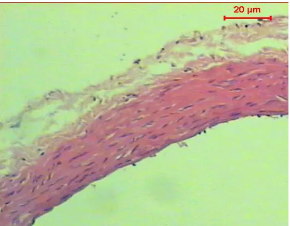
[image:4.595.166.449.478.698.2]
Figure (2): Micrograph of transverse section of aorta of diabetic rat shows a significant atherosclerosis, vacuolation in the cells of the tunica media (H & E, Scale Bar: 20 µm)
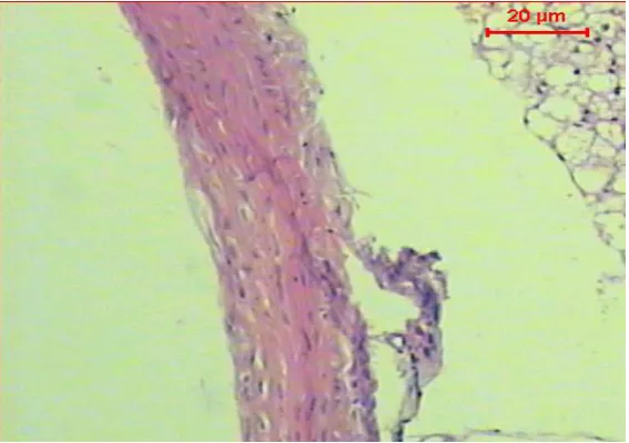
Figure (3): Micrograph of transverse section of aorta of diabetic rat treated with Myrtle extract shows the normal histological structure of the tunica intima, tunica media & tunica adventitia (H & E, Scale Bar:20µm)
RESULTS AND DISCUSSION
Diabetes mellitus is considered as a cluster of metabolic disorders characterized by high levels of blood sugar over a prolonged period of time. The prospect development of type 2 diabetes mellitus is probably due to integration of genetic factors and lifestyle. Although some of these factors like obesity and diet are personally controlled, the others are not. In contrast to type 1DM, type 2 diabetes mellitus is described mainly by hyperglycemia (high levels of blood sugar) and reduction in insulin level due to insulin resistance [20].
[image:5.595.164.452.67.272.2] [image:5.595.165.449.334.534.2]This study showed that Myrtle improved glucose level in both control and diabetic groups in accordance with Benkhayal et al.[21] who found that prolonged period of diabetes leads to miccrovascular injurious effects as retinopathy, nephropathy and neuropathy and macrovascular complications such as peripheral arterial disease, coronary artery disease and stroke.
Hyperglycemia has a vital role in the prospect incidence of endothelial cell dysfunction (ECD). Endothelial dysfunction which is considered as a systemic of pathological state, actually represent a state of imbalance between vasoconstricting and vasodilating substances which released by the overall endothelium functions. In a definite cell, the accessibility of NO attributed mainly to several factors as activity and expression of nitric oxide synthase (NOS), quenching of NO by reactive oxygen species (ROS), and abundance of NOS substrate, tetra- hydrobiopterin, L-arginine, and its cofactor [22].
In the present study, group of untreated-diabetic rats showing a significant reduction in the levels of NO in the plasma as well as in the aortic and liver extracts associated with a significant increase in level of malondialdehyde (MDA) in both aortic and liver extracts compared to the other groups. The latter compound was used an index of lipid oxidation under oxidative stress or imbalance in the antioxidant system. These results are in agreement with Abo-Zaid et al [23] and Claybaugh et al [24]. They suggested a possible mechanism explained the deficiency of NO is mainly due to the prolonged period of hyperglycemia in patients with type 2 DM. It has been proved the capability of insulin to activate the production of NO by endothelial cells. Increased the glucose serum levels can enhance NO oxidative degradation forming peroxynitrite and so reducing its bioavailability [22]. Several studies proved that the production of NO may be controlled by the endogenous inhibitors NOS, particularly asymmetric dimethylarginine (ADMA). Deficiency in NO in several pathological diseases may be due to the change that occurred in the level of NOS inhibitors in various physiological and pathological conditions [25]. Prolonged period of hyperglycemia can down regulate dimethylaminohydrolase (DDAH) and dimethyl-arginine activities, accordingly accumulation of asymmetric dimethylarginine (ADMA) will occur. Increased ADMA levels stimulate uncoupling mechanism of endothelial nitric oxide synthase (eNOS), which causes a significant reduction, in the bioavailability of NO and increased in hydrogen peroxide production [26].
Several clinical and experimental results indicated that even slight modulations of ADMA can change significantly the production of vascular NO, systemic vascular resistance and vascular tone. This finding supports the hypothesis that ADMA candidates are biomarkers of endothelial dysfunction [27, 28, 29].
In this regard, the present study showed that serum level of ADMA is significantly raised in parallel with decreasing in the endothelium-dependent relaxation factor (NO) in STZ diabetic compared to the control, while treatment with Myrtle improved these results. As mentioned previously, NO level is reduced in the aortic and liver homogenate in diabetic group comparing to control. Our results are in agreement with Maritim et al [30] and Majithiya et al [31]. Their studies recorded that 2 mechanisms may be responsible for the significant reduction in the level of NO in aorta; the first assumed that a decrease in the production or secretion of NO from eNOS exist in the endothelium. Second possible mechanism suggested that NO inactivation and oxidative degradation may be elevated in the vessels of such rats since lipid peroxidation increased significantly whereas the endogenous antioxidants levels were significantly decreased in STZ-diabetic group.
In case of AST and ALT enzymes, significant decrease was observed in the Myrtle extract receiving diabetic group compared to the corresponding diabetic group. Hepatocytes damage occurs in diabetes leads to release these enzymes in large quantities into the blood. These results are in agreement with Johari et al [32]. Also, Matsuura et al.[33] found that flavonoid compounds like rosmarinic acid which present in Myrtle leaf have an antioxidant feature to inhibit these damaging effects.
Histopathological examination of the normal control rat aorta revealed a normal histological structure of the tunica intima, tunica media and tunica adventitia while, the STZ-induced diabetic rats displayed a significant atherosclerosis and vacuolation in the cells of the tunica media. Treating with Myrtle revealed no injuries on aorta tissue in diabetic rats.
CONCLUSION
Our study concluded that Myrtle extract acts as a natural antioxidant as well as hepatoprotective agent in diabetic rats.
Acknowledgment
This research was funded by National Research Centre project no. 10010310- Cairo, Egypt.
.
REFERENCES
[1]SH Tawfik; BF Mahmoud; MI Saad; M Shehata; MA Kamel; MH Helmy, Research International Volume 2015, Article ID 567945, 1-8.
[2]E PoniedziaBek-Czajkowska; R MierzyNski; D Szymula; BL Gorzelak; J Oleszczuk, Journal of Diabetes
Research Volume 2016, Article ID 1342643, 1-5. http://dx.doi.org/10.1155/2016/1342643
[3]A Avogaro; M Albiero; L Menegazzo; S De Kreutzenberg; GP Fadini, Diabetes Care 2011,.34 (2): S285- S290. [4]Asgarpanah and A Ariamanesh,Indian Journal of Traditional Knowledge 2015, 14 (1): 82- 87.
[5]C Messaoud; A Laabidi; M Boussaid, Journal of Food Science 2012,77: Nr. 9.C941-C 947.
[6]G Hardya; S Vergnaudb; J Lunardic; M Peoc’hd, G Bessarda; F Stanke-Labesquea, Prostaglandins & other Lipid Mediators 2005, 75: 91–103.
[7]H Passing and WA Bablok, Journal of Clinical Chemistry and Clinical Biochemistry 1983; 21:709-720. [8]CC Allain;LS Poon; CS Chan; W Richmond; PC Fu, Clin Chem. Apr 1974, 20(4):470-475.
[9]MF Lopez-Virella; P Stone; S Ellis; JA Colwell, Clin Chem. May 1977, 23(5):882-884. [10]D Steinberg. Elsevier North Holland, 1981, 1 (2): 31-34.
[11]MR Glick; KW Ryder; SA Jackson, Clin Chem, 1986, 32: 470-474. [12]JE Deanfield; JP Halcox; TJ Rabelink , Circulation, 2007,115:1285–95.
[13]H Moshage; B Kok; JR Huizenga; PL Jansen, Clin Chem, 1995 Jun; 41(6 Pt 1):892-6. [14]H Esterbauer; KH Cheeseman, The FASEB Journal ,1990, 21:846 – 848.
[15]F Yallow; WA Bawman, Diabetes Mellitus ,1983, 119-150. [16]RJ Marklund; G Marklund, Eur. J. Biochem, 1974, 47:469–471. [17]AK.Sinha, Anal. Biochem, 1972, 47: 389–394.
[18]W Thefeld; H Hffmiester; EW Busch; PU Koller; J Volmer; Deut Med Wochenchr, 1974, 99(8):343–351. [19]R.A Drury; EAC Wallington, Coreleton's Histological technique 4th edition 1980, Oxford, Oxford University Press.
[20]E Selvin; MW Steffes; H Zhu; K Matsushita; L Wagenknecht; J Pankow; J Coresh; FL Brancati, N. Engl. J. Med 2010, 362 (9): 800–11.
[21]FA Benkhayal; E Musbah; S Ramesh; DWH El-Ageeli, J Tamilnadu, Vet. Anim. Sci 2009, 5: 155-156.
[22]KY Lin; T Asagami; PS Tsao; S Adimoolam , M Kimoto; H Tsuji; GM Reaven, Circulation. Aug 2002, 106(8):987-92.
[23]OAR Abo-zaid; S E Hussain Ahmed, Benha Veterinary Medical Journal 2013, 24(1): 309-319.
[24]T Claybaugh; S Decker; K McCall; Y Slyvka; J Steimle; A Wood; et al., Int J Endocrinol., 2014:171546-171553.
[25]J Bełtowski; A Kedra,Pharmacol Rep, 2006, 58(2):159-78.
[26]K Toutouzas; M Riga; E Stefanadi; C Stefanadis, Horm Metab Res, 2008, 40(9):655-9. [27]Y Xiong; YF Fu; SH Fu; HH Zhou, J Cardiovasc Pharmacol,2003, 42(2):191-6.
[28]ZC Yang; K Xia; L Wang; SJ Jia; D Li , Z Zhang , et al., Microvasc Res, 2007, 73 (2):131-6. [29]M Cakirca, C Karatoprak, M Zorlu, M Kiskac, M Kanat, et al., Drug Des Devel Ther, 2014, 8:239-43. [30]AC Maritim; RA Sanders; JB Watkins, J Biochem Mol Toxicol, 2003, 17:24–8.
[31]JB Majithiya , AN Paramar , R Balaraman . Cardiovasc Res, 2005, 66(1):150-61. [32]H Johari; M Nozari; M Moghtari; Z Zamani; M Yazdani, ZJRMS, 2014, 16(10): 12-17. [33]H Matsuura; C Chiji; C Asakawa; et al., Biotech Biochem, 2003, 67(11): 2311-2316.