INTRODUCTION
Ovules are seed precursors and typically have two sheathing structures, termed the inner and outer integuments, that form the seed coat following fertilization. Numerous genes involved in integument patterning and growth have been identified from forward genetic screens (Skinner et al., 2004; Colombo et al., 2008). One such gene, ABERRANT TESTA SHAPE (ATS) encodes a KANADI (KAN) transcription factor (TF). KANgenes provide patterning and growth cues during embryogenesis (Izhaki and Bowman, 2007), lateral root formation (Hawker and Bowman, 2004), adaxial-abaxial leaf polarity establishment (Eshed et al., 2001; Kerstetter et al., 2001; Eshed et al., 2004) and integument formation (Eshed et al., 2001; McAbee et al., 2006; Kelley et al., 2009). Loss of ATSfunction leads to congenital fusion of the inner and outer integuments and abnormal seed formation (Leon-Kloosterziel et al., 1994; McAbee et al., 2006). ATSplays dual roles during ovule development, providing boundary maintenance and promoting laminar growth of the inner integument (McAbee et al., 2006). Similarly, loss of KAN1and KAN2disrupts laminar growth of the outer integument (Eshed et al., 2001; McAbee et al., 2006). Thus, these genes play similar roles but ATSacts in the inner
integument whereas KAN1and KAN2act redundantly in the outer integument (Leon-Kloosterziel et al., 1994; Eshed et al., 2001; McAbee et al., 2006).
In vegetative organs there is substantial functional overlap between KAN family members [KAN1, KAN2, KAN3 and ATS (KAN4)] (Eshed et al., 2001; Izhaki and Bowman, 2007). During embryo development, KAN1,KAN2and ATSare required to restrict polar PIN-FORMED 1 (PIN1) expression from the hypocotyl and subsequently maintain unidirectional auxin flows towards the cotyledon primordia (Izhaki and Bowman, 2007). Additionally, genetic data suggest that this regulation of auxin signaling might occur by the cooperative action of KAN and AUXIN RESPONSE FACTOR (ARF) proteins (Pekker et al., 2005). Specifically, the suppression of effects of ectopic KAN1 activity by loss of ETTIN (ETT, also known as ARF3) function and the resemblance of ett arf4 double mutants to kan1 kan2 double mutants suggests that these different TFs have similar functions during organogenesis (Pekker et al., 2005). Because kan mutants and ett arf4 mutants display both unique and shared phenotypes, KAN and ETT/ARF4 appear to act both in concert and in independent roles.
Although many of the genes involved in plant development are known to encode TFs, our current understanding of the transcriptional complexes that are active during organ formation is incomplete. This is especially true for organs that are small and/or difficult to mechanically isolate from other tissues, such as ovules. Definition of protein-protein interactions during ovule development is required to further our understanding of how TFs act at the molecular level to integrate hormone signaling and organogenesis. The substantial overlap in genes active during ovule and leaf development (Kelley and Gasser, 2009) suggests that transcription partners might be conserved, providing genetic modules that act repetitively throughout plant development. Here we show that ETT and ATS form a functional complex active in ovule development Development 139, 1105-1109 (2012) doi:10.1242/dev.067918
© 2012. Published by The Company of Biologists Ltd
Department of Molecular and Cellular Biology, University of California, Davis, CA 95616, USA.
*Present address: Howard Hughes Medical Institute, University of California San Diego, La Jolla, CA 92093, USA
‡Present address: Department of Genetics, Lineberger Comprehensive Cancer Center, University of North Carolina at Chapel Hill, NC 27599, USA §Present address: Department of Molecular & Cellular Biology, University of California, Berkeley, CA 94720, USA
¶Author for correspondence ([email protected])
Accepted 6 January 2012
SUMMARY
KANADI (KAN) transcription factors promote abaxial cell fate throughout plant development and are required for organ formation during embryo, leaf, carpel and ovule development. ABERRANT TESTA SHAPE(ATS, or KAN4) is necessary during ovule development to maintain the boundary between the two ovule integuments and to promote inner integument growth. Yeast two-hybrid assays identified ETTIN (ETT, or AUXIN RESPONSE FACTOR 3) as a transcription factor that could physically interact with ATS. ATS and ETT were shown to physically interact in vivo in transiently transformed tobacco epidermal cells using bimolecular fluorescence complementation. ATSand ETTwere found to share an overlapping expression pattern during Arabidopsisovule development and loss of either gene resulted in congenital fusion of the integuments and altered seed morphology. We hypothesize that in wild-type ovules a physical interaction between ATS and ETT allows these proteins to act in concert to define the boundary between integument primordia. We further show protein-protein interaction in yeast between ETT and KAN1, a paralog of ATS. Thus, a direct physical association between ETT and KAN proteins underpins their previously described common role in polarity establishment and organogenesis. We propose that ETT-KAN protein complex(es) constitute part of an auxin-dependent regulatory module that plays a conserved role in a variety of developmental contexts.
KEY WORDS: Ovule, Seed, Organ fusion, Transcription factor, Abaxial, Arabidopsis
ETTIN (ARF3) physically interacts with KANADI proteins to
form a functional complex essential for integument
development and polarity determination in
Arabidopsis
Dior R. Kelley*, Alexandra Arreola‡, Thomas L. Gallagher§and Charles S. Gasser¶
D
E
V
E
LO
P
M
E
N
and provide evidence that a similar complex between ETT and other KAN proteins underlies the cooperative activity of these proteins in leaf development.
MATERIALS AND METHODS
Plasmids and cDNA clones
ATScDNA was amplified from POP:KAN4 (Hawker and Bowman, 2004) by PCR, digested with NdeI/NcoI and inserted into pGBKT7 (Clontech), creating pDK8 [GAL4 DNA-binding domain (BD)-ATS]. ATS cDNA without the stop codon was amplified from POP:KAN4 and inserted into pENTR/D-Topo (Invitrogen), creating pAA34. ATScDNA was amplified from POP:KAN4 and inserted into pENTR4 (Invitrogen), creating pDK23. pDK23 was Gateway cloned into pDEST-GADT7 (Rossignol et al., 2007) to create pDK131 [GAL4 activation domain (AD)-ATS]. KAN2cDNA and
ETTcDNA were amplified from wild-type Columbia (Col) leaf cDNA and inserted into pENTR/D-Topo, creating pAA29 and pDK132, respectively.
ETTcDNA without the stop codon was amplified from leaf cDNA and cloned into pENTR/D-Topo to create pDK74. pAA29 was Gateway cloned into pDEST-GBKT7 (Rossignol et al., 2007) creating pAA36 (BD-KAN2). pDK132 was Gateway cloned into pDEST-GADT7 creating plasmid pDK136 (AD-ETT). ETTcDNA was cloned as an NdeI/XhoI fragment from pDK136 into pGBKT7 using NdeI/SalI to create pDK137 (BD-ETT). ARF4
cDNA was amplified from Col leaf cDNA and cloned into pENTR/D-Topo, creating pAA30. pAA30 was Gateway cloned into pDEST-GADT7 to create pAA42 (AD-ARF4). KAN1 cDNA was amplified and inserted as a
BamHI/PstI fragment into pLITMUS28 to create pLMK37 and transferred as a BamHI/PstI fragment into: (1) pGAD424, creating pLMK44 (AD-KAN1); and (2) pAS2, creating pLMK46 (BD-KAN1).
A 700 bp subclone of ETT(pDK73) cDNA was generated from pAS13 as described (Sessions et al., 1997). ATS-eGFP (pDK77) was created by Gateway cloning pAA34 into pDH51-GW-eGFP (Zhong et al., 2008). ETT-eGFP (pDK80) was created by Gateway cloning pDK74 into pDH51-GW-eGFP. Subclones for ATS-YFPc (pCG51) and ETT-YFPn (pCG54) were created by amplifying ATSor ETTcDNA without stop codons and inserting the resulting fragments into pJET1.2 (Fermentas) to form pCG45 and pCG46, respectively. The cDNAs were inserted into 2X35S-SPYCE or 2X35S-SPYNE vectors (Walter et al., 2004), respectively, as XhoI/XmaI fragments into these same sites forming pCG47 and pCG50. The resulting expression cassettes were transferred into pMLBART (Gleave, 1992) as
NotI fragments, producing pCG51and pCG54. Gateway reactions were performed using LR Clonase II (Invitrogen). All plasmids were sequence verified. Primers are listed in supplementary material Table S1.
Yeast two-hybrid assays
Yeast transformations, matings and cDNA library preparation were performed according to the Matchmaker Yeast Transformation System 2 manual (Clontech). Tissue for a cDNA library was prepared from
Arabidopsispistils from stage 9-12 flowers (Smyth et al., 1990). Total RNA
was extracted from the harvested tissue and used for cDNA library construction using the BD Matchmaker Library Construction and Screening Kit (Clontech); the cDNA library was transformed into AH109. For the initial screen, pDK8 (BD-ATS) was transformed into Y187 (Clontech) and then mated with the pistil library. Mating mixtures were screened on yeast media lacking leucine (LEU), tryptophan (TRP), adenine (ADE) and histidine (HIS). Inserts were amplified from yeast cultures using the Illustra TempliPhi Amplification Kit (GE Healthcare) and directly sequenced. Plasmids were extracted from yeast using the Zymo II Yeast Plasmid Minipreparation Kit (Zymo). Pairwise interaction tests were performed by directly transforming plasmids into AH109 and replica plating the transformants on yeast media lacking (1) LEU and TRP and (2) LEU, TRP, ADE and HIS. Colonies were restreaked on similar media at least twice for verification.
Plant material
Arabidopsisplants were grown under long-day conditions as previously
described (McAbee et al., 2006). The alleles used in this study in a Col-0 background were ats-2(McAbee et al., 2006) and arf3-2 (Okushima et al., 2005). Other alleles examined are in a Ler(ats-1, ett-3) or a Ws-2 (arf4-1,
ett-1,ett-2) background (Sessions et al., 1997; Pekker et al., 2005).
Transient expression studies and bimolecular fluorescence complementation (BiFC) assays
Transient expression of protein fusions ATS-eGFP (pDK77) and ETT-eGFP (pDK80) in onion epidermal cells was performed as described (Skinner et al., 2001). A 35S:-glucuronidase control plasmid (pHK17) (Norris et al., 1993) was co-bombarded as a positive control for cell transformation. After imaging for fluorescence, staining for GUS activity was performed as described (Skinner et al., 2001) and the positively transformed cells were counted.
BiFC assays were performed in triplicate as described (Szemenyei et al., 2008). The abaxial surfaces of infiltrated tobacco leaves were imaged 2-5 days after inoculation.
In situ hybridization
Tissue preparation, in situ hybridization and ATSprobe synthesis were performed as described (Kelley et al., 2009). The ETTprobe was generated by digesting pDK73 with BamHI and transcribing with T7 RNA polymerase (Promega) and DIG labeling mix (Roche).
Microscopy
Scanning electron microscopy and ovule clearings were performed as described (Kelley et al., 2009). Seeds were imaged using a SPOT RT Slider camera system (SPOT Imaging Solutions) on a Leica MZ FLIII dissecting scope. Bright-field and fluorescence images of onion cells were acquired using a Kodak MDS290 camera on a Zeiss Axioplan microscope with an Endow GFP bandpass emission 41017 filter set (Chroma Technology). Bright-field and fluorescence images of tobacco cells were acquired using a SPOT RT Slider camera system on a Leica DM5000B microscope with a GFP filter set. Images were edited in Adobe Photoshop CS2.
RESULTS AND DISCUSSION
Yeast two-hybrid screen with ATS identifies ETTIN To identify protein complex(es) in which ATS might participate during ovule development we performed a yeast two-hybrid screen using full-length ATS as bait and a pistil cDNA library as prey. We obtained a number of positive clones, including a clone encoding the DNA-binding domain of ETT (amino acids 12-34) (Ulmasov et al., 1999). We subsequently produced fusions of full-length ETT to both the GAL4 DNA-binding domain (BD) and activation domain (AD) and showed that full-length ATS and ETT interact in yeast irrespective of the GAL4 fusion protein orientation (supplementary material Fig. S1). We examined interactions of ATS paralogs KAN1 and KAN2 with ETT and also of these proteins with the ETT paralog ARF4 (supplementary material Fig. S1). These studies revealed an interaction between KAN1 and ETT, but BD-KAN2 alone showed auto-activation in this yeast system, masking any possible interactions (supplementary material Fig. S1). None of the other tested fusion proteins showed auto-activation of the reporter genes (supplementary material Fig. S1). The yeast assays did not provide evidence for interaction between ARF4 and KAN proteins. Thus, at least two different KAN proteins can bind ETT in yeast. Differences in the protein-protein interactions of ETT and ARF4 might result from structural differences between these proteins, as ETT lacks the conserved domains III and IV that are present in ARF4.
ATS and ETT can physically interact in the plant cell nucleus
In onion epidermal cells, transient expression of translational fusions of ATS or ETT to eGFP led to fluorescence that was observed only in nuclei (Fig. 1B,D), confirming nuclear localization of these two TFs. Transient co-expression of a translational fusion of ATS to the C-terminal portion of yellow fluorescent protein (YFP) (ATS-YFPc) and of ETT to the N-terminal portion of YFP (ETT-YFPn) showed BiFC (Kerppola,
D
E
V
E
LO
P
M
E
N
2006) in the nuclei of tobacco epidermal cells (Fig. 1E,F). As a control, we performed similar tests for protein interaction between ATS and another ARF family member, MONOPTEROS (MP), which has been shown to be nuclear localized and to interact with a TOPLESS fusion protein in the same BiFC assay (Szemenyei et al., 2008). ATS-YFPc and MP-YFPn (Fig. 1G,H) did not show fluorescence complementation in tobacco cells, confirming that the observed BiFC between ATS and ETT was specific. These results demonstrate that ATS and ETT can directly interact in planta.
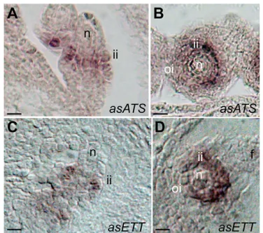
ATSand ETTare co-expressed in the inner
integument during ovule development
We examined ATS and ETTexpression during ovule development by in situ hybridization to establish the biological relevance of the ATS-ETT physical interaction. ATSexpression is restricted to the abaxial region of the inner integument in both young and mature ovules (Fig. 2A,B) (McAbee et al., 2006). ETTwas expressed in the same pattern as ATS during ovule development, with ETT
transcript first appearing during inner integument initiation and persisting throughout ovule development (Fig. 2C,D). This finding is consistent with prior transcriptional profiling (Skinner and Gasser, 2009) and hybridization studies (Ng et al., 2009). The coincident expression patterns of ATSand ETTin ovules shows that the interaction between these proteins as observed in yeast and in transgenic plant cells could occur during the normal expression of these genes.
Ovule and seed phenotypes of ett mutants
resemble those of ats
During wild-type ovule development the outer integument forms a hood-like structure covering the inner integument and the nucellus (Fig. 3A,E). In atsmutant ovules the inner and outer integument cell layers grow as a unit, producing a single fused structure (Leon-Kloosterziel et al., 1994; McAbee et al., 2006) (compare Fig. 3B,F with 3A,E). As a result of this fusion, ats seeds are abnormally rounded and variable in size (Fig. 3J) (McAbee et al., 2006) compared with the uniformly elongate mature wild-type Arabidopsisseeds (Fig. 3I). Examination ofettmutant ovules (Fig. 3C,G) and seeds (Fig. 3K and supplementary material Fig. S2) revealed that they phenotypically resemble atsovules and seeds (compare Fig. 3C with 3B, 3G with 3F and 3K with 3J; seeds in supplementary material Fig. S2). ats ettdouble-mutant ovules and seeds showed no phenotypic differences to either single mutant (Fig. 3D,H,L). Thus, loss of either ATS or ETT is sufficient to disrupt a common regulatory pathway that is mediated by both TFs. The severely compromised gynoecia of arf4-1 ett-1 double mutants preclude examination of the ovules of this mutant combination. However, the wild-type morphology of arf4 ett/+ ovules and seeds (supplementary material Fig. S2) indicates that ARF4is not required for integument development.
A model for ATS-ETT action during ovule development
[image:3.612.56.291.59.438.2]KAN, ETT and ARF4 have been proposed to act as transcriptional repressors (Tiwari et al., 2003; Wu et al., 2008; Causier et al., 2011). We therefore speculate that an ATS-ETT TF complex acts
[image:3.612.344.532.60.227.2]Fig. 1. ATS and ETT are nuclear localized and physically interact in vivo in a BiFC assay. (A-D)ATS-eGFP (A,B) and ETT-eGFP (C,D) are nuclear localized. (E,F)Fluorescence indicates direct interaction in vivo between ATS and ETT that is restricted to nuclei of transformed tobacco cells. (G,H)Co-transformation of ATS and a different ARF protein, MP, fails to show fluorescence complementation. Scale bars: 20mm in A-D; 50mm in E-H.
Fig. 2. ATSand ETTdisplay overlapping mRNA accumulation patterns during ovule development. (A-D)ATS (A,B) and ETT(C,D) in situ hybridizations on young (A,C) and mature (B,D) wild-type
Arabidopsisovules show an inner integument-specific signal. f, funiculus; n, nucellus; ii, inner integument; oi, outer integument. Scale bars: 5mm in A,C; 10mm in B,D.
D
E
V
E
LO
P
M
E
N
to repress the expression of specific genes in the abaxial domains of developing inner integuments. Furthermore, the action of such an ATS-ETT module might be directly linked to auxin signaling. PIN1-dependent auxin maxima have been shown to occur in ovule and integument primordia (Benkova et al., 2003). KAN proteins play a role in restricting PIN activity and thus auxin flow during embryogenesis (Pekker et al., 2005; Izhaki and Bowman, 2007; Ilegems et al., 2010). PIN proteins regulate auxin flow, and auxin has been shown to control PIN gene expression as well as PIN cellular polarity via the TIR1-Auxin/IAA-ARF pathway (Schrader et al., 2003; Vieten et al., 2005; Sauer et al., 2006).
Based on these observations, we propose a model for ATS-ETT action during ovule development (Fig. 4). Initially, an auxin maximum occurs at the apex of the nucellus (Fig. 4A). Following
integument initiation, the ATS-ETT complex accumulates in the abaxial layer of the inner integument and refines auxin action in the chalaza through negative regulation of PIN1 and hence auxin transport (Fig. 4B). Mutation in either protein eliminates this resolution, leading to the formation of a single broad integument (Fig. 4C). Auxin is also known to positively regulate ETTactivity (Tiwari et al., 2003). Positive feedback from auxin levels on ETT combine with ATS-ETT suppression of PIN (and hence auxin levels) to establish and homeostatically maintain the appropriate level of ETT, PIN1 and auxin activity necessary for lateral organ outgrowth (Fig. 4D).
[image:4.612.52.334.58.287.2]Our model for inner integument outgrowth parallels related models for KAN, PIN and auxin interaction proposed for the development and polarity establishment of leaf lamina, the embryo
Fig. 3. Loss of ATSor ETTresults in integument fusion and altered seed morphology. (A-I)Wild-type (A,E,I), ats
(B,F,J) ett(C,G,K) and ats ett(D,H,L) ovules (A-H) and seeds (I-L). Wild-type ovules initiate two distinct integument primordia (A). Integument formation in ats, ettand ats ettleads to a single integument primordium (B-D). The outer integument grows over the inner integument in wild-type ovules (E), whereas the fused integuments in ats, ettand ats ettmutants grow in a single plane (F-H). Abnormal integument formation in ats, ettand ats ettmutants gives rise to aberrant seed morphology (compare J-L with I). n, nucellus; ii, inner integument; oi, outer integument; i, integument. Scale bars: 5mm in A-D; 10mm in E-H; 200mm in I-L.
Fig. 4. Model for ATS-ETT action during ovule development.
(A,B)Benkova et al. (Benkova et al., 2003) have shown that PIN1-mediated auxin transport in an ovule primordium initially creates an auxin maximum at the nucellar tip (A) and subsequently two auxin maxima in the chalaza corresponding to the two integument primordia (B). Inner integument initiation coincides with the formation of an active ATS-ETT complex, which we hypothesize restricts the domain of PIN1 and thus enables the formation of the two auxin maxima at the chalaza (B). (C)In the absence of an active ATS-ETT complex (i.e. in ats or ettmutant ovules) PIN1 fails to be restricted and auxin remains distributed across the chalaza. (D)Formation of ATS-ETT heterodimers restricts PIN1 activity, which controls auxin efflux, while auxin positively regulates both ETT and PIN1 function.
D
E
V
E
LO
P
M
E
N
[image:4.612.50.417.468.732.2]axis, carpels and vascular tissues (Pekker et al., 2005; Izhaki and Bowman, 2007; Ilegems et al., 2010). Our observation that KAN1 also interacts with ETT suggests the possibility that ETT potentiates KAN function in these structures. Further protein-protein interaction tests will evaluate all possible KAN-ARF interactions that might occur in vivo. Although ETT and ARF4 act redundantly in leaves, we were unable to detect interactions between KAN proteins and ARF4 (supplementary material Fig. S1), but it is possible that an in planta interaction requires other factor(s). Thus, it is possible that KAN-ETT protein complexes act differently in organs other than inner integuments, consistent with the distinct evolutionary origins of leaves and the inner integument (Endress, 2011).
Acknowledgements
We thank Lauren Kotow and Robert Meister for plasmids; Patricia Zymbryski for pAS13; Jeff Long for MP-YFPn and assistance with BiFC assays; the Arabidopsis Biological Resource Center at Ohio State for seeds, pDEST-GBKT7 and pDEST-GADT7; Keiko Yamada, Debra Skinner and Marissa Simon for technical assistance; and John Bowman for helpful comments.
Funding
Supported by a National Science Foundation grant [IOS-0920618 to C.S.G.]; a National Institutes of Health traineeship [T32M007377 to D.R.K.]; and fellowships from the Summer Undergraduate Research Program (U. C. Davis) and McNair Scholars Program to A.A. Deposited in PMC for release after 12 months.
Competing interests statement
The authors declare no competing financial interests.
Supplementary material
Supplementary material available online at
http://dev.biologists.org/lookup/suppl/doi:10.1242/dev.067918/-/DC1
References
Benkova, E., Michniewicz, M., Sauer, M., Teichmann, T., Seifertova, D., Jurgens, G. and Friml, J.(2003). Local, efflux-dependent auxin gradients as a common module for plant organ formation. Cell115, 591-602.
Causier, B., Ashworth, M., Guo, W. and Davies, B.(2011). The TOPLESS interactome: a framework for gene repression in Arabidopsis. Plant Physiol. 158, 423-438.
Colombo, L., Battaglia, R. and Kater, M. M.(2008). Arabidopsis ovule development and its evolutionary conservation. Trends Plant Sci. 13, 444-450.
Endress, P. K.(2011). Angiosperm ovules: diversity, development, evolution. Ann. Bot. 107, 1465-1489.
Eshed, Y., Baum, S. F., Perea, J. V. and Bowman, J. L.(2001). Establishment of polarity in lateral organs of plants. Curr. Biol. 11, 1251-1260.
Eshed, Y., Izhaki, A., Baum, S. F., Floyd, S. K. and Bowman, J. L.(2004). Asymmetric leaf development and blade expansion in Arabidopsis are mediated by KANADI and YABBY activities. Development131, 2997-3006.
Gleave, A. P.(1992). A versatile binary vector system with a T-DNA organisational structure conducive to efficient integration of cloned DNA into the plant genome. Plant Mol. Biol. 20, 1203-1207.
Hawker, N. P. and Bowman, J. L.(2004). Roles for Class III HD-Zip and KANADI genes in Arabidopsis root development. Plant Phys. 135, 2261-2270.
Ilegems, M., Douet, V., Meylan-Bettex, M., Uyttewaal, M., Brand, L., Bowman, J. L. and Stieger, P. A.(2010). Interplay of auxin, KANADI and Class III HD-ZIP transcription factors in vascular tissue formation. Development137, 975-984.
Izhaki, A. and Bowman, J. L.(2007). KANADI and class III HD-Zip gene families regulate embryo patterning and modulate auxin flow during embryogenesis in Arabidopsis. Plant Cell19, 495-508.
Kelley, D. R. and Gasser, C. S.(2009). Ovule development: genetic trends and evolutionary considerations. Sex. Plant Reprod. 22, 229-234.
Kelley, D. R., Skinner, D. J. and Gasser, C. S.(2009). Roles of polarity determinants in ovule development. Plant J. 57, 1054-1064.
Kerppola, T. K.(2006). Design and implementation of bimolecular fluorescence complementation (BiFC) assays for the visualization of protein interactions in living cells. Nat. Protoc. 1, 1278-1286.
Kerstetter, R. A., Bollman, K., Taylor, R. A., Bomblies, K. and Poethig, R. S.
(2001). KANADI regulates organ polarity in Arabidopsis. Nature411, 706-709.
Leon-Kloosterziel, K. M., Keijzer, C. J. and Koornneef, M.(1994). A seed shape mutant of Arabidopsis that is affected in integument development. Plant Cell6, 385-392.
McAbee, J. M., Hill, T. A., Skinner, D. J., Izhaki, A., Hauser, B. A., Meister, R. J., Venugopala Reddy, G., Meyerowitz, E. M., Bowman, J. L. and Gasser, C. S.(2006). ABERRANT TESTA SHAPEencodes a KANADI family member, linking polarity determination to separation and growth of Arabidopsis ovule integuments. Plant J. 46, 522-531.
Ng, K. H., Yu, H. and Ito, T.(2009). AGAMOUS controls GIANT KILLER, a multifunctional chromatin modifier in reproductive organ patterning and differentiation. PLoS Biol. 7, e1000251.
Norris, S. R., Meyer, S. E. and Callis, J.(1993). The intron of Arabidopsis thaliana
polyubiquitin genes is conserved in location and is a quantitative determinant of chimeric gene expression. Plant Mol. Biol. 21, 895-906.
Okushima, Y., Overvoorde, P. J., Arima, K., Alonso, J. M., Chan, A., Chang, C., Ecker, J. R., Hughes, B., Lui, A., Nguyen, D. et al.(2005). Functional genomic analysis of the AUXIN RESPONSE FACTORgene family members in Arabidopsis thaliana: unique and overlapping functions of ARF7 and ARF19.
Plant Cell17, 444-463.
Pekker, I., Alvarez, J. P. and Eshed, Y.(2005). Auxin response factors mediate Arabidopsis organ asymmetry via modulation of KANADI activity. Plant Cell 17, 2899-2910.
Rossignol, P., Collier, S., Bush, M., Shaw, P. and Doonan, J. H.(2007). Arabidopsis POT1A interacts with TERT-V(I8), an N-terminal splicing variant of telomerase. J. Cell Sci. 120, 3678-3687.
Sauer, M., Balla, J., Luschnig, C., Wisniewska, J., Reinohl, V., Friml, J. and Benkova, E.(2006). Canalization of auxin flow by Aux/IAA-ARF-dependent feedback regulation of PIN polarity. Genes Dev. 20, 2902-2911.
Schrader, J., Baba, K., May, S. T., Palme, K., Bennett, M., Bhalerao, R. P. and Sandberg, G.(2003). Polar auxin transport in the wood-forming tissues of hybrid aspen is under simultaneous control of developmental and environmental signals. Proc. Natl. Acad. Sci. USA100, 10096-10101.
Sessions, A., Nemhauser, J. L., McColl, A., Roe, J. L., Feldmann, K. A. and Zambryski, P. C.(1997). ETTIN patterns the Arabidopsis floral meristem and reproductive organs. Development124, 4481-4491.
Skinner, D. J. and Gasser, C. S.(2009). Expression-based discovery of candidate ovule development regulators through transcriptional profiling of ovule mutants.
BMC Plant Biol. 9, 29.
Skinner, D. J., Baker, S. C., Meister, R. J., Broadhvest, J., Schneitz, K. and Gasser, C. S.(2001). The Arabidopsis HUELLENLOSgene, which is essential for normal ovule development, encodes a mitochondrial ribosomal protein. Plant Cell13, 2719-2730.
Skinner, D. J., Hill, T. A. and Gasser, C. S.(2004). Regulation of ovule development. Plant Cell16 Suppl., S32-S45.
Smyth, D. R., Bowman, J. L. and Meyerowitz, E. M.(1990). Early flower development in Arabidopsis. Plant Cell2, 755-767.
Szemenyei, H., Hannon, M. and Long, J. A.(2008). TOPLESS mediates auxin-dependent transcriptional repression during Arabidopsis embryogenesis. Science 319, 1384-1386.
Tiwari, S. B., Hagen, G. and Guilfoyle, T.(2003). The roles of auxin response factor domains in auxin-responsive transcription. Plant Cell15, 533-543.
Ulmasov, T., Hagen, G. and Guilfoyle, T. J.(1999). Dimerization and DNA binding of auxin response factors. Plant J. 19, 309-319.
Vieten, A., Vanneste, S., Wisniewska, J., Benkova, E., Benjamins, R., Beeckman, T., Luschnig, C. and Friml, J.(2005). Functional redundancy of PIN proteins is accompanied by auxin-dependent cross-regulation of PINexpression.
Development132, 4521-4531.
Walter, M., Chaban, C., Schutze, K., Batistic, O., Weckermann, K., Nake, C., Blazevic, D., Grefen, C., Schumacher, K., Oecking, C. et al.(2004). Visualization of protein interactions in living plant cells using bimolecular fluorescence complementation. Plant J. 40, 428-438.
Wu, G., Lin, W. C., Huang, T., Poethig, R. S., Springer, P. S. and Kerstetter, R. A.(2008). KANADI1 regulates adaxial-abaxial polarity in Arabidopsis by directly repressing the transcription of ASYMMETRIC LEAVES2. Proc. Natl. Acad. Sci. USA105, 16392-16397.
Zhong, S., Lin, Z., Fray, R. G. and Grierson, D.(2008). Improved plant transformation vectors for fluorescent protein tagging. Transgenic Res.17, 985-989.