N A N O E X P R E S S
Open Access
Synapsable quadruplex-mediated fibers
Miguel Angel Mendez
1,2and Veronika A Szalai
1,3*Abstract
We have fabricated a DNA-based nanofiber created by self-assembly of guanine quadruplex (Hoogsteen base pairing) and double-stranded DNA (Watson-Crick base pairing). When duplexes containing a long stretch of contiguous guanines and single-stranded overhangs are incubated in potassium-containing buffer, the preformed duplexes create high molecular weight species that contain quadruplexes. In addition to observation of these larger species by gel electrophoresis, solutions were analyzed by atomic force microscopy to reveal nanofibers. Analysis of the atomic force microscopy images indicates that fibers form with lengths ranging from 250 to 2,000 nm and heights from 0.45 to 4.0 nm. This work is a first step toward the creation of new structurally heterogeneous
(quadruplex/duplex), yet controllable, DNA-based materials exhibiting novel properties suitable for a diverse array of nanotechnology applications.
Keywords:Guanine quartet, Guanine quadruplex, Atomic force microscopy, Nanowires, Nanofibers, DNA nanomaterials, Synapsable quadruplex
Background
Programmable self-assembly from deoxyribonucleic acid (DNA) building blocks has led to a myriad of nanoscale structures, including 3D architectures [1-8]. At the core, construction of ever more complicated and elegant DNA nanoshapes relies on the self-recognition proper-ties of DNA. In DNA-based wires, tiles (double or triple crossover) [8-11], and DNA origami structures, canon-ical Watson-Crick base pairing drives and stabilizes for-mation of the desired structure. Non-canonical base pairing schemes are not typically exploited to create novel DNA-based materials [12], even though such in-teractions are in the lexicon of nucleic acid self-interactions observed in biological systems [13-23].
Several years ago, Watson-Crick self-recognition was combined with non-canonical base pairing to create
‘synapsable’ DNA [24]. Synapsable DNA is fashioned
from two duplex DNA precursors that connect to form a four-stranded DNA unit with blunt ends. Each DNA strand in the unit created originally by Sen's group con-tains an internal run of eight guanines, which creates a
region of guanine-guanine mismatches in the duplex precursor. Introduction of potassium ions induces the
guanine-rich tracts in the duplex precursors to
Hoogsteen base pair, creating a DNA element called a guanine quartet. In the final structure, the central six guanines are involved in creating the guanine quartets [24], and four duplex ‘tails,’ two at each end, project from the quadruplex core.
In addition to the Hoogsteen base pairing in synapsable DNA mimicking interactions and structures found in biology [13,15,19,20,25], synapsable DNA also has been suggested to be an attractive tool for nanofabrication [1,26] although there are no reports of specific examples utilizing synapsable DNA in such a capacity. For the first time, we report the assembly of synapsable DNA-based nanofibers that constitute a novel DNA molecular manufacturing element. Our structure is likely stiffer than canonical DNA-based structures, which potentially improves its ease of use in patterning and other nanotechnology applications. Fur-ther, our unique strategy is expected to create DNA building blocks with a broad temperature response range that can be modulated additionally by sequence control. Finally, our novel design permits future integration with other established and emerging programmable self-assembly methods such as DNA origami or tiles to cre-ate new multi-functional nanomcre-aterials.
* Correspondence:[email protected]
1Department of Chemistry and Biochemistry, University of Maryland,
Baltimore County, 1000 Hilltop Drive, Baltimore, MD 21250, USA
3Center for Nanoscale Science and Technology, National Institute of
Standards and Technology, 100 Bureau Drive, Gaithersburg, MD 20899-6204, USA
Full list of author information is available at the end of the article
Methods
Certain commercial entities, equipment, or materials may be identified in this document in order to describe an experimental procedure or concept adequately. Such identification is not intended to imply recommendation or endorsement by the National Institute of Standards and Technology, nor is it intended to imply that the en-tities, materials, or equipment are necessarily the best available for the purpose.
All DNA oligonucleotides were purchased from Midland Oligos (Midland, TX, USA). DNA was resuspended in purified water with a total organic content of less than 3.4 × 10⁻⁵ kg m−3 (34 μg/L) and a resistivity of 18.2
MΩ·cm. DNA was ethanol-precipitated using a slightly
modified version of a previously reported protocol and resuspended in purified water [27]. Tetramethylammonium chloride (TMACl), ammonium persulfate, mercaptoethanol,
MgCl2, KCl, tris(hydroxymethyl) aminomethane (Tris),
boric acid, andN-methylmesoporphyrin IX were
biochem-ical grade or equivalent reagents purchased from commer-cial suppliers. To separate and isolate DNA in some cases, microcentrifugal filter units (3,000 or 10,000 molecu-lar weight cutoff) and hydrophilic polyvinylidene fluoride filters (0.45-μm pore size) were used. A solution of a mixture of 19 equivalents of acrylamide to 1 equivalent bisacrylamide with an acrylamide mass fraction of 40% was used for gel electrophoresis. Three types of buffer were used and are given here and listed in Table S1 in Additional
file 1: 0.01 KMgTB, which is 1.0 × 10⁻² mol/L (10 mM)
KCl, 1.0 × 10⁻³ mol/L (1.0 mM) MgCl2, 0.05 mol/L (50 mM) Tris-borate, pH 8.0; 0.01 TMgTB, which is 1.0 × 10⁻² mol/L (10 mM) TMACl, 1.0 × 10⁻³mol/L (1.0 mM) MgCl2, 0.05 mol/L (50 mM) Tris-borate, pH 8.0; and 1 KMgTB, which is 1.0 mol/L (1 M) KCl, 1.0 × 10⁻³mol/L (1.0 mM) MgCl2, 0.05 mol/L (50 mM) Tris-borate, pH 8.0. A silicon wafer substrate for atomic force microscopy was obtained from Silicon Valley Microelectronics, Inc. (Santa Clara, CA, USA). Sybr Green I Nucleic Acid Gel Stain 10 000 X was purchased from Lonza (Rockland, MA, USA).
Standard DNA handling and purification
Oligonucleotide sequence information is in Table 1. Syn-thetic oligonucleotide pellets resuspended in water were
ethanol-precipitated using 2.5 mol/L (2.5 M) TMACl. Typically, an equal volume of 2.5 mol/L (2.5 M) TMACl and oligonucleotide (typically 1 × 10⁻³mol/L to 3 × 10⁻³ mol/L (1 mM to 3 mM)) in water were combined and vortexed. A volume of ethanol/water with a volume frac-tion of 95% ethanol (2.5 times the initial sample volume) was added, and the sample was stored at−13°C for 1 h or
−80°C for 30 min. Samples were centrifuged for 90 to 100
min at 14,000 ×g. The ethanol supernatant was removed
using a pipette, and the pellet was resuspended in purified water. Extinction coefficients for the single-stranded oligo-nucleotides were calculated by the nearest neighbor method and are included in Table 1 [28]. The strand con-centration was determined spectrophotometrically. Com-parisons of experimentally measured spectra and spectra predicted using nearest neighbor-derived extinction coeffi-cients [29] generate overall root mean square deviations of 0.013 for single-stranded DNA.
Double-stranded DNA was purified by native poly-acrylamide gel electrophoresis (PAGE) in TMACl prior to use in assembling larger structures. Complementary single-stranded DNA sequences were hybridized in 0.01 TMgTB by heating to 90°C for 10 min followed by slow cooling to 25°C. TMACl inhibits guanine quadruplex formation [30]. Duplex DNA was stored at 4°C prior to further purification by native PAGE. In most cases, du-plex DNA precursor was prepared immediately before gel electrophoresis. Duplex DNA requiring storage for longer than 12 h prior to electrophoresis was stored at
−17°C or −80°C. Duplex DNA was purified by native
[image:2.595.57.541.628.724.2]PAGE (acrylamide mass fraction of 12%) run at 250 to 300 V. The electrophoresis running buffer was 0.01 TMgTB. All solutions containing TB were prepared from a TB stock solution consisting of 0.5 mol/L (0.5 M) Tris and 0.5 mol/L (0.5 M) boric acid at pH 8.0. The DNA in the gel was visualized by UV shadowing, and the gel was imaged using a digital camera. Duplex DNA was excised from the gel and recovered following stand-ard procedures [31]. DNA was either isolated and con-centrated in 0.05 mol/L (50 mM) TMACl using microfuge filtration devices (10,000 molecular weight cutoff ) or ethanol-precipitated using 2.5 M TMACl as described above and resuspended in 0.01 TMgTB buffer.
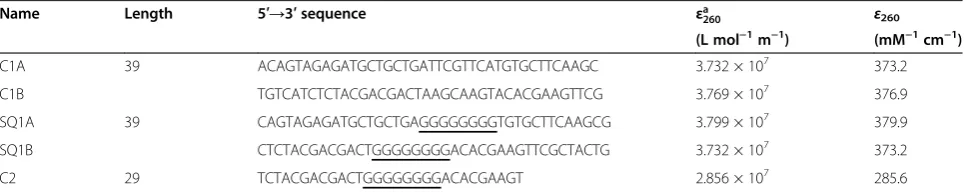
Table 1 Oligonucleotide sequences
Name Length 5′→3′sequence εa
260 ε260
(L mol−1m−1) (mM−1cm−1)
C1A 39 ACAGTAGAGATGCTGCTGATTCGTTCATGTGCTTCAAGC 3.732 × 10⁷ 373.2
C1B TGTCATCTCTACGACGACTAAGCAAGTACACGAAGTTCG 3.769 × 10⁷ 376.9
SQ1A 39 CAGTAGAGATGCTGCTGAGGGGGGGGTGTGCTTCAAGCG 3.799 × 10⁷ 379.9
SQ1B CTCTACGACGACTGGGGGGGGACACGAAGTTCGCTACTG 3.732 × 10⁷ 373.2
C2 29 TCTACGACGACTGGGGGGGGACACGAAGT 2.856 × 10⁷ 285.6
Non-denaturing PAGE of synapsable G-quadruplexes Duplex precursors were incubated in high potassium ion-containing buffers to form quadruplexes. Control samples of the homoquadruplexes formed by SQ1A, SQ1B, or C2 were prepared by heating a single-stranded oligonucleotide to 95°C in 1 KMgTB buffer for 10 min
followed by slow cooling to room temperature. For N
-methylmesoporphyrin IX (NMM)-staining experiments, samples were incubated with NMM for at least 30 min at room temperature prior to gel loading.
Non-denaturing PAGE for gels with an acrylamide mass fraction of 15% was performed at 4°C at 300 V; gels containing an acrylamide mass fraction of 12% were run at 4°C and 250 V. The electrophoresis running buf-fer was either 0.01 TMgTB bufbuf-fer or 0.01 KMgTB bufbuf-fer. Gels were UV-shadowed, imaged by UV transillumin-ation, or stained with Sybr Green I dye by soaking the gels for 10 to 20 min. All gels were wrapped in plastic wrap prior to imaging. UV shadowing was accomplished using a handheld UV lamp and standard digital imaging device. Transillumination to visualize NMM fluores-cence was performed using a standard UV transillumi-nator device equipped with an ethidium bromide photographic filter. Images were processed (background subtraction, contrast adjustment) using ImageJ software. Sybr Green I-stained gels were scanned on a laser-based fluorescence imaging device and analyzed using the instrument-supplied software.
Atomic force microscopy
For the preparation of atomic force microscopy (AFM) substrates, small squares of silicon wafer were washed at 65°C for 30 min in a cleaning solution (piranha) made of three parts sulfuric acid to one part H2O2in H2O (H2O2 mass fraction of 30%) followed by rinsing three times with purified water. Cleaned silicon wafers were stored under purified water. Immediately prior to use, cleaned silicon wafer substrate squares were dried under a stream of nitrogen gas. One drop of 2 mol/L (2 M)
MgCl2 in water (enough to cover the surface) was
dropped on the silicon wafer. The substrate was washed extensively with purified water until cloudy spots were no longer visible on the surface. The wafer was then dried under a stream of nitrogen. The washing and
dry-ing process was repeated twice. At this point, 2 μL of
the sample was applied to the surface and allowed to dry for 5 min. The surface was washed with purified water and dried under nitrogen three times.
We imaged mixtures of higher order structures and monomers by AFM. Three sets of sample preparation conditions were used. In the first set, samples were pre-pared from native PAGE-purified duplex DNA solutions that had been incubated at 4°C for 12 h with 1 KMgTB buffer. Note that this condition does not involve thermal
treatment. Samples for the second set of conditions were heated at 90°C for 5 min and incubated at 50°C for 12 or 72 h. In this second strategy, the precursor synapsable DNA was heated to 90°C, which should not affect the G-quadruplex structure but should affect the duplex re-gion. The third procedure was more involved and was chosen to test if under mild conditions of heating the synapsable DNA fiber formation was improved or resulted in significantly different structures than under the other two conditions tested. Gel-purified comple-mentary strands were annealed in the presence of TMACl to obtain precursor duplex DNA. These du-plexes were exchanged into the 1 KMgTB buffer using microcentrifugal filters and then incubated at 30°C for 10 min followed by slow cooling to 4°C at a rate of 0.5°C/min. Fibers formed from this protocol are shown in Figures S1 and S2 in Additional file 1.
In summary, the prepared DNA solutions were incu-bated at different temperatures prior to deposition on the AFM substrate. In the first and second protocols, DNA samples were prepared to test duplex-mediated synapsable quadruplex formation. In many cases, the same stock solutions, or the same samples used for na-tive PAGE, were used for AFM, but they were diluted so that the final DNA concentration applied to the silicon wafer was 1.6 × 10⁻⁴kg m−3(0.16 ng/μL). Images were collected in air in tapping mode.
To calculate the average height of the fiber, a trajectory along the fiber was traced to obtain cross sections of the images. This method gives the values of heights along the trajectory of the fiber. A number of points,N, were obtained for the fibers in the image being analyzed, and the average and standard deviation of these values were calculated. One fiber representative of those found in each image was used and the value reported. In general, there was a height distribution between fibers and also within each fiber depending on the direction of the cross section. Nevertheless, the distribution was tight (within 1 to 2 nm of the total height depending on the sample). An explanation of the factors that created height vari-ability will be discussed further below. One of those fi-bers was selected per method of preparation to be reported here.
the longer fibers, six images were analyzed for a total of 30 fibers.
Results and discussion
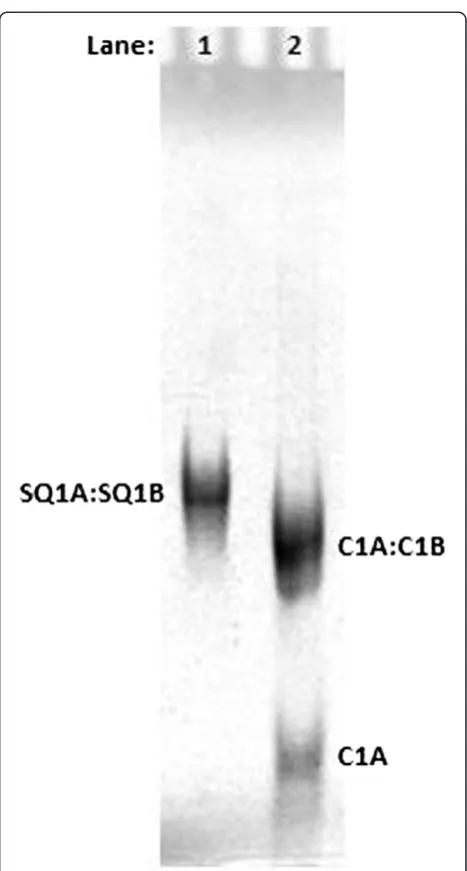
Duplex precursors form synapsable DNA nanofibers Single-stranded DNA sequences (Table 1) were annealed in TMACl-containing buffer (0.01 TMgTB). The resulting duplexes were purified by native PAGE using standard methods [31]. Figure 1 shows that the SQ1A:SQ1B duplex runs slightly more slowly than the random sequence, blunt-end C1A:C1B duplex control, which is of the same length (39 bases). The C1A:C1B duplex control was used as a migration standard because it shows reproducible gel mobility that is not affected by the presence of overhangs or secondary structure. This result is reproducible over a dozen replicates.
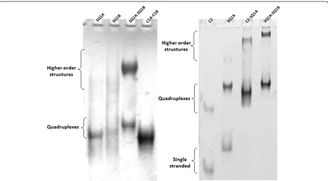
Upon incubation in potassium-containing buffer, the SQ1A:SQ1B duplex assembles into a‘synapsed’quadruplex, (SQ1A:SQ1B)2. In addition to observation of the (SQ1A: SQ1B)2quadruplex, a much slower mobility species is also observed (Figure 2, higher order structures). These slower migrating species form at the high duplex concentrations used in the UV-shadowing gel experiments (Figure 2, left) as well as in SYBR Green-stained gels loaded with lower DNA concentration samples (Figure 2, right). To test if the assembly of larger species is specific to the SQ1A:SQ1B du-plex sequence, we used the C2:SQ1A dudu-plex. This dudu-plex is generated by hybridizing C2, a 29-mer complementary strand, to SQ1A, which results in a duplex with a smaller molecular mass and shorter overall length than the SQ1A: SQ1B duplex. As shown in Figure 2, both the SQ1A:SQ1B and SQ1A:C2 duplexes incubated in potassium-containing buffer form species that migrate more slowly in the gel than the 39-mer homoquadruplexes of C2 and SQ1A.
Higher order species contain quadruplexes
When referenced to the control C1A:C1B duplex, the SQ1A:SQ1B duplex in TMACl (Figure 1) migrates
with about the same mobility as the (SQ1A:SQ1B)2
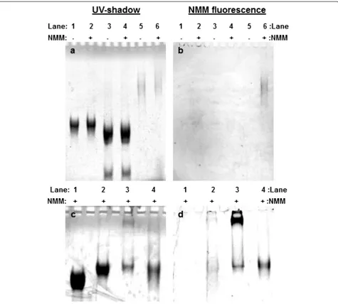
quadruplex in KCl (Figure 2). This observation raises the possibility that the bands we ascribe to higher order structures are either simple quadruplexes (i.e., not linked together) or duplexes that link together without quadruplex formation. To test this possibility, gel elec-trophoresis was performed on samples incubated with NMM, a dye that exhibits increased fluorescence only upon binding quadruplex DNA [34-37]. Figure 3 shows gel images of samples incubated with NMM and ana-lyzed by gel electrophoresis in TMACl (Figure 3a,b) or KCl (Figure 3c,d). Figure 3a shows that incubation of NMM with our samples does not generate new species; a slight shift in band mobility is observed, which is due to NMM binding. Figure 3b,d shows NMM fluorescence intensity recorded for each gel. The control sequence is
the preformed SQ1A homoquadruplex, which causes NMM to fluoresce in either buffer (Figure 3b, lane 6; Figure 3d, lane 4). The SQ1A:SQ1B duplex in TMACl does not induce NMM fluorescence (Figure 3b, lane 2),
while the synapsed (SQ1A:SQ1B)2 quadruplex in KCl
[image:4.595.305.539.84.520.2]clearly does (Figure 3d, lane 3). There is a slight amount of NMM fluorescence for the SQ1A:SQ1B duplex pre-pared in TMACl and run on the KCl gel (Figure 3d, lane 2), which is an expected result because exposure of the
SQ1A:SQ1B duplex to KCl during gel electrophoresis should shift the structure from duplex to quadruplex. The strongest NMM fluorescence is observed for
the slowly migrating species formed by (SQ1A:SQ1B)2
(Figure 3d, lane 3), indicating that quadruplex is present in this structure.
Morphology of the synapsable DNA nanofibers by AFM On the basis of the gel electrophoresis results indicating that slowly migrating species form quadruplex DNA, we
examined solutions of (SQ1A:SQ1B)2 using AFM. We
observed that fibers form under several conditions with varying morphology depending on the preparation method. Gel-purified duplex DNA precursors formed very long fibers (>2μm) when incubated at 4°C for 12 h in 1 KMgTB (Figure 4, left). The average height of the nanofiber in Figure 4 is 0.45 ± 0.04 nm. When synapsed samples were heated to 90°C and then incubated at 50°C for 72 h in 1 KMgTB, more fibers were observed by AFM and some of these fibers form bundles with lengths longer than 2μm (Figure 4, right). The height above the background for these bundles is 0.9 ± 0.4 nm.
The AFM images show that fibers form with lengths ranging from 250 to 2,000 nm and heights from 0.45 to 4.0 nm. The variation in height is most likely due to the existence of the two different regions in the structure:
the quadruplex box and the duplex arms. G-quadruplexes have a similar diameter to B-form DNA on the basis of AFM measurements [38], although there is a difference in G-quadruplex height depending on whether the quadruplex is unimolecular (1.0 ± 0.2 nm [39] or 1.5 ± 0.3 nm [40]) or tetramolecular (2.2 ± 0.2 nm [39,41]). In our final suprastructures, the duplex arms could be stacked on one another, which could ex-plain the considerable height variation because duplex DNA height depends on the thickness of the hydration layer [38]. Up to a 0.6-nm increase can be observed as a function of hydration [38]. Figures S1 and S2 in Additional file 1 show the existence of at least two height distributions, which are likely due to G-quadruplex and duplex arm regions. We estimate a persistence length, depending on the treatment, that ranges from 161 ± 20 nm for the longest fibers (i.e., Figure 4, left panel). For the shortest fibers, the average persistence length is 203 ± 70 nm, which is within error of the persistence length of the longest fibers. We consistently observe a long persistence length in our fibers, suggesting that this reflects the stiff-ness of our nanofibers.
Previously, duplex DNA containing a mismatched G-box region has been used to form an unusual G-quadruplex
termed ‘synapsable DNA.’ These G-quadruplexes are
[image:5.595.59.538.88.352.2]as-sembled from duplex precursors and therefore contain two Figure 2Native PAGE showing higher order species formed by SQ1A:SQ1B duplex incubated in potassium-containing buffer.Left: Sample concentrations are 1.0 × 10⁻⁴mol/L (100
μM) per strand SQ1A or SQ1B, 5.0 × 10⁻⁵mol/L (50
μM) SQ1A:SQ1B duplex, and 5.0 × 10⁻⁵ mol/L (50μM) C1A:C1B duplex. Gel (acrylamide mass fraction 12%) was run in 0.01 KMgTB buffer and then UV-shadowed. Right: Sample concentrations are 2.0 × 10⁻⁶mol/L (2
μM) strand C2, 2.0 × 10⁻⁶mol/L (2
μM) strand SQ1A, 1.0 × 10⁻⁶mol/L (1
μM) duplex C2:SQ1A, and 1.0 × 10⁻⁶mol/L (1
pairs of antiparallel strands. This is unusual as, typically, intermolecular G-quadruplexes containing four separate strands of DNA tend to adopt a parallel strand alignment [42]. The unique structural features of the synapsed quadruplexes have led to the suggestion that they are suit-able for building nanostructures [26]. Actual preparation of nanostructures using this strategy has not been demon-strated, however.
We aimed to exploit synapsable quadruplex DNA to create a novel, addressable material by adding additional
[image:6.595.61.537.88.516.2]Figure 4AFM images of the (SQ1A:SQ1B)2nanofiber.Left panel: The synapsable DNA nanofiber was prepared by dilution of purified SQ1A: SQ1B duplex originally diluted from 0.05 mol/L (50 mM) TMACl into 1 KMgTB buffer. The quadruplex sample was incubated for 12 h at 4°C prior to depositing it on the silicon wafer for imaging. The average height of the nanofiber is 0.45 ± 0.04 nm. Right panel: Gel-purified SQ1A:SQ1B duplex was heated to 90°C for 5 min and kept at 50°C for 72 h. The concentration was 6.7 × 10⁻⁹mol/L (6.7 nM) quadruplex. A drop of sample was placed on the silicon wafer substrate, evaporated for 10 min at room temperature, and then washed with purified water three times prior to drying at room temperature for 1 to 2 h. Average height above the background of the bundles is 0.9 ± 0.4 nm.
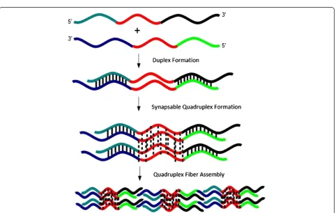
[image:7.595.58.541.409.715.2]quadruplex contains four DNA strands: two strands are oriented 5′to 3′and the other two oriented from 3′to 5′(Figure 5). The synapsed quadruplex is assigned using gel electrophoresis on the basis of comparison to control sequences and through quadruplex-specific dye staining experiments. We note that there are several duplex ar-rangements possible as a result of the orientations in which the duplex precursors can come together. In our design, each synapsed quadruplex contains four duplex
‘arms’ flanking the G-rich region, and each arm has a
short single-stranded overhang. To explain fiber forma-tion, we propose that the duplex regions in the quadruplexes partially melt, thereby allowing linking of synapsed quadruplexes together into a larger structure.
Our tentative model for association of (SQ1A:SQ1B)2
quadruplexes into fibers involves partial duplex melting, which allows individual quadruplex units to associate into larger fibers (Figure 5). The G-quadruplex region, which contains eight guanines, does not melt at the salt concentrations used in our work [24,27]. After the du-plex is incubated in potassium to form a quadrudu-plex, a considerable amount of crowding is introduced at the ends of each G-quadruplex. Under these conditions, it might be more favorable for a (partially) melted duplex region to base pair with a complementary strand in an-other synapsed quadruplex. Because four strands are available at each end of the G-quadruplex region, the likelihood of occurrence of a single event (base pairing with a strand in another synapsable quadruplex unit) is greatly increased. We observed by AFM that increasing the annealing temperature increases fiber formation, which is consistent with our assembly model. The in-creased annealing temperature melts the duplex regions more completely, thereby increasing the likelihood that two arms on separate synapsed quadruplex molecules will pair. This model allows for formation of branched structures. This working hypothesis is currently under investigation in our laboratories to test its validity.
Our work is one of the first in which a macromolecu-lar structure is assembled actively via cooperation of Hoogsteen and Watson-Crick base pairing [12]. In con-trast, structures such as G4-DNA [44-48], frayed wires [49-51], and G-wires [46] are driven only by Hoogsteen hydrogen bonding in G-quartets. Canonical base pairing has been used to create duplex DNA branches on the ends of frayed wires [49], but initial assembly of the frayed wires exploits only Hoogsteen hydrogen bonding and used a single DNA sequence, which does not allow significant variability/flexibility [49]. Finally, structures
created by acid-dependent assembly of d(CGG)4also
de-pend mainly on Hoogsteen hydrogen bonding [52]. In contrast, all of the main DNA fabrication methods using DNA tiles/origami rely on canonical base pairing, with the exception of a structure in which building blocks are
connected by quadruplexes rather than duplexes [12]. The presence of duplex and quadruplex elements in our final structures results in distinct recognition sites for in-corporation of additional elements [53]. Future work will measure the accessibility and selectivity of these address-able sites in both precursor units and final structures.
Conclusions
We present a novel strategy to generate fibers with morphologies that differ from duplex-only-based wires. Our method uses hybridization of DNA strands to form duplexes followed by cation-mediated assembly of quadruplexes. The dimensions and quantities of our fi-bers vary depending on the preparation conditions, but the final assemblies contain quadruplexes. We have shown here the proof of concept for mixed duplex-quadruplex fiber fabrication that we believe holds prom-ise for organized control of fiber assembly.
Additional file
Additional file 1:PDF document containing buffer formulations and abbreviations, tapping mode AFM images of
duplex-quadruplex nanofibers, and a gel electrophoresis image of a control duplex with overhangs.
Abbreviations
AFM:Atomic force microscopy; NMM:N-methylmesoporphyrin IX; PAGE: Polyacrylamide gel electrophoresis; TMACl: Tetramethylammonium chloride.
Competing interests
The authors declare that they have no competing interests.
Authors’contributions
MAM designed the sequences, carried out the gel electrophoresis and AFM measurements, and wrote initial drafts of the manuscript. VAS conducted gel electrophoresis experiments, supervised the design and completion of the work, and wrote the final version of the manuscript. Both authors read and approved the final manuscript.
Authors’information
VAS is a project leader in the CNST Energy Research Group. She received an A.B. in Chemistry from Bryn Mawr College and a Ph.D. in inorganic chemistry from Yale University, where her thesis work centered on biophysical measurements of water oxidation chemistry in photosynthesis. After completing post-doctoral work at the University of North Carolina at Chapel Hill, VAS moved to the Department of Chemistry and Biochemistry at the University of Maryland, Baltimore County, where she advanced to the rank of associate professor with tenure. During that time, she and her group elucidated the biophysical chemistry of copper in Alzheimer's disease fibrils and developed methods to create quadruplex-based DNA nanomaterials. VAS joined the CNST in 2010 and is leading projects focused on nanofabrication tools based on biomacromolecular nanomaterials and fundamental measurements of nanostructured catalysts for solar fuels applications.
computational methods for modeling non-canonical DNA structures, and development of biosensors based on the nanosystems under investigation in his laboratory.
Acknowledgments
We thank Y. Zhang for assistance with early AFM measurements and D. Fabris and M. Scalabrin for mass spectrometry measurements. This work was supported by an NSF CAREER award to VAS (CHE-0346066).
Author details
1
Department of Chemistry and Biochemistry, University of Maryland, Baltimore County, 1000 Hilltop Drive, Baltimore, MD 21250, USA.2Universidad
San Francisco de Quito, Vía Interoceánica Km 2 1/2, Cumbayá, Quito 17-1200-84, Ecuador.3Center for Nanoscale Science and Technology,
National Institute of Standards and Technology, 100 Bureau Drive, Gaithersburg, MD 20899-6204, USA.
Received: 25 February 2013 Accepted: 20 April 2013 Published: 3 May 2013
References
1. Aldaye FA, Palmer AL, Sleiman HF:Assembling materials with DNA as the guide.Science2008,321(5897):1795–1799.
2. Lin C, Liu Y, Rinker S, Yan H:DNA tile based self-assembly: building complex nanoarchitectures.Chemphyschem2006,7(8):1641–1647. 3. Dietz H, Douglas SM, Shih WM:Folding DNA into twisted and curved
nanoscale shapes.Science2009,325(5941):725–730.
4. Bath J, Turberfield AJ:DNA nanomachines.Nat Nanotechnol2007,
2(5):275–284.
5. Sugimoto N:Designable DNA functions toward new nanobiotechnology.
Bull Chem Soc Jpn2009,82:1–10.
6. McLaughlin CK, Hamblin GD, Aldaye FA, Yang H, Sleiman HF:A facile, modular and high yield method to assemble three-dimensional DNA structures.Chem Commun2011,47(31):8925–8927.
7. Howorka S:DNA nanoarchitectonics: assembled DNA at interfaces.
Langmuir2013. doi:10.1021/la3045785.
8. Seeman NC:Nanomaterials based on DNA.Annu Rev Biochem2010,
79:1545–4509.
9. Rothemund PWK:Folding DNA to create nanoscale shapes and patterns.
Nature2006,440(7082):297–302.
10. Ke Y, Sharma J, Liu M, Jahn K, Liu Y, Yan H:Scaffolded DNA origami of a DNA tetrahedron molecular container.Nano Lett2009,9(6):2445–2447. 11. Ke Y, Voigt NV, Gothelf KV, Shih WM:Multilayer DNA origami packed on
hexagonal and hybrid lattices.J Am Chem Soc2011,134(3):1770–1774. 12. Dutta K, Fujimoto T, Inoue M, Miyoshi D, Sugimoto N:Development of
new functional nanostructures consisting of both DNA duplex and quadruplex.Chem Commun2010,46(41):7772–7774.
13. Nair DT, Johnson RE, Prakash S, Prakash L, Aggarwal AK:Replication by human DNA polymerase-ιoccurs by Hoogsteen base-pairing.Nature 2004,430(6997):377–380.
14. Hermann T, Westhof E:Non-Watson-Crick base pairs in RNA-protein recognition.Chem Biol1999,6(12):R335–R343.
15. Leontis NB, Stombaugh J, Westhof E:The non-Watson-Crick base pairs and their associated isostericity matrices.Nucl Acids Res2002,30(16): 3497–3531.
16. Potaman VN:Applications of triple-stranded nucleic acid structures to DNA purification, detection and analysis.Expert Rev Mol Diagn2003,3(4): 481–496.
17. Biffi G, Tannahill D, McCafferty J, Balasubramanian S:Quantitative visualization of DNA G-quadruplex structures in human cells.Nat Chem 2013,5:182–186.
18. Jain A, Wang G, Vasquez KM:DNA triple helices: biological consequences and therapeutic potential.Biochimie2008,90(8):1117–1130.
19. Nair DT, Johnson RE, Prakash L, Prakash S, Aggarwal AK:Hoogsteen base pair formation promotes synthesis opposite the 1, N6-ethenodeoxyadenosine lesion by human DNA polymeraseι.Nat Struct Mol Biol2006,13(7):619–625. 20. Johnson RE, Prakash L, Prakash S, Radding CM:Biochemical evidence for
the requirement of Hoogsteen base pairing for replication by human DNA polymeraseι.PNAS2005,102(30):10466–10471.
21. Johnson RE, Haracska L, Prakash L, Prakash S:Role of Hoogsteen edge hydrogen bonding at template purines in nucleotide incorporation by human DNA polymerase {iota}.Mol Cell Biol2006,26(17):6435–6441. 22. Wells RD:Non-B DNA conformations, mutagenesis and disease.Trends
Biochem Sci2007,32(6):271–278.
23. Ghosal G, Muniyappa K:Hoogsteen base-pairing revisited: resolving a role in normal biological processes and human diseases.Biochem Biophys Res Comm2006,343(1):1–7.
24. Venczel EA, Sen D:Synapsable DNA.J Mol Biol1996,257(2):219–224. 25. Anuradha S, Muniyappa K:Meiosis-specific yeast Hop1 protein promotes
synapsis of double-stranded DNA helices via the formation of guanine quartets.Nucl Acids Res2004,32(8):2378–2385.
26. Fahlman RP, Sen D:“Synapsable”DNA double helices: self-selective modules for assembling DNA superstructures.J Am Chem Soc1999,
121(48):11079–11085.
27. Evans S, Mendez M, Turner K, Keating L, Grimes R, Melchoir S, Szalai V: End-stacking of copper cationic porphyrins on parallel-stranded guanine quadruplexes.J Biol Inorg Chem2007,12(8):1235–1249.
28. Borer PN:Optical properties of nucleic acids, absorption, and circular dichroism spectra. InHandbook of Biochemistry and Molecular Biology.3rd edition. Edited by Fasman G. Cleveland: CRC Press; 1975:589.
29. Tataurov AV, You Y, Owczarzy R:Predicting ultraviolet spectrum of single stranded and double stranded deoxyribonucleic acids.Biophys Chem 2008,133:66–70.
30. Mendez MA, Szalai VA:Fluorescence of unmodified oligonucleotides: a tool to probe G-quadruplex DNA structure.Biopolymers2009,91(10):841–850. 31. Sambrook J, Russell D:Molecular Cloning a Laboratory Manual. Volume 2. 3rd
edition.Cold Spring Harbor Laboratory Press: Cold Spring Harbor; 2001. 32. Bloomfield VA, Crothers DM, Ignacio Tinoco J:Nucleic Acids: Structures,
Properties, and Functions.Sausalito: University Science Books; 2000. 33. Frontali C, Dore E, Ferrauto A, Gratton E, Bettini A, Pozzan MR, Valdevit E:An
absolute method for the determination of the persistence length of native DNA from electron micrographs.Biopolymers1979,18:1353–1373. 34. Arthanari H, Basu S, Kawano TL, Bolton PH:Fluorescent dyes specific for
quadruplex DNA.Nucleic Acids Res1998,26:3724–3728.
35. Nicoludis JM, Barrett SP, Mergny J-L, Yatsunyk LA:Interaction of human telomeric DNA withN-methyl mesoporphyrin IX.Nucleic Acids Res2012,
40:5432–5447.
36. Nicoludis JM, Miller ST, Jeffrey PD, Barrett SP, Rablen PR, Lawton TJ, Yatsunyk LA:Optimized end-stacking provides specificity ofN-methyl mesoporphyrin IX for human telomeric G-quadruplex DNA.J Am Chem Soc2012,134:20446–20456.
37. Ragazzon P, Chaires JB:Use of competition dialysis in the discovery of G-quadruplex selective ligands.Methods2007,43:313–323.
38. Armstrong T, Root J, Vesenka J:Hydration layer scanning tunneling microscopy of“G-wire”DNA. InAIP Conference Proceedings.Melville: American Institute of Physics; 2004:59–64.
39. Borovok N, Iram N, Zikich D, Ghabboun J, Livshits GI, Porath D, Kotlyar AB:
Assembling of G-strands into novel tetra-molecular parallel G4-DNA nanostructures using avidin-biotin recognition.Nucl Acids Res2008,
36:5050–5060.
40. Pisano S, Varra M, Micheli E, Coppola T, De Santis P, Mayol L, Savino M:
Superstructural self-assembly of the G-quadruplex structure formed by the homopurine strand in a DNA tract of human telomerase gene promoter.Biophys Chem2008,136(2–3):159–163.
41. Marsh TC, Vesenka J, Henderson E:A new DNA nanostructure, the G-wire, imaged by scanning probe microscopy.Nucleic Acids Res1995,23:696– 700.
42. Fahlman RP, Sen D:Cation-regulated self-association of "synapsable" DNA duplexes.J Mol Biol1998,280(2):237–244.
43. Delrow JJ, Heath PJ, Fujimoto BS, Schurr JM:Effect of temperature on DNA secondary structure in the absence and presence of 0.5 M
tetramethylammonium chloride.Biopolymers1998,45:503–515. 44. Cohen H, Sapir T, Borovok N, Molotsky T, Di Felice R, Kotlyar AB, Porath D:
Polarizability of G4-DNA observed by electrostatic force microscopy measurements.Nano Lett2007,7(4):981–986.
45. Di Felice R, Calzolari A, Garbesi A, Alexandre SS, Soler JM: Strain-dependence of the electronic properties in periodic quadruple helical G4-wires.J Phys Chem B Condens Matter Mater Surf Interfaces Biophys2005,
46. Marsh TC, Henderson E:G-wires: self-assembly of a telomeric oligonucleotide, d(GGGGTTTGGGG), into large superstructures.
Biochemistry1994,33:10718–10724.
47. Kotlyar AB, Borovok N, Molotsky T, Cohen H, Shapir E, Porath D:Long monomolecular guanine-based nanowires.Adv Mater2005,17:1901–1905. 48. Shapir E, Sagiv L, Borovok N, Molotski T, Kotlyar AB, Porath D:
High-resolution STM imaging of novel single G4-DNA molecules.J Phys Chem B2008,112(31):9267–9269.
49. Protozanova, E, Macgregor, RB Jr:Transient association of the DNA-ligand complex during gel electrophoresis.Electrophoresis1999,
20(10):1950–1957.
50. Poon K, Macgregor RB:Formation and structural determinants of multi-stranded guanine-rich DNA complexes.Biophys Chem2000,84(3):205–216. 51. Protozanova E, Macgregor RB Jr:Analysis of the electrophoretic migration
of DNA frayed wires.Biophys Chem1998,75(3):249–257.
52. Chen F-M:Acid-facilitated supramolecular assembly of G-quadruplexes in d(CGG)β4.J Biol Chem1995,270(39):23090–23096.
53. Zheng L, Wang X, Zhang JL, Li W:DNA nanotechnology based on polymorphic G-quadruplex.Progress in Chemistry2011,23(5):974–982.
doi:10.1186/1556-276X-8-210
Cite this article as:Mendez and Szalai:Synapsable quadruplex-mediated
fibers.Nanoscale Research Letters20138:210.
Submit your manuscript to a
journal and benefi t from:
7Convenient online submission 7Rigorous peer review
7Immediate publication on acceptance 7Open access: articles freely available online 7High visibility within the fi eld
7Retaining the copyright to your article