Copyright © 2003, American Society for Microbiology. All Rights Reserved.
Evolution of
sfbI
Encoding Streptococcal Fibronectin-Binding Protein
I: Horizontal Genetic Transfer and Gene Mosaic Structure
Rebecca J. Towers,
1,2,3Peter K. Fagan,
1,3Susanne R. Talay,
1Bart J. Currie,
3Kadaba S. Sriprakash,
3,4Mark J. Walker,
2and Gursharan S. Chhatwal
1*
GBF-German Research Centre for Biotechnology, Braunschweig, Germany,1and University of Wollongong, Wollongong,2
Menzies School of Health Research, Darwin,3and Queensland Institute of Medical Research, Brisbane,4Australia
Received 24 June 2003/Returned for modification 30 July 2003/Accepted 28 August 2003
Streptococcal fibronectin-binding protein is an important virulence factor involved in colonization and
invasion of epithelial cells and tissues by Streptococcus pyogenes. In order to investigate the mechanisms
involved in the evolution ofsfbI, thesfbIgenes from 54 strains were sequenced. Thirty-four distinct alleles were
identified. Three principal mechanisms appear to have been involved in the evolution of sfbI. The
amino-terminal aromatic amino acid-rich domain is the most variable region and is apparently generated by intergenic recombination of horizontally acquired DNA cassettes, resulting in a genetic mosaic in this region. Two distinct and divergent sequence types that shared only 61 to 70% identity were identified in the central
proline-rich region, while variation at the 3ⴕend of the gene is due to deletion or duplication of defined repeat
units. Potential antigenic and functional variabilities in SfbI imply significant selective pressure in vivo with
direct implications for the microbial pathogenesis ofS. pyogenes.
Streptococcal fibronectin-binding protein I (SfbI), also known as protein F1, is probably the most extensively charac-terized fibronectin-binding protein in group A streptococcus (GAS; Streptococcus pyogenes). Identified simultaneously in two independent laboratories (15, 39), SfbI is a membrane-anchored surface protein shown to be a major adhesin of GAS (13, 15, 28, 29, 39, 42). In addition, SfbI binds to fibrinogen via a domain in the amino terminus (20) and human immunoglob-ulin (IgG) via the carboxy-terminal fibronectin-binding repeat domain (25). SfbI is protective in mice (14, 34) and a mucosal adjuvant (26). According to various reports, it is present in 64 to 82% of strains (13, 22, 28). Beside its ability to mediate adherence to host cells via binding to fibronectin as a bridging molecule, SfbI is a potent invasin of epithelial cells (16, 27). While efficient adhesion is mediated through the fibronectin-binding repeat region, internalization into host cells is gov-erned by the fibronectin-binding spacer region in a cooperative manner (41). Recent work demonstrates that SfbI-induced in-tegrin clustering initiates caveola recruitment and subsequent caveola-mediated uptake of the pathogen into epithelial and endothelial cells (33). The interaction between the fibronectin-binding repeat region and the N-terminal F1 modules of fi-bronectin has been elegantly resolved by defining the structure of the complex that is formed (35). Another important and recently identified function of SfbI is the ability to recruit collagen via prebound fibronectin. SfbI thereby mediates bac-terial aggregation, colonization of the collagen matrix, and phagocyte evasion (9). SfbI, like many membrane-associated streptococcal proteins, has a repetitive structure and exhibits decreasing variability from the amino terminus to the carboxy
terminus (40). The protein is synthesized as a propeptide with a signal sequence at the amino terminus of the protein which is subsequently cleaved to form the mature protein. The amino terminus of the mature protein contains a domain rich in aromatic amino acids, adjacent to which is a proline-rich do-main. The proline-rich domain is made up of two regions: the proline-rich repeats and the proline-rich upstream region en-compassing the region previously described as proline-rich re-peats 1 and 2 (40). Toward the carboxy terminus lie two func-tional fibronectin-binding domains: the fibronectin-binding repeat domain and a second fibronectin-binding domain which lies upstream of the fibronectin-binding repeats and which has been designated spacer 2 (40) or the upstream fibronectin-binding domain (36). These domains are highly conserved and display variability only in the number of fibronectin-binding repeats, which may range from one to six (28, 42). Variable numbers of proline-rich repeats have also been reported; how-ever, no correlation between variation in this domain and the numbers of fibronectin-binding repeats has been found (42). The carboxy terminus consists of a wall-associated region, a membrane anchor motif (LPATGD), and a membrane-associ-ated region rich in hydrophobic residues, all of which are typical of surface proteins of gram-positive cocci (11).
Several groups have described the arrangement of the genomic region containing the gene encoding SfbI (6, 30). This locus has been designated the fibronectin-collagen-T antigen (FCT) region (6) since it may contain genes encoding the fibronectin-binding protein SfbI and/or protein F2, a collagen-binding protein (Cpa), and the T antigen. Incorporation of sequences from the recently completed M3 (4), M5 (http: //www.sanger.ac.uk/Projects/S_pyogenes), and M18 (37) ge-nomes reveals a complex gene arrangement in this region which is indicative of a zone of frequent genetic rearrange-ment.
Mosaic alleles are made up of distinct DNA cassettes of different phylogenic origins which have recombined to produce
* Corresponding author. Mailing address: Department of Microbial Pathogenesis and Vaccine Research, GBF German Research Centre for Biotechnology, Mascheroder Weg 1, 38124 Braunschweig, Ger-many. Phone: 49 531 6181 297. Fax: 49 531 6181 708. E-mail: gsc @gbf.de.
5398
on May 15, 2020 by guest
http://jcm.asm.org/
encoding streptokinase (19), hyaluronidase (24), streptococcal superantigen (32), and a novel collagen-like protein called SclA (31) have been reported in GAS, the classic example in GAS are members of the M-protein family encoded by genes within the Vir (ormga) regulon (43). Horizontal transfer and intergenic recombination have played major roles in the evo-lution of theemm-like family, contributing to both sequence variation and variation in the overall architecture of the Vir regulon (44). Since different members of the M-protein family exhibit different protein-binding capabilities, this rearrange-ment may have direct ramifications for the virulence and tro-pisms of the various strains. This is supported by the observa-tion that different architectural arrangements (emm pattern types) have been associated with preferences for specific tissue sites (5).
Although evaluation of the accumulation of mutations is a valuable tool for assessment of strain relatedness, transfer of whole cassettes of DNA between strains complicates such anal-yses. Multilocus sequence typing with several presumably se-lection-neutral housekeeping genes has been used to deter-mine the extent of intergenic recombination in bacterial genomes. The lack of congruence between maximum-likeli-hood trees for each of the “neutral” genes in GAS was indic-ative of very high rates of recombination, and it was concluded that this was sufficient to “obliterate the phylogenetic signal in [individual] gene trees” (10).
Recombination at distal sites such as the Vir regulon and the FCT region is most likely independent. Evidence to support this theory includes the fact that T types do not always conform to M types, with several T types reported for isolates of the same M type and vice versa (2, 17). In addition, while the distribution ofsfbIwithin M types (28) or Vir types (VTs) (13) is generally consistent, there is evidence of heterogeneity of
sfbI species within M types, as the number of fibronectin-binding repeats detected in M8 and M28 serotype strains is variable (28). Significant heterogeneity in sfbIsequences en-coding the amino-terminal half of the aromatic amino acid-rich domain from M4, M12, M15, and M18 strains has also been reported (20).
The aim of this study was to examine precisely the genetic variability in the entire sfbI in order to determine possible mechanisms involved in the evolution of this important strep-tococcal adhesin and invasin. Additionally, we have examined whether particular sfbI sequence types (STs) are associated with the clinical source of the isolates.
(Part of this work was presented at the XVth Lancefield International Symposium on Streptococci and Streptococcal Disease, October 2002, Goa, India [R. Towers, M. J. Walker, and G. S. Chhatwal, Lancefield Int. Symp. Streptococci Strep-tococcal Dis. abstr. O5.3, 2002].)
MATERIALS AND METHODS
Bacterial strains.Isolates were obtained from the Menzies School of Health Research streptococcal collection. This collection contains isolates from patients of the Royal Darwin Hospital and the surrounding Aboriginal communities. In addition, an M13 GAS reference strain from the United Kingdom (Cathy13) and the homologoussfbIIstrain (strain 75401) from Germany were included (22, 38). Background information regarding clinical details, VTs (12), andemmSTs (3), when available, are included in Table 1. All streptococcal strains were grown on agar plates containing 5% sheep blood (Sigma) at 37°C overnight.
(Bio-Rad) according to the instructions of the manufacturer, except that due to the small size of the streptococcal colonies, 10 to 20 streptococcal colonies were used. The sequencing template was amplified with primerssfbI-F1 andsfbI-R4. Each 50-l PCR mixture contained 5l of InstaGene preparation. The PCR parameters used were an initial denaturing step at 94°C for 2 min and then 35 cycles of denaturation at 94°C for 1 min, annealing at 49°C for 1 min, and extension at 72°C for 2 min 30 s, with a final extension step of 72°C for 5 min.
Screening forsfbIandrofA.Many of the data on the distribution ofsfbIin the GAS strains used in this study have been published elsewhere (8, 13). For the purposes of this study all strains were retested for the presence ofsfbIby PCR, and the results were compared with previous results. PCR screening was per-formed with primerssfbI-F1 andsfbI-R3. The number of fibronectin-binding repeats was determined with primerssfbI-F4 andsfbI-R3. Isolates were also screened for the presence ofrofAwith primersrofA-F androfA-R. The arrange-ment ofrofA, relative to that ofsfbI, was determined with primersrofA-R and
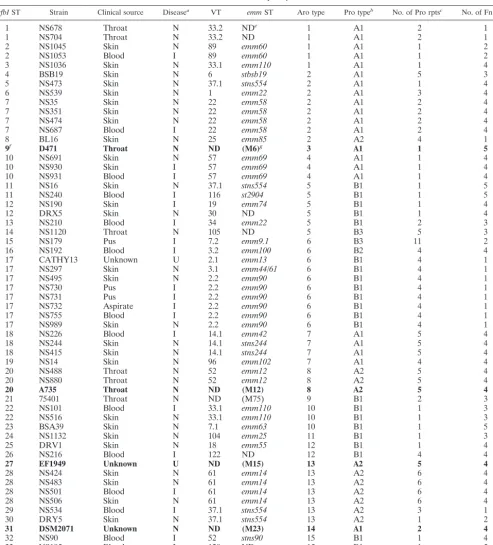
sfbI-R6. All primer sequences are shown in Table 2, while their positions relative to those of the respective genes are shown in Fig. 1A.
DNA sequence analysis.The PCR product was used as the template for sequencing reactions. This template was generated with primerssfbI-F1 and
sfbI-R4 orsfbI-R3. A total of 5l of the PCR product was visualized in an ethidium bromide-stained 1% agarose gel to determine its purity and relative concentration. The remainder was purified with QIAquick columns (Qiagen), eluted in 30l of sterile distilled water, and stored at⫺20°C. The primers used for the sequence reactions are shown in Table 2.
Sequence alignment was performed with a range of DNASTAR programs. Applied Biosystems electropherograms were aligned to generate DNA contigs by using SeqManII, and then DNA contigs were translated to amino acid sequences with the MegAlign program and were subsequently aligned by the Clustal V method. The alignment was then modified by eye before being converted back to the DNA sequence alignment.
Nucleotide sequence accession numbers.The nucleotide sequences for the 54
sfbIgenes can be found in the GenBank database under accession numbers AJ347791 to AJ347844.
RESULTS
Screening forsfbIandrofA. The presence ofsfbIwas
con-firmed by PCR for 54 GAS isolates. Representative PCR re-sults are shown in Fig. 1B. PCR was also used to determine the number of fibronectin-binding repeats as an initial indicator of variation (Fig. 1C). In addition, the relative distribution and arrangement ofrofAwith respect to those ofsfbIwere estab-lished by PCR. All strains were tested for the presence ofrofA
with primersrofA-F androfA-R (30) (Fig. 1D), while primers
rofA-R andsfbI-R6 were used to detect adjacentrofAandsfbI
genes (Fig. 1E). rofA was present in all 54 strains and was situated immediately upstream of sfbI, but in an orientation opposite that shown in Fig. 1A.
Sequence analysis ofsfbI.The entiresfbIgene was amplified
by PCR for use as the sequencing template from most strains by using primers sfbI-F1 andsfbI-R4. Initially, thesfbIPCR products from 20 strains were sequenced in their entirety in both directions. It was determined from these data that the 3⬘ end ofsfbIwas highly conserved, apart from the various num-bers of proline-rich and fibronectin-binding repeats. As the number of fibronectin-binding repeats could be readily deter-mined by PCR (Fig. 1C) and these results were consistent with preliminary DNA sequence data, no attempt was made to sequence the fibronectin-binding repeat domains for the addi-tional 34 strains examined in this study, and conclusions as to the relatedness of the genes were drawn from the sequence and PCR results combined.
The nucleotide sequences were aligned with publishedsfbI
sequences, including those encoding full-length SfbI (Gen-Bank accession no. X67947) (2); protein F1 (Gen(Gen-Bank
on May 15, 2020 by guest
http://jcm.asm.org/
TABLE 1. Summary ofsfbISTs
sfbIST Strain Clinical source Diseasea VT emmST Aro type Pro typeb No. of Pro rptsc No. of Fn rptsd
1 NS678 Throat N 33.2 NDe 1 A1 2 1
1 NS704 Throat N 33.2 ND 1 A1 2 1
2 NS1045 Skin N 89 emm60 1 A1 1 2
2 NS1053 Blood I 89 emm60 1 A1 1 2
3 NS1036 Skin N 33.1 emm110 1 A1 1 4
4 BSB19 Skin N 6 stbsb19 2 A1 5 3
5 NS473 Skin N 37.1 stns554 2 A1 1 4
6 NS539 Skin N 1 emm22 2 A1 3 4
7 NS35 Skin N 22 emm58 2 A1 2 4
7 NS351 Skin N 22 emm58 2 A1 2 4
7 NS474 Skin N 22 emm58 2 A1 2 4
7 NS687 Blood I 22 emm58 2 A1 2 4
8 BL16 Skin N 25 emm85 2 A2 4 1
9f D471 Throat N ND (M6)g 3 A1 1 5
10 NS691 Skin N 57 emm69 4 A1 1 4
10 NS930 Skin I 57 emm69 4 A1 1 4
10 NS931 Blood I 57 emm69 4 A1 1 4
11 NS16 Skin N 37.1 stns554 5 B1 1 5
11 NS240 Blood I 116 st2904 5 B1 1 5
12 NS190 Skin I 19 emm74 5 B1 1 4
12 DRX5 Skin N 30 ND 5 B1 1 4
13 NS210 Blood I 34 emm22 5 B1 2 3
14 NS1120 Throat N 105 ND 5 B3 5 3
15 NS179 Pus I 7.2 emm9.1 6 B3 11 2
16 NS192 Blood I 3.2 emm100 6 B2 4 4
17 CATHY13 Unknown U 2.1 emm13 6 B1 4 1
17 NS297 Skin N 3.1 emm44/61 6 B1 4 1
17 NS495 Skin N 2.2 emm90 6 B1 4 1
17 NS730 Pus I 2.2 emm90 6 B1 4 1
17 NS731 Pus I 2.2 emm90 6 B1 4 1
17 NS732 Aspirate I 2.2 emm90 6 B1 4 1
17 NS755 Blood I 2.2 emm90 6 B1 4 1
17 NS989 Skin N 2.2 emm90 6 B1 4 1
18 NS226 Blood I 14.1 emm42 7 A1 5 4
18 NS244 Skin N 14.1 stns244 7 A1 5 4
18 NS415 Skin N 14.1 stns244 7 A1 5 4
19 NS14 Skin N 96 emm102 7 A1 4 4
20 NS488 Throat N 52 emm12 8 A2 5 4
20 NS880 Throat N 52 emm12 8 A2 5 4
20 A735 Throat N ND (M12) 8 A2 5 4
21 75401 Throat N ND (M75) 9 B1 2 3
22 NS101 Blood I 33.1 emm110 10 B1 1 3
22 NS516 Skin N 33.1 emm110 10 B1 1 3
23 BSA39 Skin N 7.1 emm63 10 B1 1 5
24 NS1132 Skin N 104 emm25 11 B1 1 3
25 DRV1 Skin N 18 emm55 12 B1 1 4
26 NS216 Blood I 122 ND 12 B1 4 4
27 EF1949 Unknown U ND (M15) 13 A2 5 4
28 NS424 Skin N 61 emm14 13 A2 6 4
28 NS483 Skin N 61 emm14 13 A2 6 4
28 NS501 Blood I 61 emm14 13 A2 6 4
28 NS506 Skin N 61 emm14 13 A2 6 4
29 NS534 Blood I 37.1 stns554 13 A2 3 1
30 DRY5 Skin N 37.1 stns554 13 A2 1 2
31 DSM2071 Unknown N ND (M23) 14 A1 2 4
32 NS90 Blood I 52 stns90 15 B1 1 4
33 NS195 Blood I 120 ND 15 B1 1 5
34 NS1042 Skin N 77 stck401 15 B1 1 2
aInvasive or noninvasive disease: I, invasive; N, noninvasive; U, unknown. bPro type, SfbI proline-rich region sequence subtype.
cPro rpts, “true” proline-rich repeats. dFn rpts, fibronectin-binding repeats. eND, not determined.
fBoldface indicates information published previously. gM serotype, as stated in relevant publications.
on May 15, 2020 by guest
http://jcm.asm.org/
sion no. L10919) (20); protein F1.12 (GenBank accession no. AF447492) (6); protein F15 and 12 partial sfbI sequences (GenBank accession nos. AF009908 to AF009920) (20); and
gfbA, an sfbI homolog from GGS (GenBank accession no. U131115) (21). The nucleotide sequence alignment can be found at GenBank alignment accession number ALIGN _000212 (data not shown).
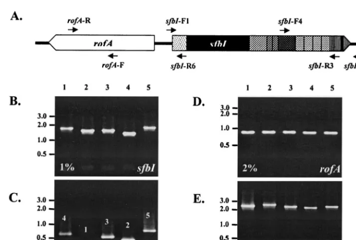
matic of the variation observed in thesfbIsequences. As noted previously (40), variation insfbI decreases in the 5⬘-to-3⬘ di-rection. The variation at the 3⬘ end of the gene is due to intragenic recombination, i.e., rearrangement of repetitive el-ements within genes through homologous recombination. The deletion or duplication of repeat units (narrower bars in Fig. 2) has resulted in a variable number of proline-rich and fibronec-tin-binding repeats. There is no relationship between the num-bers of proline-rich repeats and the numnum-bers of fibronectin-binding repeats, nor is there an association between the numbers of repeats and the upstream sequence variation. Therefore, it can be assumed that intragenic recombination is a frequent phenomenon within these regions.
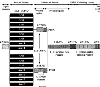
Two distinct and divergent STs which exhibited 61 to 70% identity in pairwise alignments of the DNA sequences were identified within the central proline-rich region. These have been designated ProA and ProB. In addition, defined internal sequences have been duplicated or deleted, thereby generating two to three subtypes within each proline-rich region type (Fig. 2 and 3). In the case of ST ProB, in which up to two copies of the internal sequences were present, these repetitive structures were designated the ProB repeats.
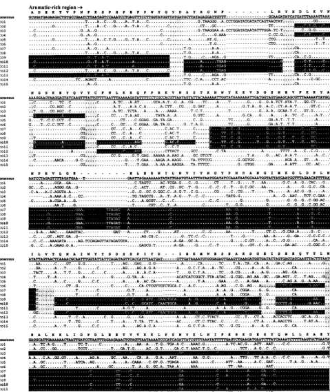
The 5⬘end ofsfbI, which encodes the aromatic amino acid-rich domain, is by far the most variable region of the gene. Fifteen distinct aromatic amino acid-rich domain sequences types (Aro types), designated Aro1 to Aro15, were identified. In pairwise alignments, a minimum 98.8% identity was seen within Aro types, while 50.3 to 97.6% identity was observed between types. While some of this variation is due to point
Primer direction
and primer Sequence
Forward
sfbI-F1 ...5⬘-GTCTTTCTTGACAATAACGTGGTAAGCTC-3⬘
sfbI-F2 ...5⬘-GTAGCCTATGCTGCCGATGAGAAG-3⬘
sfbI-F3 ...5⬘-TTTGTACCAGAAAATCCCCCTAAACCTG-3⬘
sfbI-F4 ...5⬘-GACTTACCTATTGAAGATCCTCGTTATGAG-3⬘
sfbI-F5 ...5⬘-GCATGCGCGGGTGCTATCG-3⬘
sfbI-F7 ...5⬘-GTACAGAATATGTACAAGATAATCC-3⬘
sfbI-S1F ...5⬘-GTGCTGAATATGTACCTGATAGTCC-3⬘
PCRP1 ...5⬘-TATCAAAATCTTCTAAGTGCTGAG-3⬘
rofA-F ...5⬘-GCCAATAACTGAGGTAGC-3⬘
Reverse
sfbI-R1...5⬘ -CTCATAACGAGGATCTTCAATAGGTAAGTC-3⬘
sfbI-R2...5⬘-CACTCCTGGCTCTTTCGTATCTTCTG-3⬘
sfbI-R3...5⬘-GTATCTTCAACAATGGTCACTGTTTCACTG-3⬘
sfbI-R4...5⬘-CAGTTATCCACTATTCAGCATATTTGCGC-3⬘
sfbI-R5...5⬘-CGATAGCACCCGCGCATGC-3⬘
sfbI-R6...5⬘-GCAGCATAGGCTACTTGACCAAAAC-3⬘
sfbI-R7...5⬘-GGATTATCTTGTACATATTCTGTAC-3⬘
sfbI-R8...5⬘-CAGGTCTAGGGGGATCTTCTGGTACAAA-3⬘
sfbI-S1R ...5⬘-GGACTATCAGGTACATATTCAGCAC-3⬘
PCRP1R ...5⬘-CTCAGCACTTAGAAGATTTTG-3⬘
rofA-R ...5⬘-GGTTTTGCTCTTTTAGGT-3⬘
FIG. 1. (A) Positions of primers relative to the two genes. The shading of the SfbI domains is as described in the legend to Fig. 2. The schematic is not drawn to scale. (B to E) Ethidium bromide-stained 1 or 2% agarose gels showing the results of PCR screening forsfbIandrofAfor five representative strains. (B) Screening forsfbIwith primerssfbI-F1 andsfbI-R3; (C) determining the number of fibronectin-binding repeats in SfbI with primerssfbI-F4 andsfbI-R3; (D) screening forrofAwith primersrofA-F androfA-R; (E) determining arrangement ofrofArelative to that of
sfbIwith primersrofA-R andsfbI-R6. DNA marker (GeneRuler DNA Ladder Mix, MBI Fermentas) sizes (in kilobases) are shown at the left. Lanes 1 to 5, GAS strains DSM2071, NS732, NS1132, DRY5, and BSA39, respectively.
on May 15, 2020 by guest
http://jcm.asm.org/
[image:4.603.112.469.426.667.2]mutations, which are easily identifiable as single nucleotide changes within otherwise conserved regions, the majority ap-pear to be due to intergenic recombination. Similar to theemm
gene, this intergenic DNA transfer has generated a gene mo-saic structure insfbI. The overall effects at the protein level can be seen in the amino acid sequence alignments of the various aromatic amino acid-rich domains (Fig. 4). Aro10 has been
used to illustrate how distinct DNA cassettes, flanked by highly conserved junction sequences, are shared between otherwise divergent STs. This type of mosaic allele can be generated only by horizontal gene transfer between strains.
Conserved structure ofsfbI.Despite the high degree of
[image:5.603.126.463.71.364.2]vari-ation observed, the overall structural properties of SfbI were conserved. Within the aromatic amino acid-rich domain, the
FIG. 2. Overall schematic of variation seen in SfbI. Decreasing variability of SfbI occurs from the amino-terminal aromatic amino acid-rich domain (solid black) through the proline-rich region (thin diagonal stripe) and to the proline-rich repeats (thick diagonal stripe) and the functional fibronectin (Fn)-binding domains (stippled). Narrower bars indicate the deletion or duplication of repeat units. The percent identities observed in pairwise DNA alignments is shown above each domain. Gaps introduced to aid alignment have been treated as single base changes. The diagram is not drawn to scale. UFBD, upstream fibronectin-binding domain.
FIG. 3. Alignment of amino acid sequences corresponding to the proline-rich domain of SfbI. The consensus sequence is shown at the top in boldface with underlining. The proline-rich region has two STs, ProA and ProB. Deletion or duplication events within these STs define the proline-rich region subtypes. Dots represent identical nucleotides relative to the consensus sequence, and hyphens indicate the positions of insertions found in particular isolates. The ProB repeats are indicated, and the first true proline-rich repeat is boxed.
on May 15, 2020 by guest
http://jcm.asm.org/
[image:5.603.97.489.543.685.2]FIG. 4. Alignment of nucleotide sequences corresponding to the aromatic amino acid-rich domain STs. The overall consensus sequence is shown in boldface with underlining above the alignment, and the translated protein sequence is also shown. Dots represent identical nucleotides relative to the consensus sequence, and hyphens indicate the positions of insertions found in particular isolates. The gene mosaic structure is illustrated by using Aro10 as an example (boldface), putative horizontally transferred DNA cassettes are shaded in black, while predicted junctions are shaded in gray.
on May 15, 2020 by guest
http://jcm.asm.org/
distribution of aromatic amino acids remained relatively un-changed, and similarly, the distribution of proline residues within the proline-rich domain remained relatively unchanged. Over the entire length of the deduced amino acid sequence, hydrophobicity, average charge, and surface probability plots were generally conserved among the SfbI proteins from differ-ent strains. By restriction of comparisons to the aromatic amino acid- and proline-rich domains, the positively charged regions remained clustered in the aromatic amino acid-rich domain, despite sequence variation, with a few peaks in the proline-rich region before the negatively charged proline-rich repeats (Fig. 4). The periodicity of the repeat sequences is apparent in hydrophobicity, charge, and surface probability plots (data not shown). The theoretical pI of the putative SfbI proteins ranged between 4.11 and 4.81 (mean⫾standard de-viation, 4.51 ⫾ 0.13), indicating that SfbI would have a net negative charge at physiological pH.
Summary of sfbI STs.Thirty-five individual sfbI STs were
identified on the basis of combinations of aromatic amino acid-and proline-rich domain STs acid-and the numbers of proline-rich and fibronectin-binding repeats (Table 1). The strains within some VTs (VT2.2, VT14.1, VT22, VT33.1, VT33.2, VT52, VT57, and VT61) appeared to be clonal and were of the same
emmandsfbISTs. M12 strain A735 (6) is also likely a clone of VT52, since all three strains are M12 and have identical sfbI
genes. By contrast, VT37.1 was highly heterogeneous and in-cluded strains exhibitingsfbISTs 5, 11, 30, and 31. Conversely, other strains had virtually identicalsfbIgenes but belonged to different VTs; for example, sfbI ST18 included strains of VT2.1, VT2.2, and VT3.1. SomesfbISTs were identical except in the variable numbers of repeats; for example,sfbIST30 and
sfbIST31 were identical except that the former has three pro-line-rich repeats and one fibronectin-binding repeat, while the latter has one and two repeats, respectively. This demonstrates that while the presence of sfbI is associated with VT, the species of thesfbIallele may be independent.
No correlation between the relative numbers of proline-rich and fibronectin-binding repeats was observed. There was also no apparent relationship between the numbers of repeats and aromatic amino acid- or proline-rich STs; for example, com-paresfbISTs 4 to 8 within Aro2 (Table 1). Proline-rich domain STs were associated with particular aromatic amino acid-rich domain STs; however, several proline-rich domain sequence subtypes could be seen to be associated with a single aromatic amino acid-rich domain ST. For example, within Aro7, sfbI
ST16, sfbIST17, and sfbIST18 exhibit proline-rich subtypes ProB3, ProB2, and ProB1, respectively.
Phylogenic analysis of sfbI. The aromatic amino acid-rich
domain was considered to be the greater indicator of related-ness, since the high degree of variability of this domain ap-peared to have been generated by many individual mutations, while the variability in the numbers of repeats was due to a smaller number of mutation events that occurred at a relatively high frequency but that involved large fragments of highly conserved DNA. This is probably best illustrated by the Aro6 group, the sequences of which are highly conserved in the aromatic amino acid-rich domain but which contain 0, 1, or 2 internal ProB repeats; 4 or 11 proline-rich repeats; and 1, 2, or 4 fibronectin-binding repeats. This suggests that the repetitive units are frequently shuffled and that this has little or no true
bearing on the phylogeny of the strain. Variation insfbIhas arisen through genetic exchange evolution of different alleles and is not independent. Therefore, a phylogenic tree was not generated, since the principal assumption of such analysis is that the alleles have evolved independently.
Is the sfbI ST associated with the clinical source of the
isolate?Information on the clinical sources of the isolates was
available. Clonal distribution indicated that the skin was the principal reservoir for invasive disease isolates in the Northern Territory of Australia. SomesfbISTs were exclusively throat isolates (1, 9, 14, 21, 22); however, this association did not extend to Aro or proline-rich types. Further investigation re-vealed that all throat isolates were cultured from samples from nonindigenous people; therefore, this observation was proba-bly due to the differences in the strains circulating in the two populations rather than to the tissue tropism that might be attributed to the SfbI variants. Overall, there was no common trend to indicate that particular or related STs were associated with the clinical sources of the isolates.
DISCUSSION
Components of both the Vir regulon and the FCT region have undergone frequent genetic rearrangement. These re-gions encode important virulence factors, principally adhesins with affinities for different human proteins such as fibronectin, plasminogen, collagen, fibrinogen, and immunoglobulins. Hor-izontal gene transfer has played a major role in the generation of variability within both regions, resulting in the mosaic alleles seen inemm/ennandsfbI.
The advantages of increased variability through horizontal gene transfer include the generation of antigenic variation and variable binding capabilities. As a mechanism of generating genetic diversity, horizontal gene transfer has the unique ad-vantage of allowing incorporation of cassettes or domains which have already been checked for fitness in another genetic context. This bypasses the less efficient trial-and-error strategy and is particularly useful in altering the functional capabilities of a protein in a relatively short time frame.
The mosaic structure of the genes encoding the M-protein family has resulted in a divergent group of proteins exhibiting variable binding abilities and serotype-specific epitopes. The role that such variation plays in the pathogenesis of GAS is purely speculative; however, clear links have been shown be-tween Vir regulon arrangements (emm patterns) and tissue tropism (5). We have shown thatsfbIalso has a gene mosaic structure especially apparent in the amino-terminal aromatic amino acid-rich domain and have identified 35 distinct sfbI
STs. The biological function of the aromatic amino acid-rich domain is not known. To date, the reported binding functions of this domain include fibrinogen binding (20) and a putative carbohydrate-binding motif (40) which, from our data, appears to be highly conserved (Fig. 3A). The conserved distribution of aromatic amino acids within this domain, as well as the appar-ent intragenomic translocation of a highly homologous domain into Cpa (30), implies that the domain may have a significant function. In addition, the fact that the entire domain is present as a defined unit in Cpa implies that the function(s) and/or properties of the aromatic amino acid-rich domain require the whole domain. Despite the high degree of amino acid
on May 15, 2020 by guest
http://jcm.asm.org/
Since the proline-rich domain is predominantly negatively charged, we speculate that interaction between the two do-mains may be pivotal to the overall structure or folding of the native protein.
The other possibility is that variability has arisen in response to selective pressure from the immune system. Anti-M-protein antibodies are protective yet are serotype specific (23), as the protective epitopes reside in the variable amino terminus of the M protein (1, 7, 18). SfbI is a protective antigen when used in animal infection models (14); however, unlike M protein the protective epitope(s) resides in the conserved carboxy termi-nus and can provide protection from heterologous challenge (34). Natural infection in individuals from populations in which streptococcal infections are endemic also elicits signifi-cant levels of anti-SfbI IgG antibodies (13). Since these indi-viduals are still infected, despite the presence of anti-SfbI IgG antibodies, it appears that anti-SfbI antibodies either are not protective in humans or are not present in significantly high enough concentrations to be effective. While the fibronectin-binding domain is protective and conserved, variability in the amino terminus may represent a mechanism of immune eva-sion, thereby directing the immune response away from the carboxy terminus.
Recombination within selection-neutral genes in GAS is common to the point that it masks phylogenic signals (10). As a consequence, phylogenic analyses ofsfbIdo not clarify evo-lutionary relationships between strains but clarify only the sim-ilarities betweensfbIalleles. In looking at possible correlations between thesfbISTs and the clinical sources of the isolates, we did not find any associations. As with T-agglutination patterns, specific sfbISTs were consistently associated with emmSTs; however, like T types, they were not restricted to singularemm
STs. This seems to indicate thatemmandsfbIand not closely linked. Given that theteeandsfbIgenes both reside in the FCT region, it would be interesting to determine if T types andsfbI
STs are more closely associated with each other than withemm
STs. Such an analysis would require determination of the T types of the 54 GAS strains examined in this study.
The 34 individualsfbISTs identified in this study seem to have evolved through a combination of different evolutionary mechanisms working independently on distinct regions of the
sfbIgene. The majority of divergence in the Aro domain ap-pears to be due to intergenic recombination of horizontally transferred DNA cassettes, resulting in a genetic mosaic; two distinct and divergent STs are apparent in the central proline-rich region of sfbI; and the deletion or duplication of repeat units has resulted in a variable number of ProB, proline-rich, and fibronectin-binding repeats.
SfbI is an important virulence factor involved in colonization and internalization in fibronectin-rich epithelial tissues such as the upper respiratory mucosa and injured skin epithelium. Without clear associations between SfbI variants and function or tissue tropism, it is impossible to draw conclusions regarding the selective factors influencing these evolutionary mecha-nisms; however, the variability observed implies significant se-lective pressure on this locus in vivo. In light of the role played by intergenic transfer in the generation of variability in ligand-binding capacities in members of the M-protein family, the
and antigenic variability in SfbI warrant further investigation.
ACKNOWLEDGMENTS
R.J.T. received postgraduate awards from the University of Wollon-gong and the German Centre for Biotechnology (Braunschweig, Ger-many). This work was partially supported by the National Health and Medical Research Council of Australia (grant 9936609) and BMBF Network Pathogenomics.
We thank those people who have contributed to the accumulation of data on isolates in the Northern Territory Streptococcal Collection, including M. Hibble, D. E. Bessen, J. Carapetis, A. Delvecchio, M. L. Edwards, and A. M. Goodfellow.
REFERENCES
1. Beachey, E. H., J. M. Seyer, J. B. Dale, W. A. Simpson, and A. H. Kang.1981. Type-specific protective immunity evoked by synthetic peptide of Streptococ-cus pyogenesM protein. Nature292:457–459.
2. Beall, B., R. R. Facklam, J. A. Elliott, A. R. Franklin, T. Hoenes, D. Jackson, L. Laclaire, T. Thompson, and R. Viswanathan.1998. Streptococcalemm
types associated with T-agglutination types and the use of conservedemm
gene restriction fragment patterns for subtyping group A streptococci. J. Med. Microbiol.47:893–898.
3. Beall, B., R. Facklam, and T. Thompson.1996. Sequencingemm-specific PCR products for routine and accurate typing of group A streptococci. J. Clin. Microbiol.34:953–958.
4. Beres, S. B., G. L. Sylva, K. D. Barbian, B. Lei, J. S Hoff, N. D. Mammarella, M. Y. Liu, J. C. Smoot, S. F. Porcella, L. D. Parkins, D. S. Campbell, T. M. Smith, J. K. McCormick, D. Y. Leung, P. M. Schlievert, and J. M. Musser.
2002. Genome sequence of a serotype M3 strain of group A Streptococcus: phage-encoded toxins, the high-virulence phenotype, and clone emergence. Proc. Natl. Acad. Sci. USA99:10078–10083.
5. Bessen, D. E., M. W. Izzo, T. R. Fiorentino, R. M. Caringal, S. K. Hollings-head, and B. Beall.1999. Genetic linkage of exotoxin alleles andemmgene markers for tissue tropism in group A streptococci. J. Infect. Dis.179:627– 636.
6. Bessen, D. E., and A. Kalia.2002. Genomic localization of a T serotype locus to a recombinatorial zone encoding extracellular matrix-binding proteins in
Streptococcus pyogenes.Infect. Immun.70:1159–1167.
7. Dale, J. B., and E. H. Beachey.1986. Localization of protective epitopes of the amino terminus of type 5 streptococcal M protein. J. Exp. Med.163:
1191–1202.
8. Delvecchio, A., B. J. Currie, J. D. McArthur, M. J. Walker, and K. S. Sriprakash.2002.Streptococcus pyogenes prtFII, but notsfbI,sfbIIorfbp54, is represented more frequently among invasive-disease isolates of tropical Australia. Epidemiol. Infect.128:391–396.
9. Dinkla, K., M. Rohde, W. T. M. Jansen, J. R. Carapetis, G. S. Chhatwal, and S. R. Talay.2003.Streptococcus pyogenesrecruits collagen via surface bound fibronectin: a novel colonisation and immune evasion mechanism. Mol. Mi-crobiol.47:861–869.
10. Feil, E. J., E. C. Holmes, D. E. Bessen, M. S. Chan, N. P. Day, M. C. Enright, R. Goldstein, D. W. Hood, A. Kalia, C. E. Moore, J. Zhou, and B. G. Spratt.
2001. Recombination within natural populations of pathogenic bacteria: short-term empirical estimates and long-term phylogenetic consequences. Proc. Natl. Acad. Sci. USA98:182–187.
11. Fischetti, V. A., V. Pancholi, and O. Schneewind.1980. Conservation of a hexapeptide sequence in the anchor region of surface proteins from gram-positive cocci. Mol. Microbiol.4:1603–1605.
12. Gardiner, D., J. Hartas, B. Currie, J. D. Mathews, D. J. Kemp, and K. S. Sriprakash.1995. Vir typing: a long-PCR typing method for group A strep-tococci. PCR Methods Appl.4:288–293.
13. Goodfellow, A. M., M. Hibble, S. R. Talay, B. Kreikemeyer, B. J. Currie, K. S. Sriprakash, and G. S. Chhatwal.2000. Distribution and antigenicity of fibronectin binding proteins (SfbI and SfbII) ofStreptococcus pyogenes clin-ical isolates from the Northern Territory, Australia. J. Clin. Microbiol.38:
389–392.
14. Guzma´n, C. A., S. R. Talay, G. Molinari, E. Medina, and G. S. Chhatwal.
1999. Protective immune response againstStreptococcus pyogenesin mice after intranasal vaccination with the fibronectin-binding protein SfbI. J. In-fect. Dis.179:901–906.
15. Hanski, E., and M. Caparon.1992. Protein F, a fibronectin-binding protein, is an adhesin of the group A streptococcusStreptococcus pyogenes.Proc. Natl. Acad. Sci. USA89:6172–6176.
16. Jadoun, J., V. Ozeri, E. Burstein, E. Skutelsky, E. Hanski, and S. Sela.1998. Protein F1 is required for efficient entry ofStreptococcus pyogenes into epithelial cells. J. Infect. Dis.178:147–158.
17. Johnson, D. R., and E. L. Kaplan.1995. A review of the correlation of T-agglutination patterns and M-protein typing and opacity factor production in the identification of group A streptococci. J. Med. Microbiol.38:311–315.
on May 15, 2020 by guest
http://jcm.asm.org/
18. Jones, K. F., B. N. Manjula, K. H. Johnston, S. K. Hollingshead, J. R. Scott, and V. A. Fischetti.1985. Location of variable and conserved epitopes among the multiple serotypes of streptococcal M protein. J. Exp. Med.161:623–628. 19. Kapur, V., S. Kanjilal, M. R. Hamrick, L. L. Li, T. S. Whittam, S. A. Sawyer, and J. M. Musser.1995. Molecular population genetic analysis of the strep-tokinase gene ofStreptococcus pyogenes: mosaic alleles generated by recom-bination. Mol. Microbiol.16:509–519.
20. Katerov, V., A. Andreev, C. Schalen, and A. A. Totolian.1998. Protein F, a fibronectin-binding protein ofStreptococcus pyogenes, also binds human fi-brinogen: isolation of the protein and mapping of the binding region. Mi-crobiology144:119–126.
21. Kline, J. B., S. Xu, A. L. Bisno, and C. M. Collins.1996. Identification of a fibronectin-binding protein (GfbA) in pathogenic group G streptococci. In-fect. Immun.64:2122–2129.
22. Kreikemeyer, B., S. R. Talay, and G. S. Chhatwal.1995. Characterization of a novel fibronectin-binding surface protein in group A streptococci. Mol. Microbiol.17:137–145.
23. Lancefield, R. C.Current knowledge of the type specific M antigens of group A streptococci. J. Immunol.89:307–313.
24. Marciel, A. M., V. Kapur, and J. M. Musser.1997. Molecular population genetic analysis of aStreptococcus pyogenesbacteriophage-encoded hyal-uronidase gene: recombination contributes to allelic variation. Microb. Pathog.22:209–217.
25. Medina, E., G. Molinari, M. Rohde, B. Haase, G. S. Chhatwal, and C. A. Guzman.Fc-mediated nonspecific binding between fibronectin-binding pro-tein I ofStreptococcus pyogenesand human immunoglobulins. J. Immunol.
163:3396–3402.
26. Medina, E., S. R. Talay, G. S. Chhatwal, and C. A. Guzma´n.1998. Fibronec-tin-binding protein I ofStreptococcus pyogenesis a promising adjuvant for antigens delivered by mucosal route. Eur. J. Immunol.28:1069–1077. 27. Molinari, G., S. R. Talay, P. Valentin-Weigand, M. Rohde, and G. S.
Chhat-wal.1997. The fibronectin-binding protein ofStreptococcus pyogenes, SfbI, is involved in the internalization of group A streptococci by epithelial cells. Infect. Immun.65:1357–1363.
28. Natanson, S., S. Sela, A. E. Moses, J. M. Musser, M. G. Caparon, and E. Hanski.1995. Distribution of fibronectin-binding proteins among group A streptococci of different M types. J. Infect. Dis.171:871–878.
29. Okada, N., A. P. Pentland, P. Falk, and M. G. Caparon.1994. M protein and protein F act as important determinants of cell-specific tropism of Strepto-coccus pyogenesin skin tissue. J. Clin. Investig.94:965–977.
30. Podbielski, A., M. Woischnik, B. A. Leonard, and K. H. Schmidt.1999. Characterization ofnra, a global negative regulator gene in group A strep-tococci. Mol. Microbiol.31:1051–1064.
31. Rasmussen, M., A. Eden, and L. Bjorck.2000. SclA, a novel collagen-like surface protein ofStreptococcus pyogenes.Infect. Immun.68:6370–6377. 32. Reda, K. B., V. Kapur, D. Goela, J. G. Lamphear, J. M. Musser, and R. R.
Rich.1996. Phylogenetic distribution of streptococcal superantigen SSA allelic variants provides evidence for horizontal transfer ofssawithin Strep-tococcus pyogenes.Infect. Immun.64:1161–1165.
33. Rohde, M., E. Mu¨ller, G. S. Chhatwal, and S. R. Talay.2003. Host cell caveolae act as an entry-port for group A streptococci. Cell. Microbiol.
5:323–342.
34. Schulze, K., E. Medina, S. R. Talay, R. J. Towers, G. S. Chhatwal, and C. A. Guzman.2001. Characterization of the domain of fibronectin-binding pro-tein I ofStreptococcus pyogenesresponsible for elicitation of a protective immune response. Infect. Immun.69:622–625.
35. Schwarz-Linek, U., J. M. Werner, A. Pickford, S. Gurusiddappa, J. H. Kim, E. S. Pilka, J. A. Briggs, T. S. Gough, M. Hook, I. D. Campbell, and J. R. Potts.2003. Pathogenic bacteria attach to human fibronectin through a tandem beta-zipper. Nature423:177–181.
36. Sela, S., A. Aviv, A. Tovi, I. Burstein, M. G. Caparon, and E. Hanski.1993. Protein F: an adhesin ofStreptococcus pyogenesbinds fibronectin via two distinct domains. Mol. Microbiol.10:1049–1055.
37. Smoot, J. C., K. D. Barbian, J. J. Van Gompel, L. M. Smoot, M. S. Chaussee, G. L. Sylva, D. E. Sturdevant, S. M. Ricklefs, S. F. Porcella, L. D. Parkins, S. B. Beres, D. S. Campbell, T. M. Smith, Q. Zhang, V. Kapur, J. A. Daly, L. G. Veasy, and J. M. Musser.2002. Genome sequence and comparative microarray analysis of serotype M18 group A Streptococcus strains associ-ated with acute rheumatic fever outbreaks. Proc. Natl. Acad. Sci. USA
99:4668–4673.
38. Talay, S. R., E. Ehrenfeld, G. S Chhatwal, and K. N. Timmis.1991. Expres-sion of the fibronectin-binding components ofStreptococcus pyogenes in
Escherichia colidemonstrates that they are proteins. Mol. Microbiol.5:1727– 1734.
39. Talay, S. R., P. Valentin-Weigand, P. G. Jerlstrom, K. N. Timmis, and G. S. Chhatwal.1992. Fibronectin-binding protein ofStreptococcus pyogenes: se-quence of the binding domain involved in adherence of streptococci to epithelial cells. Infect. Immun.60:3837–3844.
40. Talay, S. R., P. Valentin-Weigand, K. N. Timmis, and G. S. Chhatwal.1994. Domain structure and conserved epitopes of Sfb protein, the fibronectin-binding adhesin ofStreptococcus pyogenes.Mol. Microbiol.13:531–539. 41. Talay, S. R., A. Zock, M. Rohde, G. Molinari, M. Oggioni, G. Pozzi, C. A.
Guzman, and G. S. Chhatwal.2000. Cooperative binding of human fibronec-tin to SfbI protein triggers streptococcal invasion into respiratory epithelial cells. Cell. Microbiol.2:521–535.
42. Valentin-Weigand, P., S. R. Talay, A. Kaufhold, K. N. Timmis, and G. S. Chhatwal.1994. The fibronectin binding domain of the Sfb protein adhesin ofStreptococcus pyogenesoccurs in many group A streptococci and does not cross-react with heart myosin. Microb. Pathog.17:111–120.
43. Whatmore, A. M., and M. A. Kehoe.1994. Horizontal gene transfer in the evolution of group A streptococcalemm-like genes: gene mosaics and vari-ation in Vir regulons. Mol. Microbiol.11:363–374.
44. Whatmore, A. M., V. Kapur, J. M. Musser, and M. A. Kehoe.1995. Molec-ular population genetic analysis of theennsubdivision of group A strepto-coccalemm-like genes: horizontal gene transfer and restricted variation amongenngenes. Mol. Microbiol.15:1039–1048.