Copyright © 1999, American Society for Microbiology. All Rights Reserved.
Phylogenetic Analysis of Ara
1
and Ara
2
Burkholderia pseudomallei
Isolates and Development of a Multiplex PCR Procedure
for Rapid Discrimination between the Two Biotypes
TARARAJ DHARAKUL,1BOONRATN TASSANEETRITHEP,1SUWANNA TRAKULSOMBOON,2ANDSIRIRURG SONGSIVILAI1*
Laboratory of Cellular and Molecular Immunology, Department of Immunology,1and Division of Infectious Disease,
Department of Medicine,2Faculty of Medicine, Siriraj Hospital, Mahidol University, Bangkok 10700, Thailand Received 9 October 1998/Returned for modification 8 December 1998/Accepted 17 March 1999
ABurkholderia pseudomallei-like organism has recently been identified among some soil isolates ofB.
pseu-domalleiin an area with endemic melioidosis. This organism is almost identical toB. pseudomalleiin terms of
morphological and biochemical profiles, except that it differs in ability to assimilateL-arabinose. These Ara1 isolates are also less virulent than the Ara2isolates in animal models. In addition, clinical isolates ofB.
pseu-domalleiavailable to date are almost exclusively Ara2. These features suggested that these two organisms may
belong to distinctive species. In this study, the 16S rRNA-encoding genes from five clinical (four Ara2and one
Ara1) and nine soil isolates (five Ara2and four Ara1) ofB. pseudomalleiwere sequenced. The nucleotide
sequences and phylogenetic analysis indicated that the 16S rRNA-encoding gene of the Ara1biotype was
sim-ilar to but distinctively different from that of the Ara2soil isolates, which were identical to the classical clinical
isolates ofB. pseudomallei. The nucleotide sequence differences in the 16S rRNA-encoding gene appeared to be specific for the Ara1or Ara2biotypes. The differences were, however, not sufficient for classification into a new
species within the genus Burkholderia. A simple and rapid multiplex PCR procedure was developed to dis-criminate between Ara2and Ara1B. pseudomalleiisolates. This new method could also be incorporated into
our previously reported nested PCR system for detectingB. pseudomalleiin clinical specimens.
Burkholderia pseudomallei is a causative agent of melioid-osis, a severe and fatal infectious disease in humans which is known to be endemic in Southeast Asia and northern Australia (8). Sporadic cases are also reported throughout the world. Melioidosis is one of the most important causes of fatality in community-acquired septicemia in northeastern Thailand (6). The highest mortality occurs in patients with the septicemic form of melioidosis, which is characterized by dissemination of the bacteria in circulation and isolation of the bacteria from blood and from various organs. The clinical course of septice-mic melioidosis often includes rapid deterioration and death that occurs within a few days after hospitalization. Rapid di-agnosis and prompt treatment with appropriate antibiotics can significantly reduce the mortality (27).
B. pseudomalleiis found in soil and water, especially in rice paddy fields (21). Most of the melioidosis patients in the areas of endemicity are rice farmers or others with direct contact with soil (14). Infection in humans is acquired by soil contam-ination through skin abrasions or wounds or by ingestion and inhalation. Recently, detailed comparative analysis of soil iso-lates and clinical isoiso-lates ofB. pseudomalleirevealed that there are two biotypes of the organism, which are almost identical in terms of phenotypes and biochemical profiles. The main dis-tinctive characteristic of the two groups is the difference in their ability to assimilateL-arabinose (20). Almost all clinical isolates ofB. pseudomalleiare unable to assimilate arabinose as a single substrate (Ara2), while the soil isolates included
both Ara1and Ara2biotypes. Both biotypes were found in soil
in the area of endemic melioidosis. In addition, evidence from our group and other groups demonstrated that the Ara1
iso-lates are much less virulent than the Ara2classicalB.
pseudo-mallei isolates. In animals experimentally infected with these organisms, the 50% lethal dose (LD50) for the Ara1biotype was much higher than that for the Ara2biotype (3, 20). Their
ribotyping patterns were also found to be distinctively different (24). These accumulated data suggest that the two organisms may belong to closely related but distinctive species. They also lead to a hypothesis that severe clinical melioidosis occurs following exposure to the virulent Ara2 B. pseudomallei
iso-lates but not to the nonvirulent Ara1B. pseudomalleiisolates.
They may also provide an insight into the pathogenesis of clinical melioidosis and development of vaccines. In addition, a simple and rapid system to differentiate the two biotypes should be useful for epidemiological study and for confirma-tion of true infecconfirma-tion or contaminaconfirma-tion of the clinical samples. The present study describes the comparative analysis of nu-cleotide sequences of the 16S rRNA-encoding genes of the clinical and soil isolates ofB. pseudomallei. Phylogenetic anal-ysis was performed to demonstrate the relationship between the two biotypes, in relation to otherBurkholderiaspecies and other related bacteria. The signature biotype-specific nucleo-tides were identified. A simple and rapid multiplex PCR pro-cedure for identification of and discrimination between the two biotypes ofB. pseudomalleiwas also established. This system has been used for bacterial identification and also for detection of the virulent B. pseudomallei DNA in clinical specimens collected from acute melioidosis patients.
MATERIALS AND METHODS
Bacterial isolates.Five clinical isolates and nine soil isolates ofB. pseudomallei
were used in this study. Four clinical isolates (S94004, S94017, K94050, and K96243) and five soil isolates (TRF684, TRF685, TRF688, TRF824, and TRF830) were of Ara2biotype. The other clinical isolate (S95019) was one of
* Corresponding author. Mailing address: Laboratory of Cellular and Molecular Immunology, Department of Immunology, Faculty of Medicine, Siriraj Hospital, Mahidol University, 2 Prannok Rd., Bangkok 10700, Thailand. Phone: 66-2-4197066. Fax: 66-2-4181636. E-mail: [email protected].
1906
on May 15, 2020 by guest
http://jcm.asm.org/
the extremely rare isolates collected from a human which can assimilate arabi-nose (Ara1). This isolate was obtained from the sputum of a melioidosis patient
in Bangkok for whom the diagnosis of melioidosis was confirmed based on clinical characteristics and the response to treatment. The possibility of bacterial contamination was excluded. The other four soil isolates were arabinose assim-ilators (TRF681, TRF682, TRF683, and TRF686). The clinical isolates were obtained from the bacterial collection bank of T. Dharakul and S. Sirisinha, and the soil isolates were obtained from the recent survey ofB. pseudomalleiin the environment sponsored by the Thailand Research Fund. All isolates were iden-tified asB. pseudomalleibased on biochemical characteristics and antimicrobial susceptibility patterns. The analysis of arabinose assimilation by these isolates was performed as described (28). In addition, bacterial strains ofBurkholderia cepacia,Pseudomonas putida,Pseudomonas aeruginosa,Haemophilus influenzae,
Escherichia coli(ATCC 25922),Acinetobacter anitratus,Klebsiella pneumoniae,
Enterobacter aerogenes,Proteussp.,Serratia marcescens,Aeromonas hydrophila,
Staphylococcus aureus, group A and group B streptococci, and group D entero-cocci were provided by the Department of Microbiology, Faculty of Medicine, Siriraj Hospital, Bangkok, Thailand. ThePseudomonas fluorescenstype strain DMS2589 and theStenotrophomonas maltophiliatype strain DMS904 were pro-vided by the Department of Medical Sciences, Ministry of Public Health, Thai-land.
Preparation of bacterial DNA for PCR.Bacterial DNA was prepared by using a modified proteinase K digestion technique as previously described (11). Briefly, bacterial DNA (from 104viable bacteria) was extracted in a total volume of 200
ml containing 10 mM Tris-HCl (pH 7.8), 5 mM EDTA, 0.5% sodium dodecyl sulfate, 0.5% Tween-20, and 0.2 mg of proteinase K/ml. The tube was incubated at 56°C for 2 h, and proteinase K was inactivated by heat denaturation at 95°C for 10 min. Twenty microliters of the reaction mixture was then taken for PCR amplification.
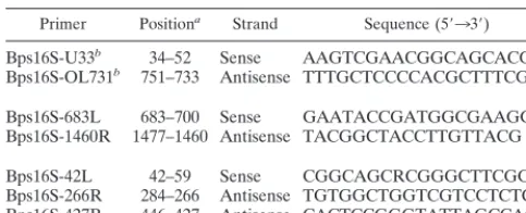
Nucleotide sequencing and phylogenetic analysis of the 16S rRNA gene.The 16S rRNA-encoding gene was amplified from the bacterial DNA by using two sets of overlapping oligonucleotide primers. Primers U33 and Bps16S-OL731 were used for amplifying the 59end of the 16S rRNA, and primers Bps16S-683L and Bps16S-1460R were used for amplifying the 39end (Table 1). Fragments of 717 and 795 bp in size were purified and subjected to cycle sequencing in both directions. Nucleotide sequencing of the PCR-amplified products was performed in an automated nucleotide sequencer (model 310; Applied Biosystems, Foster City, Calif.), using a BigDye terminator sequencing system (Perkin-Elmer, Foster City, Calif.). Nucleotide sequences obtained were analyzed by using MacVector software (Oxford Biomolecular Group, Oxford, United Kingdom) and were compared to the sequences deposited in the Gen-Bank database through the National Center for Biotechnology Information com-puter server (18a).
Phylogenetic analysis was performed by using the DNAdist, Neighbor, and Drawtree software in PHYLIP (phylogenetic inference package), version 3.572 (J. Felsenstein, University of Washington, Seattle, Wash.). The nucleotide sequences of the 16S rRNA-encoding genes of 44 other bacteria including 16 members ofBurkholderiaspecies, for which the complete or almost complete 16S rRNA gene sequences were available, were used for nucleotide sequence com-parison and phylogenetic analysis. The species (and their GenBank accession numbers) were as follows:B. pseudomallei1026b (U91839),Burkholderia thai-landensisE264 (U91838),Burkholderia plantarii(U96933),Burkholderia glumae
(U96931),Burkholderia vietnamensis(U96928),B. cepacia(U96927), Burkhold-eria cocovenenans(U96934),Burkholderia pyrrocinia(U96930),Burkholderia gla-thei(U96935),Burkholderia gladioli(X67038),Burkholderia graminis(U96939),
Burkholderia phenazinium(U96936),Burkholderia vandii(U96932),Burkholderia caryophylli(X67039),Burkholderia andropogonis(X67037),Burkholderia caribien-sis(Y17009),Herbaspirillum seropedicae(Y10146),Ralstonia eutropha(AF027407),
Bordetella bronchiseptica(X57026),Ideonella dechloratans(X72724),Bordetella holmesiiCDC F5101 (U04820),Zoogloea ramigera(X74914),Oxalobacter form-igenes(U49754),Janthinobacterium lividum(Y08846),Pseudomonas syzygii
R001 (U28237), denitrifying Fe-oxidizing bacteria (U51102),Duganella
zoo-gloeoides(D14256),Delftia acidovorans(AF078774), the ultramicrobacterium ND5 (AB008506),Rubrivivax gelatinosus(D16214),Bordetella parapertussis
ATCC 15311 (U04949),Leptothrix discophoraSS-1 (L33975),Bordetella pertussis
ATCC 9797 (U04950),Brachymonas denitrificans(D14320),Bordetella avium
ATCC 35086 (U04947),Alcaligenes xylosoxidans(D88005),Azoarcus indigens
(AF011345),Aquaspirillum sinuosum(AF078754),Alcaligenes faecalis(M22508),
S. maltophilia(X95923),P. aeruginosaLMG1242T (Z76651),P. putida(Z76667),
Salmonella typhiSTRNA16 (Z47544), andE. coliEHEC ATCC 43895 (Z83205). The nucleotide sequences were aligned by using MacVector software, and the evolutionary distances were calculated by using the program DNAdist based on the maximum-likelihood algorithm. The phylogenetic trees were constructed by the Drawtree program from the distance matrix by using the neighbor-joining method.
Primer design.A new set of multiplex PCR amplification primers was de-signed from the variable region of the 16S rRNA gene of the Ara1and Ara2 B. pseudomalleiisolates obtained from this study. Primer Bps16S-42L is homol-ogous to the sequences of both biotypes ofB. pseudomalleibut not to those of other organisms (Fig. 1). Primer Bps16S-427R was complementary to the se-quence of only Ara2B. pseudomalleiisolates and not to that of Ara1B. pseu-domalleiisolates or other bacteria. Primer Bps16S-266R could bind to both organisms as well as to a few other organisms. PCR amplification of Ara2 B. pseudomalleiisolate DNA yields two amplified fragments, of 405 and 243 bp in length, but only the 243-bp fragment is obtained from the amplification of Ara1B. pseudomalleiisolate DNA. The primers do not amplify the 16S rRNA
gene of other organisms.
These multiplex oligonucleotide primers were also designed to be located internally to the outer primer set for amplifying the 16S rRNA gene and there-fore were compatible with our previously described nested PCR procedure for detecting B. pseudomallei isolate DNA in clinical specimens (11). The new multiplex primers could be used as the nested primers by replacing the primers BS3L and BS4R with this new primer set.
Extraction of bacterial DNA from buffy-coat samples.The bacterial DNA was extracted from buffy-coat samples collected from seven patients with suspected melioidosis, using the heat treatment and proteinase K digestion protocol as previously described (11). The diagnosis of melioidosis was confirmed by isola-tion ofB. pseudomalleifrom five patients. For the other two patients culture was negative for B. pseudomallei, and these patients therefore served as negative controls.
PCR amplification.PCR amplification was carried out in a total volume of 50
ml containing 10ml of DNA extract, 5ml of 103amplification buffer (containing 500 mM KCl, 100 mM Tris-HCl [pH 8.3], 15 mM MgCl2, and 0.01% [wt/vol] gelatin), 4ml of deoxynucleoside triphosphates (2.5 mM each), 20 pmol of each primer, and 2.5 U ofTaqDNA polymerase (Perkin-Elmer). The tube was subjected to thermal cycling for 35 cycles, each comprised of 95°C for 1 min, 60°C for 1 min, and 72°C for 1 min, and then subjected to a final extension step for 10 min at 72°C, in a GeneAmp DNA Thermal Cycler 2400 (Perkin-Elmer). Tris-EDTA buffer was used as the negative control to exclude amplicon contamina-tion. The presence of amplified DNA product was analyzed on a 2% agarose gel, stained with ethidium bromide and visualized under a UV light. Strict laboratory precautions were carefully exercised to avoid contamination (13). For nested PCR amplification of DNA obtained from clinical specimens, the nested PCR procedure described previously (11) was performed. The three multiplex primers were used instead of the primers BS3L and BS4R.
Nucleotide sequence accession numbers.The nucleotide sequences of 16S rRNA genes of the nine Ara2B. pseudomalleiisolates and five Ara1B. pseu-domalleiisolates obtained from this study have been deposited in GenBank under accession no. AF093047 to AF093060.
RESULTS
Nucleotide sequence analysis. Nucleotide sequences ob-tained from PCR-amplified products of 14 B. pseudomallei isolates were compared (Fig. 2). The sequences of four Ara2
clinical isolates and five Ara2soil isolates were almost
identi-cal, indicating that they were isolates of the same species, the classical B. pseudomallei. The only nucleotide difference among the classical isolates was located at position 157, where six B. pseudomalleiisolates (three clinical and three soil iso-lates) had A but the other three (one clinical isolate and two soil isolates) had G. All of the Ara1isolates in this study had
G at position 157, and the published sequences ofB. pseudo-mallei 1026b (Ara2) and B. thailandensis E264 (Ara1) also
included G at position 157. In addition, the nucleotide se-quences of the nine classical Ara2 B. pseudomallei isolates
[image:2.612.53.295.93.191.2]obtained from this study included C at position 1292, which is different from the nucleotide (T) found at this position for B. pseudomallei1026b.
TABLE 1. Nucleotide sequences of PCR primers for amplifying the 16S rRNA genes of Ara1and Ara2B. pseudomalleiisolates Primer Positiona Strand Sequence (59339)
Bps16S-U33b 34–52 Sense AAGTCGAACGGCAGCACGG
Bps16S-OL731b 751–733 Antisense TTTGCTCCCCACGCTTTCG
Bps16S-683L 683–700 Sense GAATACCGATGGCGAAGG Bps16S-1460R 1477–1460 Antisense TACGGCTACCTTGTTACG
Bps16S-42L 42–59 Sense CGGCAGCRCGGGCTTCGG Bps16S-266R 284–266 Antisense TGTGGCTGGTCGTCCTCTC Bps16S-427R 446–427 Antisense CACTCCGGGTATTAGCCAGA
aAccording to reference 4.
bPreviously described in reference 11.
on May 15, 2020 by guest
http://jcm.asm.org/
The 16S rRNA gene sequences were virtually identical among all five Ara1B. pseudomallei isolates at all positions
sequenced. These sequences were also identical to the pub-lished sequence ofB. thailandensisE264 except at two posi-tions (G replaced A at posiposi-tions 1020 and 1141).
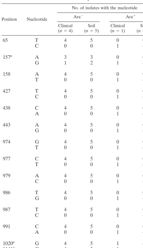
Among a total of 1,488 nucleotides analyzed, differences among isolates ofB. pseudomallei were observed at 15 posi-tions, 11 of which appeared to be biotype specific, as they were present in all Ara2biotypeB. pseudomalleiisolates but not in
those with Ara1biotype, and vice versa. The biotype-specific
nucleotides identified are listed in Table 2. These sequences were different from those of the 16S rRNA genes of other Burkholderiaspp., includingB. vietnamensis, which was initially isolated from the rhizosphere of a rice plant from a region of Vietnam in close proximity to northeast Thailand (17).
Phylogenetic analysis. Phylogenetic analysis demonstrated that the two biotypes ofB. pseudomalleiwere related but were clearly distinguishable (Fig. 3). The two biotypes were more
FIG. 1. Specificity of the PCR primers. Comparison of nucleotide sequences of the 16S rRNA genes corresponding to primers Bps16S-42L (A) and Bps16S-427R (B). The nucleotide sequence of Ara2B. pseudomalleiisolates was compared with those of Ara1B. pseudomalleiisolates, otherBurkholderiaspp.,H. seropedicae,R. eutropha,P. aeruginosa,P. putida,S. typhi, andE. coli. The sequences at the primer sites are underlined.
on May 15, 2020 by guest
http://jcm.asm.org/
closely related to each other than to other members of the genusBurkholderia(Fig. 4). It should be noted thatB. vietna-mensiswas more closely related toB. cepaciathan toB. pseu-domallei.
Discrimination of Ara2and Ara1B. pseudomalleiisolates by
multiplex PCR.A new set of multiplex PCR primers was de-signed to differentiate the two biotypes ofB. pseudomalleifrom other organisms. The forward primers Bps16S-42L bound only to the sequences of Ara2and Ara1B. pseudomalleiisolates.
The reverse primer Bps16S-427L was complementary to the region covering nucleotides T 427 C (T substituted for C at position 427), C 438 A, and A 443 G, which were used as the targets for discrimination between Ara2and Ara1B.
pseudo-mallei. In the presence of Ara2B. pseudomalleiisolate DNA,
PCR-amplified products consisting of two bands of 405 and 243 bp could be visualized in an ethidium bromide-stained aga-rose gel (Fig. 5). However, only the 243-bp fragment was
pres-ent when the Ara1B. pseudomalleiisolate DNA was amplified.
These primers could amplify all clinical and soil isolates of B. pseudomalleiused in this study. The DNA from other or-ganisms, includingB. cepacia,P. putida,P. aeruginosa,P. fluo-rescens,S. maltophilia, E. coli,A. anitratus,K. pneumoniae, E. aerogenes, Proteus sp.,A. hydrophila, S. marcescens, H. in-fluenzae, S. aureus, group A and group B streptococci, and group D enterococci, could not be amplified by these multiplex primers (data not shown).
Detection ofB. pseudomallei DNA in buffy-coat specimens. The new set of multiplex primers was used as the inner primers in the nested PCR system, by replacing our previously de-scribed BS3L and BS4R primers (11). Buffy-coat specimens from seven clinically suspected cases of melioidosis were tested, five of which were confirmed to be melioidosis by bac-terial cultures. All of the specimens from the five culture-proven melioidosis cases were PCR positive (resulting in two
FIG. 2. Alignment of nucleotide sequences of 16S rRNA genes from four clinical isolates (K96243, K94050, S94004, and S94017) and five soil isolates (TRF684, TRF685, TRF688, TRF824, and TRF830) of Ara2B. pseudomallei, and one clinical isolate (S95019) and four soil isolates (TRF681, TRF682, TRF683, and TRF686) of Ara1B. pseudomallei(GenBank accession no. AF093047 to AF093060). The published sequences ofB. pseudomallei1026b (U91839) andB. thailandensisE264 (U91838) are also included for comparison.
on May 15, 2020 by guest
http://jcm.asm.org/
amplified bands), but the specimens from the other two pa-tients without melioidosis were PCR negative.
DISCUSSION
The genus Burkholderia was proposed in 1992, and since then a number of former Pseudomonas species have been transferred to this genus (30). Several members of this genus are present in the environment and can be isolated from soil, water, and plants. In addition, two members of this genus are human pathogens, causing both acute diseases and opportu-nistic infection, such as in patients with cystic fibrosis. These pathogenic species include B. pseudomallei and B. cepacia. B. pseudomallei is the only member of this genus that causes severe disseminated disease, i.e., melioidosis. The emergence of a B. pseudomallei-like organism which was nonvirulent or less virulent than the classicalB. pseudomallei(9, 16, 18) re-ceived much attention. A nonvirulent isolate was explored as a potential vaccine candidate for melioidosis (10).
The major biochemical difference between the virulent (clas-sical) and nonvirulent isolates is the ability to assimilate L -arabinose. About 25 to 50% of the soil isolates ofB. pseudo-malleiare Ara1(20), but these Ara1 isolates are extremely
rare in clinical specimens. Only 1 of the 400 clinical isolates in our collection and none of the 1,200 clinical isolates studied by Smith et al. (20) was an arabinose assimilator. This Ara1
clinical isolate was isolated from the sputum of a elderly dia-betic Thai patient who had pneumonitis. The patient did not respond to the usual antibiotics and, after sputum cultured positive forB. pseudomallei, he was treated with ceftazidime (an antibiotic used for treatment of melioidosis). The patient responded to the treatment and fully recovered.B. pseudomal-lei was the only organism isolated from this patient. When subsequent analysis showed that this isolate was Ara1, in
in-dependent reviews of the case two infectious-disease specialists concluded that the pneumonitis in this patient was caused by this Ara1B. pseudomallei. The isolation of Ara1B.
pseudo-malleifrom a clinical case demonstrated that it can, although rarely, cause clinical illness, particularly in an immunocompro-mised host. This isolate demonstrated an LD50of.109CFU in mice. The nonvirulent or less-virulent status of the Ara1
iso-lates has been demonstrated in several animal models of ex-perimental B. pseudomallei infection (3, 20). The arabinose assimilation characteristic of these two biotypes appeared to be stable, as demonstrated by the failure to convert the Ara phe-notypes between the Ara2and Ara1isolates by selective
cul-ture (20). The distinctive characteristics of these organisms led to the question of whether they were different variants or biotypes of the same species or belonged to a closely related but distinctive species. The 16S rRNA gene sequences are highly conserved within the same species and have been widely used for species confirmation (15, 22). Therefore, the nucleo-tide sequences of the 16S rRNA gene were compared and phylogenetic analysis was performed in the present study.
This study clearly showed that the 16S rRNA gene se-quences of the two biotypes were distinctively different, but the difference was less than the differences among other species of the genusBurkholderia(Fig. 4). Eleven biotype-specific otide positions were identified among the total of 1,488 nucle-otides (99.26% homology), and these were totally conserved within the biotype. Differences in their phenotypes (biochem-ical assimilation ofL-arabinose, adonitol, 5-ketogluconate, and D-xylose and inability to assimilate dulcitol, erythritol, and tre-halose by the nonvirulent isolates) (28), colony morphology (3), virulence as demonstrated by the clinical severity in hu-mans and experimental animals such as mice and hamsters (3, 20), genomic sequences such as their ribotyping patterns (24), and nucleotide sequences in the 16S rRNA gene have been demonstrated, and a distinctive species namedB. thailandensis has been proposed for the Ara1 biotype (4). The data
de-scribed herein provide evidence that, based on the current polyphasic taxonomy system encompassing phenotypic, geno-typic, and phylogenetic information (7, 26), the degree of nu-cleotide differences in the 16S ribosomal RNA genes of the Ara2and Ara1B. pseudomalleiisolates may not be sufficient
to warrant classification as distinctive species.
The nucleotide differences between the 16S rRNA genes of the Ara2and Ara1B. pseudomalleiisolates and those of other
[image:5.612.53.291.91.504.2]species clustered in three regions. These three hypervariable regions were also observed in other bacterial species and were therefore used for species identification (23, 29, 30). A new set of multiplex PCR primers was designed for differentiating the two biotypes from each other and from other bacteria. The primers were based on the sequences of the first two diverse regions, as used in our previously described PCR system (11). TABLE 2. Nucleotide sequence differences between Ara1
and Ara2isolates ofB. pseudomallei
Position Nucleotide
No. of isolates with the nucleotide
Ara2 Ara1
Clinical
(n54) (nSoil55) Clinical(n51) (nSoil54)
65 T 4 5 0 0
C 0 0 1 4
157a A 3 3 0 0
G 1 2 1 4
158 A 4 5 0 0
T 0 0 1 4
427 T 4 5 0 0
C 0 0 1 4
438 C 4 5 0 0
A 0 0 1 4
443 A 4 5 0 0
G 0 0 1 4
974 G 4 5 0 0
T 0 0 1 4
977 C 4 5 0 0
T 0 0 1 4
979 A 4 5 0 0
C 0 0 1 4
986 T 4 5 0 0
G 0 0 1 4
987 T 4 5 0 0
C 0 0 1 4
991 C 4 5 0 0
A 0 0 1 4
1020a G 4 5 1 4
1141a G 4 5 1 4
1292a C 4 5 1 4
aDifferent from the published sequences ofB. pseudomallei1026b (GenBank
accession no. U91839) andB. thailandensisE264 (U91838) (see text).
on May 15, 2020 by guest
http://jcm.asm.org/
This rationale allowed the generation of PCR-amplified prod-ucts of appropriate sizes (243 and 405 bp) which can be clearly detected. Detection of the rRNA gene by PCR has several advantages in terms of both sensitivity and specificity because of the multiple copies of targeted DNA sequences and the conservation of the sequences in the same species (1). The described system allowed one-step discrimination between the two biotypes. In addition, the multiplex primers could be used as the inner primers for nested PCR amplification of sequences fromB. pseudomalleiin clinical specimens. This PCR amplifi-cation system has been proven useful in clinical situations, in which the PCR results were obtained before bacterial cultures became positive. The PCR system appears to be almost as
[image:6.612.123.477.71.298.2]sensitive as standard bacterial culture for the detection of B. pseudomallei(5, 11). PCR amplification can be performed directly from the clinical samples, which may contain small numbers of the bacteria. This technique has proven valuable in identifying bacterial pathogens that are difficult to detect and identify by traditional microbiological methods (1). It should be noted that the 16S rRNA genes ofB. pseudomallei and B. malleiare identical (2, 11, 25, 30), and therefore the two species cannot be differentiated using this system. As previ-ously mentioned, this is not considered a problem in clinical situations since glanders, the disease caused byB. mallei, is a
FIG. 3. Phylogenetic tree of the 16S rRNA genes shows the relationship of the Ara2and Ara1B. pseudomalleiisolates with other bacteria. The sequences used in this analysis are from the bacteria (1) Ara2B. pseudomallei, (2) Ara1B. pseudomallei, (3)B. plantarii, (4)B. glumae, (5)B. vietnamensis, (6)B. cepacia, (7) B. cocovenenans, (8)B. pyrrocinia, (9)B. glathei, (10)B. gladioli, (11)B. graminis, (12)B. phenazinium, (13)B. vandii, (14)B. caryophylli, (15)B. andropogonis, (16) B. caribiensis, (17)R. eutropha, (18)H. seropedicae, (19)B. bronchiseptica, (20)I. dechloratans, (21)B. holmesii, (22)Z. ramigera, (23)O. formigenes, (24)J. lividum, (25) P. syzygii, (26) denitrifying Fe-oxidizing bacteria, (27)D. zoogloeoides, (28)D. acidovorans, (29) ultramicrobacterium, (30)R. gelatinosus, (31)B. parapertussis, (32) L. discophora, (33)B. pertussis, (34)B. denitrificans, (35)B. avium, (36)A. xylosoxidans, (37)A. indigens, (38)A. sinuosum, (39)A. faecalis, (40)S. maltophilia, (41) P. aerguinosa, (42)P. putida, (43)S. typhi, and (44)E. coli. The scale bar indicates a distance in substitutions per nucleotide.
FIG. 4. Phylogenetic analysis of the 16S rRNA genes of 16 members in the genusBurkholderiafor which the complete or almost-complete 16S rRNA se-quences were available. The scale bar indicates a distance in substitutions per nucleotide.
FIG. 5. Ethidium bromide-stained agarose gel electrophoresis of the ampli-fied 16S rRNA genes of Ara2and Ara1B. pseudomalleiisolates performed by
using the multiplex primer set. M, 100-bp marker; lane 1, Ara2B. pseudomallei
isolate K94050; lane 2, Ara2 B. pseudomalleiisolate K96243; lane 3, Ara1 B. pseudomalleiisolate S95019; lane 4, Ara1B. pseudomalleiisolate TRF681;
lane 5, negative control.
on May 15, 2020 by guest
http://jcm.asm.org/
disease of the horse, mule, and donkey and is epidemiologi-cally different from melioidosis (18). Only sporadic cases of glanders occur in animals in Asia, Africa, and South America (19). An extensive survey of soil and water samples collected in Thailand, an area with endemic melioidosis, failed to isolate B. mallei(unpublished data).
Our previously described sets of PCR primers for identifying B. pseudomalleihave been used in the confirmation and in the diagnosis of melioidosis (11). Primers in the 16S rRNA gene region had sensitivity approaching 100% for clinical samples, and this level is higher than that for the primers for the 23S rRNA gene and for the intergenic region between the 16S and 23S rRNA genes (12). In addition, these primers were also useful for the isolation and identification ofB. pseudomalleiin soil (reference 5 and our unpublished data). PCR of soil sam-ples has been proven to be robust and more sensitive than bacterial culture and is also a useful confirmatory test in de-termining the identity of the soil isolates where biochemical tests give inconsistent results (5). Since both Ara2and Ara1
B. pseudomalleiorganisms are present in soil samples from the same geographical region, a PCR system that can identify the species would be valuable in environmental surveys ofB. pseu-domallei.
ACKNOWLEDGMENTS
This work is a part of the SirirajBurkholderia pseudomalleigenome project, supported by the Faculty of Medicine, Siriraj Hospital, Ma-hidol University, Bangkok, Thailand. T. Dharakul and S. Songsivilai are supported by career development grants from the National Science and Technology Development Agency of Thailand and the Anand-hamahidol Foundation.
We thank V. Thamlikitkul and the Thailand Research Fund for providing the soil isolates and D. Kanistanon for technical support of phylogenetic analysis.
REFERENCES
1.Anderson, B.1994. Broad range polymerase chain reaction for detection and identification of bacteria. J. Fla. Med. Assoc.81:835–837.
2.Bauernfeind, A., C. Roller, D. Meyer, R. Jungwirth, and I. Schneider.1998. Molecular procedure for rapid detection ofBurkholderia malleiand Burk-holderia pseudomallei. J. Clin. Microbiol.36:2737–2741.
3.Brett, P. J., D. DeShazer, and D. E. Woods.1997. Characterization of Burk-holderia pseudomalleiandBurkholderia pseudomallei-like strains. Epidemiol. Infect.118:137–148.
4.Brett, P. J., D. DeShazer, and D. E. Woods.1998.Burkholderia thailandensis
sp. nov., aBurkholderia pseudomallei-like species. Int. J. Syst. Bacteriol. 48:317–320.
5.Brook, M. D., B. Currie, and P. M. Desmarchelier.1997. Isolation and identification ofBurkholderia pseudomalleifrom soil using selective culture techniques and the polymerase chain reaction. J. Appl. Microbiol.82:589– 596.
6.Chaowagul, W., N. J. White, D. A. B. Dance, Y. Wattanagoon, P. Naigowit, T. M. E. Davis, S. Looareesuwan, and N. Pitakwatchara.1989. Melioidosis: a major cause of community-acquired septicemia in northeastern Thailand. J. Infect. Dis.159:890–899.
7.Colwell, R. R.1970. Polyphasic taxonomy of the genusVibrio: numerical taxonomy ofVibrio cholerae,Vibrio parahaemolysis, and relatedVibrio spe-cies. J. Bacteriol.104:410–433.
8.Dance, D. A. B.1991. Melioidosis: the tips of the iceberg? Clin. Microbiol. Rev.4:52–60.
9.Dannenberg, A. M., Jr., and E. Scott.1958. Melioidosis: pathogenesis and immunity in mice and hamsters. II. Studies with avirulent strains of Malleo-myces pseudomallei. Am. J. Pathol.34:1099–1121.
10. Dannenberg, A. M., Jr., and E. Scott.1959. Melioidosis: pathogenesis and immunity in mice and hamsters. III. Effects of vaccination with avirulent
strains ofPseudomonas pseudomalleion the resistance to the establishment and the resistance to the progress of respiratory melioidosis caused by virulent strains: all-or-none aspects of this disease. J. Immunol.84:233–246. 11. Dharakul, T., S. Songsivilai, S. Viriyachitra, V. Luangwedchakarn, B. Tas-saneetritap, and W. Chaowagul.1996. Detection ofBurkholderia pseudomal-leiDNA in patients with septicemic melioidosis. J. Clin. Microbiol.34:609– 614.
12. Haase, A., M. Brennan, S. Barrett, Y. Wood, S. Huffam, D. O’Brien, and B. Currie.1998. Evaluation of PCR for diagnosis of melioidosis. J. Clin. Microbiol.36:1039–1041.
13. Kitchin, P. A., Z. Szotyori, C. Fromholc, and N. Almond.1990. Avoidances of false positives. Nature (London)344:201.
14. Leelarasamee, A., and S. Bovornkitti.1989. Melioidosis: review and update. Rev. Infect. Dis.11:413–425.
15. Ludwig, W., and K.-H. Schleifer.1994. Bacterial phylogeny based on 16S and 23S rRNA sequence analysis. FEMS Microbiol. Rev.15:155–173. 16. McCormick, J. B., R. E. Weaver, P. S. Hayes, J. M. Boyce, and R. A.
Feldman.1977. Wound infection by an indigenousPseudomonas pseudomal-lei-like organism isolated from the soil: case report and epidemiologic study. J. Infect. Dis.135:103–107.
17. Meyer, J. M., V. T. Van, A. Stintzi, O. Berge, and G. Winkelmann.1995. Ornibactin production and transport properties in strains ofBurkholderia vietnamensisandBurkholderia cepacia(formerlyPseudomonas cepacia). Bio-metals8:309–317.
18. Miller, W. R., L. Pannell, L. Cravitz, W. A. Tanner, and T. Rosebury.1948. Studies on certain biological characteristics ofMalleomyces malleiand Mal-leomyces pseudomallei. II. Virulence and infectivity for animals. J. Bacteriol. 55:127–135.
18a.National Center for Biotechnology Information.1 April 1999, revision date. Sequences. [Online.] National Center for Biotechnology Information, Na-tional Library of Medicine, NaNa-tional Institutes of Health, Bethesda, Md. www.ncbi.nlm.nih.gov. [6 April 1999, last date accessed.]
19. Sanford, J. P.1985.Pseudomonasspecies (including melioidosis and glan-ders), p. 1250–1254.InG. L. Mandell, R. G. Douglas, and J. E. Bennett (ed.), Principles and practice of infectious diseases, 2nd ed. John Wiley & Sons, Inc., New York, N.Y.
20. Smith, M. D., B. J. Angus, V. Wuthiekanun, and N. J. White.1997. Arabi-nose assimilation defines a nonvirulent biotype ofBurkholderia pseudomallei. Infect. Immun.65:4319–4321.
21. Smith, M. D., V. Wuthiekanun, A. L. Walsh, and N. J. White.1995. Quan-titative recovery ofBurkholderia pseudomalleifrom soil in Thailand. Trans. R. Soc. Trop. Med. Hyg.89:488–490.
22. Stackebrandt, E., and B. M. Goebel.1994. Taxonomic note: a place for DNA-DNA reassociation and 16S rRNA sequence analysis in the present species definition in bacteriology. Int. J. Syst. Microbiol.44:846–849. 23. Taghavi, M., C. Hayward, L. I. Sly, and M. Fegan.1996. Analysis of the
phylogenetic relationships of strains ofBurkholderia solanacearum, Pseudo-monas syzygii, and the blood disease bacterium of banana based on 16S rRNA gene sequences. Int. J. Syst. Bacteriol.46:10–15.
24. Trakulsomboon, S., D. A. Dance, M. D. Smith, N. J. White, and T. L. Pitt. 1997. Ribotype differences between clinical and environmental isolates of
Burkholderia pseudomallei. J. Med. Microbiol.46:565–570.
25. Tyler, S. D., C. A. Strathdee, K. R. Rozee, and W. M. Johnson.1995. Oligonucleotide primers designed to differentiate pathogenic pseudomonads on the basis of the sequencing of genes coding for 16S-23S rRNA internal transcribed spacers. Clin. Diagn. Lab. Immunol.2:448–453.
26. Vandamme, P., B. Pot, M. Gillis, P. De Vos, K. Kersters, and J. Swings.1996. Polyphasic taxonomy, a consensus approach to bacterial systematics. Micro-biol. Rev.60:407–438.
27. White, N. J., D. A. B. Dance, W. Chaowagul, Y. Wattanagoon, V. Wuthieka-nun, and N. Pitakwatchara.1989. Halving of mortality of severe melioidosis by ceftazidime. Lancetii:697–700.
28. Wuthiekanun, V., M. D. Smith, D. A. B. Dance, A. L. Walsh, T. L. Pitt, and N. J. White.1996. Biochemical characteristics of clinical and environmental isolates ofBurkholderia pseudomallei. J. Med. Microbiol.45:408–412. 29. Xiang, L., M. Dorsch, T. D. Dot, L. I. Sly, E. Stackebrandt, and A. C.
Hayward.1993. Phylogenetic studies of the rRNA II pseudomonads based on 16S rRNA gene sequences. J. Appl. Bacteriol.74:324–329.
30. Yabuuchi, E., Y. Kosako, H. Oyaizu, I. Yano, H. Hotta, Y. Hashimoto, T. Ezaki, and M. Arakawa.1992. Proposal ofBurkholderiagen. nov. and trans-fer of seven species of the genusPseudomonashomology group II to the new genus, with the type speciesBurkholderia cepacia(Palleroni and Holmes 1981) comb. nov. Microbiol. Immunol.36:1251–1275.