Copyright © 2002, American Society for Microbiology. All Rights Reserved.
Molecular Typing of Selected
Enterococcus faecalis
Isolates:
Pilot Study Using Multilocus Sequence Typing and
Pulsed-Field Gel Electrophoresis
Sreedhar R. Nallapareddy,
1,2Ruay-Wang Duh,
1,2† Kavindra V. Singh,
1,2and Barbara E. Murray
1,2,3*
Division of Infectious Diseases, Department of Internal Medicine,1Center for the Study of Emerging and
Re-emerging Pathogens,2and Department of Microbiology and Molecular Genetics,3
University of Texas Medical School, Houston, Texas 77030
Received 29 August 2001/Returned for modification 23 November 2001/Accepted 16 December 2001
The present study compared the recently developed multilocus sequence typing (MLST) approach with a well-established molecular typing technique, pulsed-field gel electrophoresis (PFGE), for subspecies
differen-tiation ofEnterococcus faecalisisolates. We sequenced intragenic regions of threeE. faecalisantigen-encoding
genes (ace, encoding a collagen and laminin adhesin; efaA, encoding an endocarditis antigen; and salA,
encoding a cell wall associated antigen) and one housekeeping gene (pyrC) of 22E. faecalisisolates chosen
largely for their temporal and geographical diversity, but also including some outbreak isolates. MLST analysis of polymorphic regions of these four genes identified 13 distinct sequence types (STs) with different allelic profiles; the composite sequences generated from the four sequenced gene fragments of individual isolates showed 98.3 to 100% identity among the 22 isolates. We also found that the allelic profiles from two
sequences,aceandsalA, were sufficient to distinguish all 13 STs of this study. The 13 STs corresponded to 12
different PFGE types, with one previously designated PFGE clone (a widespread U.S. clone of
-lactamase-producing isolates) being classified into two highly related STs which differed at 2 of 2,894 bases, both in the same allele. MLST also confirmed the clonal relationships among the isolates of two other PFGE clonal groups,
including vancomycin resistant isolates. Thus, this pilot study with representativeE. faecalisisolates suggests
that, similar to PFGE, the sequence-based typing method may be useful for differentiating isolates ofE. faecalis
to the subspecies level in addition to identifying outbreak isolates.
Enterococci, normal gut commensals, were recognized as a causative agent of endocarditis and urinary tract infections in ca. 1900 and have also been reported as a common cause of nosocomial infections since the 1970s (25, 26). Recently, accu-mulation of antibiotic resistances has made enterococcal infec-tions a life-threatening clinical challenge, and thus, the meth-ods that distinguish an outbreak from an endogenous strain have become important for designing strategies to prevent and control outbreaks (24, 26). A number of phenotypic or geno-typic typing methods (including biochemical typing, serotyping, multilocus enzyme electrophoresis [MLEE], phage typing, in-sertion sequence element-based typing, pulsed-field gel elec-trophoresis [PFGE], restriction fragment length polymorphism [RFLP] analysis, ribotyping, repetitive sequence-based PCR, arbitrary primed PCR, and random amplification of polymor-phic DNA) have been applied to the epidemiological investi-gations ofEnterococcus faecalis(2–4, 6, 14, 18, 22, 28, 35, 40, 47, 48). These molecular and epidemiological studies have provided valuable information and clarified some misconcep-tions regarding E. faecalis infections, such as demonstrating
that someE. faecalisinfections are caused by nosocomial trans-mission of outbreak strains rather than arising from the pa-tient’s own prehospitalization intestinal flora. Among the known enterococcal molecular typing methods, PFGE has proven to be a highly reproducible and accurate typing method, which can distinguish clonal populations, and hence is considered a “gold standard” for subspecies discrimination of
E. faecalisclinical isolates. However, the results obtained by
PFGE are not readily transportable, making it difficult to com-pare results among different laboratories and thus limiting studies involving interlaboratory comparisons.
Microbial genome sequencing programs have provided enormous data for easy identification of species-specific genes. This, together with rapid automated DNA sequencing, has generated considerable interest in DNA sequence-based typ-ing methods. A few recent studies have used variable intra-genic sequences (flanked by highly conserved regions) of either a single gene or two genes and reported their usefulness in strain differentiation. These include the hsp65 gene (which encodes a 65-kDa heat shock protein) of Mycobacterium
scrofulaceum, theporB gene (which encodes an outer
mem-brane porin protein) ofNeisseria meningitidis, and repeat re-gions of thespaA(which encodes protein A) as well as thecoa (which encodes coagulase) genes ofStaphylococcus aureusand theemmgene (which encodes an M protein) ofStreptococcus
pyogenes(1, 33, 38, 39, 41, 42, 44, 45).
Recently, multilocus sequence typing (MLST) was devel-oped for identification of clonal complexes within bacterial
* Corresponding author. Mailing address: Center for the Study of Emerging and Re-emerging Pathogens, Division of Infectious Dis-eases, Department of Internal Medicine, University of Texas Medical School at Houston, 6431 Fannin St., Houston, TX 77030. Phone: (713) 500-6767. Fax: (713) 500-5495. E-mail: [email protected] .edu.
† Present address: Section of Infectious Diseases, Department of Medicine, Veterans General Hospital-Taipei, Taiwan, Republic of China.
868
on May 15, 2020 by guest
http://jcm.asm.org/
populations and has been used successfully for molecular epi-demiological analysis of N. meningitidis, Streptococcus
pneu-moniae,S. aureus,S. pyogenes, andCampylobacter jejuni(7, 9,
11, 12, 21). MLST typically characterizes isolates of bacteria using⬃400 to 500 bp of intragenic sequences of six to seven housekeeping genes or loci, and thus, MLST is similar in prin-ciple to MLEE, but with greater sensitivity due to its ability to detect neutral genetic variations. This method distinguishes strains based on the observed allelic variations in the nucleo-tide sequences of several loci, rather than the degree of se-quence variation in any single gene or locus. MLST and other the DNA sequence-based typing methods have been suggested as offering advantage over other techniques because (i) the data are objective and readily comparable between laborato-ries, (ii) the data can be stored in a shared central database to provide a broader resource for epidemiological studies, and (iii) evolutionary genetic analyses can be performed. At the same time, DNA sequencing is expensive when compared to PFGE and considerable technical skill and knowledge in se-quence analysis is critical for typing the isolates (32).
In the present study, we have evaluated the discriminatory ability of a sequence-based typing method and compared the results to those obtained with PFGE. Internal fragments of three antigen-encoding genes that were detected withE.
fae-calis-infected patient sera and an internal fragment of one
pyrimidine biosynthesis gene were used for the MLST analysis.
The MLST generated sequence types were found to be com-parable to types or clones identified with PFGE.
MATERIALS AND METHODS
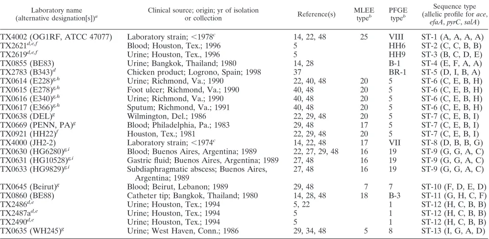
Bacterial isolates.A total of 22E. faecalisisolates from different geographic locations were chosen for this study, primarily because they had been previously studied by us using one or more other typing techniques (Table 1). A collection of eight-lactamase-producing (Bla⫹) isolates that were recovered from five cities in the United States (four isolates from the same hospital) during an 11-year period (1981 through 1991), three additional Bla⫹isolates from a single hospital in Argentina (isolated between June and September of 1989), and one Bla⫹isolate from Lebanon (isolated in 1989) that were previously analyzed by PFGE and MLEE were included (27–29, 34, 48). Six other isolates were vanco-mycin resistant, including five clinical isolates from a single hospital (of which three isolates were part of a single outbreak) in the United States (5) and one nonclinical isolate collected from Spain isolated from chicken products (37). Two isolates with high-level aminoglycoside resistance were from Thailand (14, 28), and two commonly used laboratory isolates ofE. faecalisOG1RF (ATCC 47077) and JH2-2 were also included in this study (14, 17, 22, 23, 30).
PFGE.PFGE was performed with some modifications of a previously de-scribed method (14, 28). Agarose plugs containing genomic DNA were digested withSmaI (Gibco, BRL, Gaithersburg, Md.), and electrophoresis was carried out using a clamped homogeneous electric field (CHEF-DRII device; Bio-Rad Lab-oratories, Richmond, Calif.), with ramped pulse times beginning with 5 s and ending with 45 s, at 200 V for 26 h. The gels were stained with ethidium bromide (0.4g/ml) for 30 min and photographed. The PFGE patterns were interpreted using the criteria suggested by Tenover et al. (46), with closely and possibly related patterns being designated as belonging to a single clone. PFGE pattern names that were presented in earlier publications are used here.
[image:2.587.43.542.85.329.2]Genomic DNA isolation; PCR; and DNA sequencing oface,efaA,pyrC, and
TABLE 1. Sequence-based typing results for variousE. faecalisisolates in this study
Laboratory name
(alternative designation[s])a Clinical source; origin; yr of isolationor collection Reference(s) MLEEtypeb PFGEtypeb
Sequence type (allelic profile forace,
efaA,pyrC,salA)
TX4002 (OG1RF, ATCC 47077) Laboratory strain;⬍1978c 14, 22, 48 25 VIII ST-1 (A, A, A, A)
TX2621d,e,f Blood; Houston, Tex.; 1996 5 HH6 ST-2 (C, C, B, B)
TX2619d,e,f Urine; Houston, Tex., 1996 5 HH9 ST-3 (B, C, D, E)
TX0855 (BE83) Urine; Bangkok, Thailand; 1980 14, 28 B-1 ST-4 (E, F, A, A)
TX2783 (B343)d Chicken product; Logrono, Spain; 1998 37 BR-1 ST-5 (D, I, B, A)
TX0614 (E228)g,h Urine; Richmond, Va.; 1990 22, 40, 48 20 5 ST-6 (C, E, B, H)
TX0615 (E278)g,h Foot ulcer; Richmond, Va.; 1990 40, 48 20 5 ST-6 (C, E, B, H)
TX0616 (E340)g,h Urine; Richmond, Va.; 1990 40, 48 20 5 ST-6 (C, E, B, H)
TX0617 (E366)g,h Sputum; Richmond, Va.; 1991 40, 48 20 5 ST-6 (C, E, B, H)
TX0638 (DEL)g Wilmington, Del.; 1986 22, 29, 48 20 5 ST-7 (C, E, B, I)
TX0669 (PENN, PA)g Blood; Philadelphia, Pa.; 1983 29, 48 17 5 ST-7 (C, E, B, I)
TX0921 (HH22)f Houston, Tex.; 1981 22, 29, 48 20 5 ST-7 (C, E, B, I)
TX4000 (JH2-2) Laboratory strain;⬍1974c 14, 22, 48 17 VII ST-8 (D, B, B, G)
TX0630 (HG6280)g,i Blood; Buenos Aires, Argentina; 1989 22, 27, 29, 48 16 19 ST-9 (G, G, A, C)
TX0631 (HG10528)g,i Gastric fluid; Buenos Aires, Argentina; 1989 27, 48 16 19 ST-9 (G, G, A, C)
TX0633 (HG9829)g,i Subdiaphragmatic abscess; Buenos Aires,
Argentina; 1989 27, 48 16 19 ST-9 (G, G, A, C)
TX0645 (Beirut)g Blood; Beirut, Lebanon; 1989 29, 48 7 7 ST-10 (F, D, E, D)
TX0860 (BE88) Catheter tip; Bangkok, Thailand; 1980 14, 28, 48 18 B-3 ST-11 (G, H, C, F)
TX2486d,e Urine; Houston, Tex.; 1994 5, 22 1 ST-12 (H, C, B, B)
TX2487ad,e Urine; Houston, Tex.; 1994 5 1 ST-12 (H, C, B, B)
TX2490d,e Urine; Houston, Tex.; 1994 5 1 ST-12 (H, C, B, B)
TX0635 (WH245)g Urine; West Haven, Conn.; 1986 29, 34, 48 5 8 ST-13 (I, G, A, D)
aAs designated in previous studies.
bThe original MLEE and PFGE pattern names in the earlier publication(s) are used. cIsolated at unknown time prior to the year shown.
dVancomycin resistant isolate.
eIsolates TX2619, TX2621, TX2486, TX2487a, and TX2490 were from the same hospital.
f Isolates TX2619 and TX2621 were not tested for vancomycin resistance in the previous study (5) and hence were not reported as vancomycin-resistant strains. g-Lactamase-producing isolate.
hIsolates TX0614, TX0615, TX0616, and TX0617 were from the same hospital. i Isolates TX0630, TX0631, and TX0633 were from the same hospital.
on May 15, 2020 by guest
http://jcm.asm.org/
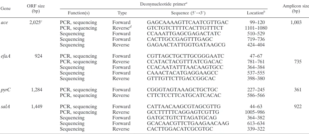
salA.Genomic DNA was extracted fromE. faecalisisolates freshly streaked from freezer vials and cultured in brain heart infusion broth. Genomic DNA was isolated by the hexadecyltrimethyl ammonium bromide method as described previously (22). We chose internal regions of threeE. faecalisantigen-encoding genes (ace,efaA, andsalA) and one housekeeping gene (pyrC) for sequencing. These four loci were chosen primarily because all these genes were well studied in our laboratory (8, 19, 31, 43), and the sequence diversity of the region coding for the A domain ofacehas been recognized in our earlier study (31). The open reading frame (ORF) sizes of the four chosen loci are listed in Table 2. The internal gene fragments oface, which encode a collagen and laminin adhesin (959 bp [890 bp for one isolate due to a 69-bp deletion] [31, 36]);efaA, which encodes an endocarditis antigen (693 bp [20]);pyrC, which encodes a dihydroo-rotase (320 bp [19]); andsalA, which encodes a cell wall-associated antigen (F. Teng, B. E. Murray, and G. M. Weinstock, unpublished data) (919 or 922 bp, due to a 3-bp in-frame deletion in some isolates) were amplified using the optimized buffer B (1⫻buffer: 60 mM Tris-HCl [pH 8.5], 15 mM ammonium sulfate, 2 mM MgCl2) obtained from Invitrogen (San Diego, Calif.). PCR was performed in
volumes of 50l, with an initial denaturation at 94°C for 2 min, followed by 30 cycles of 94°C for 1 min, 55°C for 1 min, and 72°C for 1 min 30 s (72°C for 45 s forpyrC) and a final extension of 72°C for 7 min. The PCR primers used for amplification and sequencing of all four genes are listed in Table 2. Our initial attempts withaceamplification primers were unsuccessful for three strains, namely, TX2486, TX2487a, and TX2490, subsequently found to be due to vari-ations in theacegene at the reverse primer region. Theaceamplicon for these strains was amplified usingaceforward PCR primer (Table 2) and reverse primer with the sequence 5⬘-ATTTAATTTTTGAATTGGTTCACTAAGCAG-3⬘ (lo-cated at positions 1896 to 1867, relative to the start codon). The PCR amplicons were purified using the Wizard PCR DNA Cleanup System (Promega Corpora-tion, Madison, Wis.). Sequencing of both strands of the amplified fragments was achieved using an Applied Biosystems Prism 377 automated DNA sequencer using theTaqDye-Deoxy terminator method (PE Applied Biosystems, Foster City, Calif.). Sequences were assembled using SeqMan program of DNASTAR software (Lasergene, Madison, Wis.).
Sequence analysis.Multiple sequence alignments of the 22 isolates for the four gene fragments (ace,efaA,pyrC, andsalA) were done by the Jotun Hein method (16) using the MegAlign program of DNASTAR software. A 2,894-bp (for 10 strains), 2,891-bp (for 11 strains), 2,822-bp (for one strain) nucleotide composite sequence (derived from four concatenated gene fragments) was also aligned by the MegAlign program. Phylogenetic trees (cladograms) based on the matrix of pairwise sequence divergence were constructed using the MegAlign program.
Allele and sequence type (ST) assignment.In order to identify the nucleotide variation in the above gene fragments, sequences from the different isolates were compared to the corresponding sequences in the well-studiedE. faecalisstrain OG1RF (30). Each gene sequence differing by one or more nucleotides was considered to be a different allele (no weight was given to the degree of sequence divergence between alleles, although the alleles that differed by a single nucle-otide were denoted as single nuclenucle-otide variants [SNVs]), and the distinct allelic sequences were assigned an arbitrary letter designation in the order of the number of base pairs that varied with respect to the OG1RF sequence. The alleles at the four loci provided the allelic profile, which defined the ST for each isolate. Isolates showing identical allelic profiles in the four gene fragments were assigned to the same ST, and those with a difference in their allelic profile were assigned to a different ST. The relatedness among the strains was analyzed by constructing a dendrogram based on the matrix of pairwise differences in the allelic sequences by the unweighted pair group method with arithmetic averages (UPGMA) method.
RESULTS AND DISCUSSION
Among known phylogenetic typing methods, PFGE and MLST, which are based on multiple sites and loci scattered around the chromosome, have consistently been shown to be capable of discriminating isolates at the subspecies level, al-though in different ways. PFGE is particularly useful for dis-tinguishing strains circulating within a geographical location (microvariation) and is based on the selected variable regions of the genome (9, 15). MLST is based on variations that ac-cumulate slowly and appears more suitable for long-term and global epidemiology (macrovariation) (9, 10, 15). However, in both methods, the relative rates of nucleotide substitutions and/or recombination occurring in nature set the limitations of phylogenetic analysis, and there are insufficient experiential data to establish these rates for enterococci.
Choice of loci. Since it is impractical to sequence large or
[image:3.587.44.540.85.300.2]multiple regions of the chromosome, we chose loci we had previously studied (ace, efaA, pyrC, and salA) and that had
TABLE 2. PCR and sequencing primers used in this study
Gene ORF size(bp) Deoxynucleotide primer
a
Amplicon size (bp)
Function(s) Type Sequence (5⬘33⬘) Locationb
ace 2,025c PCR, sequencing Forward GAGCAAAAGTTCAATCGTTGAC 99–120 1,003
PCR, sequencing Reversed GTCTGTCTTTTCACTTGTTTCT 1101–1080
Sequencing Forward CCAAATTGAGCGAGACTATC 510–529
Sequencing Forward CACTTGCCGAGTTTGAGC 719–736
Sequencing Reverse GAGAACTATTGGTGATAAGCG 424–404
efaA 924 PCR, sequencing Forward CGTTAGCTGCTTGCGGGAATC 47–67
PCR, sequencing Reverse CCATACTACGTTTATCGACAC 781–761 735
Sequencing Forward CCACAATATTTAACAAGTGCC 364–384
Sequencing Forward CAAACTACATGAGGAAGCC 537–555
Sequencing Reverse GTTTGTTCTTGACCGGCAC 398–380
pyrC 1,284 PCR, sequencing Forward CGGGTAGTAAAGCTGCTGC 227–245 361
PCR, sequencing Reverse CTTCTCCTTCATGCATCACAC 586–566
salA 1,449 PCR, sequencing Forward CATTAACAAGCGTAGCGTTG 44–63 922
PCR, sequencing Reverse GCCTTTTTCAGGAGTCGTTG 1005–986
Sequencing Forward GATGCTGTCTTAGATGCAG 364–382
Sequencing Forward GCACAACGTTCTGAAGAACAAG 613–634
Sequencing Reverse CACTTGGACATCGCGTGC 339–322
aace,efaA,pyrC, andsalAprimers were designed fromE. faecalisstrain V583 database sequences (The Institute for Genomic Research). bNumbering for all the primers is given relative to the start codon (ATG forace, efaA, andpyrC; TTG forsalA) for the respective genes.
caceORF size represented here is forE. faecalisstrain V583 (36).acegene size varies in differentE. faecalisstrains due to variation in the number of repeats of the B domain (31).
dSee text for the reverse primer used for the selected strains.
on May 15, 2020 by guest
http://jcm.asm.org/
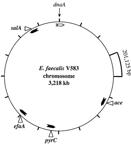
been shown to be present in allE. faecalisstrains tested; in-deed, hybridization withaceandefaAprobes had proven useful in differentiatingE. faecalisfrom other species (8, 43). On the basis of theE. faecalisV583 complete genome sequence (avail-able at http://www.tigr.org [The Institute for Genomic Re-search, Rockville, Md.]), we derived the physical map shown in Fig. 1, which illustrates that the loci chosen were spread around the chromosome and the distance between any two loci is ranged from 328 to 1,355 kb.
PFGE fingerprinting.PFGE withSmaI yielded⬍20 visible
bands for each isolate (data not shown). As per the recom-mendations described by Tenover et al. (46), including closely and possibly related patterns, a total of 12 PFGE types, 3 of which contained three or more isolates, were recognized. Seven of the twelve Bla⫹isolates were classified as belonging
to the same clonal group (PFGE pattern 5); two of these strains showed an identical pattern (TX0614 and TX0615), while others (TX0616, TX0617, TX0638, TX0669, and TX0921) showed patterns with three to six band differences, depending on which two isolates were being compared. Among three Argentinean Bla⫹isolates, TX0630 and TX0633 showed an
identical PFGE pattern, while TX0631 showed a pattern al-most identical to these two (classified as PFGE pattern 19) (27). Similarly, three of the five vancomycin-resistant Houston isolates were classified as clonally related (PFGE pattern 1). These results are in agreement with our earlier studies (5, 14, 18, 22, 28, 29, 37, 48).
MLST typing.The length of the loci used for allele
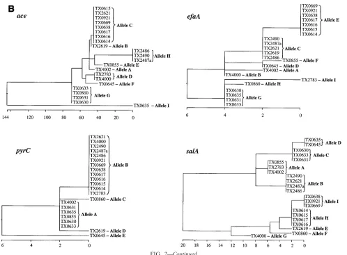
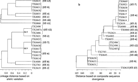
assign-ment in our MLST scheme was between 959 bp (ace) and 320 bp (pyrC). The total length of the four sequenced loci of this study approximates the total length of six loci common to several other MLST studies. Variable sites of different alleles of the four sequenced loci are presented in Fig. 2A, and the phylogenetic trees based on the matrix of pairwise divergence in these sequences are shown in Fig. 2B. Nine unique alleles were identified for three of the loci, while only five alleles were identified for the pyrC locus, likely because of the smaller region evaluated (Table 3; Fig. 2). Allelic profiles of the four loci identified 13 STs among the 22 isolates (Table 1). A den-drogram drawn from allelic profiles by the UPGMA method demonstrated four (ST-6, ST-7, ST-9, and ST-12) of the 13 STs contain more than one isolate (Fig. 3a). ST-6 and ST-7 as well as ST-2 and ST-12 differed by variation at a single locus.
The percentage of variable sites in a given locus ranged from 1.3 (efaA) to 5.6 (ace). A 69-bp in-frame deletion was detected in theacelocus of a Bla⫹isolate (allele I) from Connecticut.
Eight of nine alleles of the efaA locus differed by a single nucleotide, and they were designated SNV alleles (Table 3). Similarly, four of the five alleles of the pyrClocus are SNV alleles, and the fifth allele has five nucleotide changes (Bla⫹
isolate from Lebanon). In the salA locus, a 3-bp in-frame deletion was observed in 55% of the isolates. The nucleotide changes which alter the amino acid sequence are indicated as nonsynonymous base substitutions and the nucleotide changes which do not alter the amino acid sequence are indicated as synonymous base substitutions (Fig. 2A).
Composite sequence-based typing.In order to determine the
overall divergence of the sequenced gene fragments of the four loci studied, these sequences were spliced together to obtain a concatenated composite sequence for each of the isolates. For calculating the percentage of identity or divergence, in-frame insertions or deletions were not taken into account. A cla-dogram created from the matrix of pairwise sequence diver-gence of composite sequences identified 13 phylogenetic lin-eages (Fig. 3b), and these were identical to the 13 STs obtained from the allelic profiles. The identity between the 22 composite sequences was found to be between 98.3 and 100%. The com-posite sequence of ST-6 isolates from Richmond, Va. (TX0614, TX0615, TX0616, and TX0617, which showed 100% identity among the four isolates) have 99.93% identity (2,892 of 2,894 bases) with the composite sequence of ST-7 isolates from three different states (TX0638, TX0669, and TX0921, which were in turn 100% identical to each other).
Congruence between PFGE, MLST, and composite
se-quence-based typing methods.Analysis of the clusters in the
phylogenetic trees generated by the methods described above identified 13 different genotypes versus 12 different PFGE types among the 22E. faecalisisolates tested. Both sequence-based typing methods, i.e., MLST with four loci as well as the composite sequence alignment, confirmed the clonal relation-ships among isolates of PFGE patterns 1, 5, and 19. TX0921 (Bla⫹) obtained from Texas in 1981 showed the identical
MLST type as the Pennsylvania Bla⫹strain (TX0669) isolated
in 1983 and also strain TX0638 (Bla⫹) isolated in Delaware in
1986, confirming our previous finding of widespread dispersion of this clone in the United States (29). Sequence-based typing further confirmed that the three Bla⫹ clones from South
FIG. 1. Chromosomal locations of the sequenced loci. The loca-tions are marked on a constructed physical map ofE. faecalisV583 using the complete genome sequence (available at http://www.tigr.org/ [The Institute for Genomic Research]). The 3,218-kb genome is di-vided into 16 segments, with each segment representing 201,125 bases. The ORF coding for chromosomal replication factor (dnaA) was iden-tified based on identity withdnaA sequences ofS. pyogenes andS. aureusand positioned at nucleotide one. The arrowheads inside the circle represent the ORF orientations of the four loci and are marked at the respective positions.
on May 15, 2020 by guest
http://jcm.asm.org/
America and a single isolate from Lebanon as well as the single isolate from Connecticut were different from each other.
Although there was an agreement among all three analyses, one apparent exception was the recognition of two different STs (containing multiple isolates) by sequence-based typing among isolates characterized as belonging to PFGE pattern 5. As pointed out earlier, four of these seven Bla⫹PFGE type 5
isolates (ST-6) obtained from Richmond, Va., in 1990 to 1991 are identical at all four loci, while the other three PFGE type 5 Bla⫹isolates (isolated 5 to 10 years earlier in different states)
were classified as ST-7, which differs only by variation of two synonymous bases at thesalAlocus (i.e., these are single-locus variants [SLVs]); all other isolates differed from these Bla⫹
isolates by two or more bases at this locus, as well as differing at other loci. While most MLST studies that deal with six or more loci would have clustered these single locus variants as a single clonal group, we did not do so because of the smaller number of loci studied. The ST-6 and ST-7 group of Bla⫹
isolates has previously been considered clonally related based
on PFGE and repetitive sequence-based PCR (22). This dif-ference may be related to the use of a somewhat broader assignment of PFGE types (i.e., up to even six fragment dif-ferences were seen, depending on which two isolates were compared), while counting even a single base change in assign-ing the alleles of MLST.
Among other possibly related STs, three vancomycin-resis-tant outbreak isolates (ST-12) from Houston, Tex. (1994) are SLVs of another Houston vancomycin resistant 1996 isolate (ST-2), the only other SLV of this study. However, the ST-2 isolate has only 98.9% overall composite sequence identity with ST-12 isolates due to 33 scattered base changes in theace locus. These vancomycin-resistant SLVs also show a relatively similar PFGE pattern when compared to the other isolates of this study. This, together with the identity at three of the four loci, raises the possibility that the extensive differences inace may be due to horizontal exchange, as has been suggested to occur during conjugative transposition (49).
The other possibly related composite sequences were those
FIG. 2. Allelic variation of the four sequenced loci. (A) Variable sites identified in all the four gene fragments. The nucleotide sites that are identical in all the alleles are not shown. The nucleotides present in each of the variable sites of allele A (E. faecalisOG1RF) are shown. Only those sites that differ are shown for all the other alleles. The position of each variable site within the sequenced fragment is shown in the numbers above the nucleotide, read vertically. The consensus sequence is shown on the bottom. The variations that are synonymous (S) and nonsynonymous (N) are also shown. (B) Cladograms of the four loci sequenced. Phylogenetic trees were based on the matrix of pairwise sequence divergence in the sequences (generated by the Jotun Hein method of the DNASTAR software package). The length of each pair of branches represents the distance between sequence pairs, while the units at the bottom of the tree indicate the number of substitution events.
on May 15, 2020 by guest
http://jcm.asm.org/
of the Connecticut Bla⫹ST-13 isolate and the South American
Bla⫹ST-9 isolates; these differed by a 69-bp deletion at theace
locus and a single base change at thesalAlocus. However, the PFGE patterns of these two STs differed substantially.
Results from a prior study of MLEE found general agree-ment for most isolates tested by both PFGE and MLEE (Ta-ble 1). The MLEE study assigned the same ET to six of seven Bla⫹isolates (all ST-6 isolates and two of three ST-7 isolates of
this study), consistent with the PFGE pattern assignments. However, one ST-7 Bla⫹isolate (TX0669, also PFGE type 5)
was assigned a different ET, which was also the ET of Chile isolates in that study as well as another isolate JH2-2 (TX4000), which differed by PFGE (type VII) and MLST (ST-8). These various results, taken together, suggest the pos-sibility of cross-contamination in the MLEE study and misas-signment of the ET type of TX0669.
The phylogenetic trees of both the MLST (linkage distance derived from allelic variation [Fig. 3a]) and the composite sequence alignment (linkage distance derived from sequence divergence [Fig. 3b]) confirmed the similar clustering of clonal and related isolates, in addition to differentiating the nonre-lated (as defined by other techniques) isolates. However, an apparent difference was observed in interrelationships of pos-sibly related isolates such as ST-2 and ST-12 that differed
[image:6.587.46.544.69.436.2]extensively at a single locus. The difference in the two schemes is related to the different criteria that were used to generate linkage distances, i.e., in MLST, for a given locus, equal weight was given for an allele with a single base change or multiple base changes, while in the composite sequence-based typing weight was given to every base change. At this stage, we are not
TABLE 3. Genetic variation in four sequenced loci
Gene Fragmentsize (bp) No. ofalleles variableNo. of sites
No. of synonymousa
base substitutions
SNVballeles
ace 959c 9 54 34 B, C
efaA 693 9 9 8 A, B; B, C, D; C,
E; D, F; G, H
pyrC 320 5 8d 7 A, B, C, D
salA 919, 922e 9 21f 14 C, D
aSynonymous base substitutions are nucleotide changes which did not result in amino acid change.
bSNVs are alleles that differ by a single nucleotide. cIn strain TX0635, a 69-nt in-frame deletion was observed. dFive of eight variable sites were observed in a single allele. eA 3-bp in-frame deletion is observed in 12 of 22 strains.
[image:6.587.301.541.571.660.2]fOf 21 variable sites, 18 nt are base substitutions and the remaining 3 nt are deletions.
FIG. 2—Continued.
on May 15, 2020 by guest
http://jcm.asm.org/
able to draw any conclusion on the epidemiological signifi-cance of standard MLST approach versus the composite se-quence approach due to our inadequate knowledge on relative rates of mutations/recombinations in enterococci.
Evaluation of minimum number of loci needed for subtyp-ing.We also examined the possibility that the sequence vari-ation of fewer loci might be adequate for subspecies typing. The allelic profiles from two antigenic genes, namely,aceand salA, were found to be sufficient to distinguish all 13 STs. Similarly, the allelic profiles from a second pair of genes,ace and efaA, distinguished the 12 STs that match with the 12 PFGE types. These results indicate that the combined muta-tion rates of theace,efaA, andsalApolymorphic regions are comparable to the overall chromosome mutation rates that were detected by PFGE. Strains with differentacegenes may have the samesalAorefaAgenes, or vice versa, as a result of differences in the rates of selection pressure or horizontal recombination as suggested forporandopagenes ofNeisseria
gonorrhoeae(50). Similar to this idea, others have also applied
sequence typing to one or two genes and successfully differen-tiated most isolates ofN. meningitidis, N. gonorrhoeae,S. au-reus, and S. pyogenes(1, 33, 38, 39, 41, 42, 45, 50). A recent MLST study also used an antigen-encoding gene, in addition to housekeeping genes, to distinguish a meningococcal outbreak (13).
We also identified the most polymorphic regions by exam-ining the distribution of variable sites in the sequenced
frag-ments of all four loci. Although the variations inaceare dis-tributed evenly in the complete sequenced region, omission of 200 bases at the 3⬘end would not have affected the number of acealleles identified. Designing theacereverse PCR primer in the region between bp 910 and 880 (with reference to the start codon) would allow amplification of a PCR product in all 22 strains, thus resolving the problem of using different reverse amplification primers for some isolates (see Materials and Methods). Similarly, the first 377 bp of the sequenced region of salAhas only one nucleotide variation, and omission of this 377-bpsalAsequence did not affect the allele profile of this study.
In summary, we studied both micro- and macrovariation of
E. faecalisisolates by sequencing three antigen-encoding genes
and one housekeeping gene. Our results demonstrated that this sequence-based typing method was comparable to PFGE typing in differentiatingE. faecalisat the subspecies level, in-cluding identification of outbreak isolates. DNA sequencing of ace and salA gene fragments appears to be as efficient as sequencing of all four genes for distinguishing isolates included in this study.
ACKNOWLEDGMENT
[image:7.587.55.534.75.361.2]This work was supported by NIH grant AI47923 from the Division of Microbiology and Infectious Diseases to B. E. Murray.
FIG. 3. Phylogenetic trees of genetic relationships among 22E. faecalisstrains based on sequences of four gene fragments. (a) The dendrogram is based on the matrix of pairwise differences in the allelic sequences as determined by the UPGMA method. Linkage distances are indicated by scale at the bottom. (b) The cladogram is based on the matrix of pairwise sequence divergence in the concatenated composite sequences (generated by the Jotun Hein method of the DNASTAR software package). The length of each pair of branches represents the distance between sequence pairs, while the units at the bottom of the tree indicate the number of substitution events.
on May 15, 2020 by guest
http://jcm.asm.org/
REFERENCES
1.Beall, B., R. Facklam, T. Hoenes, and B. Schwartz.1997. Survey ofemmgene sequences and T-antigen types from systemicStreptococcus pyogenes infec-tion isolates collected in San Francisco, California; Atlanta, Georgia; and Connecticut in 1994 and 1995. J. Clin. Microbiol.35:1231–1235. 2.Caprioli, T., F. Zaccour, and S. S. Kasatiya.1975. Phage typing scheme for
group D streptococci isolated from human urogenital tract. J. Clin. Micro-biol.2:311–317.
3.Chiew, Y. F., and L. M. Hall.1998. Comparison of three methods for the molecular typing of Singapore isolates of enterococci with high-level amino-glycoside resistances. J. Hosp. Infect.38:223–230.
4.Cocconcelli, P. S., D. Porro, S. Galandini, and L. Senini.1995. Development of RAPD protocol for typing of strains of lactic acid bacteria and entero-cocci. Lett. Appl. Microbiol.21:376–379.
5.Coque, T. M., J. F. Tomayko, S. C. Ricke, P. C. Okhuysen, and B. E. Murray.
1996. Vancomycin-resistant enterococci from nosocomial, community, and animal sources in the United States. Antimicrob. Agents Chemother.40:
2605–2609.
6.Descheemaeker, P., C. Lammens, B. Pot, P. Vandamme, and H. Goossens.
1997. Evaluation of arbitrarily primed PCR analysis and pulsed-field gel electrophoresis of large genomic DNA fragments for identification of en-terococci important in human medicine. Int. J. Syst. Bacteriol.47:555–561. 7.Dingle, K. E., F. M. Colles, D. R. Wareing, R. Ure, A. J. Fox, F. E. Bolton, H. J. Bootsma, R. J. Willems, R. Urwin, and M. C. Maiden.2001. Multilocus sequence typing system forCampylobacter jejuni. J. Clin. Microbiol.39:14– 23.
8.Duh, R. W., K. V. Singh, K. Malathum, and B. E. Murray.2001.In vitro
activity of 19 antimicrobial agents against enterococci from healthy subjects and hospitalized patients and use of anacegene probe fromEnterococcus faecalisfor species identification. Microb. Drug Resist.7:39–46.
9.Enright, M. C., N. P. Day, C. E. Davies, S. J. Peacock, and B. G. Spratt.2000. Multilocus sequence typing for characterization of methicillin-resistant and methicillin-susceptible clones ofStaphylococcus aureus. J. Clin. Microbiol.
38:1008–1015.
10.Enright, M. C., and B. G. Spratt.1999. Multilocus sequence typing. Trends Microbiol.7:482–487.
11.Enright, M. C., and B. G. Spratt.1998. A multilocus sequence typing scheme forStreptococcus pneumoniae: identification of clones associated with serious invasive disease. Microbiology144:3049–3060.
12.Enright, M. C., B. G. Spratt, A. Kalia, J. H. Cross, and D. E. Bessen.2001. Multilocus sequence typing ofStreptococcus pyogenesand the relationships betweenemmtype and clone. Infect. Immun.69:2416–2427.
13.Feavers, I. M., S. J. Gray, R. Urwin, J. E. Russell, J. A. Bygraves, E. B. Kaczmarski, and M. C. Maiden.1999. Multilocus sequence typing and an-tigen gene sequencing in the investigation of a meningococcal disease out-break. J. Clin. Microbiol.37:3883–3887.
14.Gordillo, M. E., K. V. Singh, and B. E. Murray. 1993. Comparison of ribotyping and pulsed-field gel electrophoresis for subspecies differentiation of strains ofEnterococcus faecalis.J. Clin. Microbiol.31:1570–1574. 15.Goulding, J. N., J. V. Hookey, J. Stanley, W. Olver, K. R. Neal, D. A. A.
Ala’Aldeen, and C. Arnold. 2000. Fluorescent amplified-fragment length polymorphism genotyping ofNeisseria meningitidisidentifies clones associ-ated with invasive disease. J. Clin. Microbiol.38:4580–4585.
16.Hein, J.1990. Unified approach to alignment and phylogenies. Methods Enzymol.183:626–645.
17.Jacob, A. E., and S. J. Hobbs.1974. Conjugal transfer of plasmid-borne multiple antibiotic resistance in Streptococcus faecalisvar. zymogenes. J. Bacteriol.117:360–372.
18.Kuhn, I., L. G. Burman, S. Haeggman, K. Tullus, and B. E. Murray.1995. Biochemical fingerprinting compared with ribotyping and pulsed-field gel electrophoresis of DNA for epidemiological typing of enterococci. J. Clin. Microbiol.33:2812–2817.
19.Li, X., G. M. Weinstock, and B. E. Murray.1995. Generation of auxotrophic mutants ofEnterococcus faecalis. J. Bacteriol.177:6866–6873.
20.Lowe, A. M., P. A. Lambert, and A. W. Smith.1995. Cloning of an Entero-coccus faecalisendocarditis antigen: homology with adhesins from some oral streptococci. Infect. Immun.63:703–706.
21.Maiden, M. C., J. A. Bygraves, E. Feil, G. Morelli, J. E. Russell, R. Urwin, Q. Zhang, J. Zhou, K. Zurth, D. A. Caugant, I. M. Feavers, M. Achtman, and B. G. Spratt.1998. Multilocus sequence typing: a portable approach to the identification of clones within populations of pathogenic microorganisms. Proc. Natl. Acad. Sci. USA95:3140–3145.
22.Malathum, K., K. V. Singh, G. M. Weinstock, and B. E. Murray.1998. Repetitive sequence-based PCR versus pulsed-field gel electrophoresis for typing ofEnterococcus faecalisat the subspecies level. J. Clin. Microbiol.
36:211–215.
23.Miranda, A. G., K. V. Singh, and B. E. Murray.1992. Determination of the chromosomal size of three different strains ofEnterococcus faecalisand one strain ofEnterococcus faecium. DNA Cell Biol.11:331–335.
24.Mundy, L. M., D. F. Sahm, and M. Gilmore.2000. Relationships between
enterococcal virulence and antimicrobial resistance. Clin. Microbiol. Rev.
13:513–522.
25.Murray, B. E.1990. The life and times of the enterococcus. Clin. Microbiol. Rev.3:46–65.
26.Murray, B. E.2000. Vancomycin-resistant enterococcal infections. N. Engl. J. Med.342:710–721.
27.Murray, B. E., H. A. Lopardo, E. A. Rubeglio, M. Frosolono, and K. V. Singh.
1992. Intrahospital spread of a single gentamicin-resistant, beta-lactamase-producing strain ofEnterococcus faecalisin Argentina. Antimicrob. Agents Chemother.36:230–232.
28.Murray, B. E., K. V. Singh, J. D. Heath, B. R. Sharma, and G. M. Weinstock.
1990. Comparison of genomic DNAs of different enterococcal isolates using restriction endonucleases with infrequent recognition sites. J. Clin. Micro-biol.28:2059–2063.
29.Murray, B. E., K. V. Singh, S. M. Markowitz, H. A. Lopardo, J. E. Patterson, M. J. Zervos, E. Rubeglio, G. M. Eliopoulos, L. B. Rice, F. W. Goldstein, S. G. Jenkins, G. M. Caputo, N. Nasnas, L. S. Moore, E. S. Wong, and G. Weinstock. 1991. Evidence for clonal spread of a single strain of beta-lactamase-producingEnterococcus(Streptococcus)faecalisto six hospitals in five states. J. Infect. Dis.163:780–785.
30.Murray, B. E., K. V. Singh, R. P. Ross, J. D. Heath, G. M. Dunny, and G. M. Weinstock. 1993. Generation of restriction map ofEnterococcus faecalis
OG1 and investigation of growth requirements and regions encoding bio-synthetic function. J. Bacteriol.175:5216–5223.
31.Nallapareddy, S. R., K. V. Singh, R.-W. Duh, G. M. Weinstock, and B. E. Murray.2000. Diversity oface, a gene encoding a microbial surface compo-nent recognizing adhesive matrix molecules, from different strains of Entero-coccus faecalisand evidence for production of Ace during human infections. Infect. Immun.68:5210–5217.
32.Olive, D. M., and P. Bean.1999. Principles and applications of methods for DNA-based typing of microbial organisms. J. Clin. Microbiol.37:1661–1669. 33.Oliveira, D. C., I. Crisostomo, I. Santos-Sanches, P. Major, C. R. Alves, M. Aires-de-Sousa, M. K. Thege, and H. de Lencastre.2001. Comparison of DNA sequencing of the protein A gene polymorphic region with other molecular typing techniques for typing two epidemiologically diverse collec-tions of methicillin-resistantStaphylococcus aureus. J. Clin. Microbiol.39:
574–580.
34.Patterson, J. E., K. V. Singh, and B. E. Murray.1991. Epidemiology of an endemic strain of beta-lactamase-producingEnterococcus faecalis. J. Clin. Microbiol.29:2513–2516.
35.Pryce, T. M., R. D. Wilson, and J. K. Kulski.1999. Identification of entero-cocci by ribotyping with horseradish-peroxidase-labelled 16S rDNA probes. J. Microbiol. Methods36:147–155.
36.Rich, R. L., B. Kreikemeyer, R. T. Owens, S. LaBrenz, S. V. Narayana, G. M. Weinstock, B. E. Murray, and M. Hook.1999. Ace is a collagen-binding MSCRAMM fromEnterococcus faecalis. J. Biol. Chem.274:26939–26945. 37.Robredo, B., C. Torres, K. V. Singh, and B. E. Murray.2000. Molecular
analysis of Tn1546invanA-containingEnterococcusspp. isolated from hu-mans and poultry. Antimicrob. Agents Chemother.44:2588–2589. 38.Sacchi, C. T., A. P. Lemos, A. M. Whitney, C. A. Solari, M. E. Brandt, C. E.
Melles, C. E. Frasch, and L. W. Mayer.1998. Correlation between serolog-ical and sequencing analyses of the PorB outer membrane protein in the
Neisseria meningitidisserotyping system. Clin. Diagn. Lab. Immunol.5:348– 354.
39.Saunders, N. A., G. Hallas, E. T. Gaworzewska, L. Metherell, A. Efstratiou, J. V. Hookey, and R. C. George.1997. PCR-enzyme-linked immunosorbent assay and sequencing as an alternative to serology for M-antigen typing of
Streptococcus pyogenes. J. Clin. Microbiol.35:2689–2691.
40.Seetulsingh, P. S., J. F. Tomayko, P. E. Coudron, S. M. Markowitz, C. Skinner, K. V. Singh, and B. E. Murray.1996. Chromosomal DNA restric-tion endonuclease digesrestric-tion patterns of beta-lactamase-producing Entero-coccus faecalisisolates collected from a single hospital over a 7-year period. J. Clin. Microbiol.34:1892–1896.
41.Shopsin, B., M. Gomez, S. O. Montgomery, D. H. Smith, M. Waddington, D. E. Dodge, D. A. Bost, M. Riehman, S. Naidich, and B. N. Kreiswirth.1999. Evaluation of protein A gene polymorphic region DNA sequencing for typing ofStaphylococcus aureusstrains. J. Clin. Microbiol.37:3556–3563. 42.Shopsin, B., M. Gomez, M. Waddington, M. Riehman, and B. N. Kreiswirth.
2000. Use of coagulase gene (coa) repeat region nucleotide sequences for typing of methicillin-resistantStaphylococcus aureusstrains. J. Clin. Micro-biol.38:3453–3456.
43.Singh, K. V., T. M. Coque, G. M. Weinstock, and B. E. Murray.1998.In vivo
testing of anEnterococcus faecalis efaAmutant and use ofefaAhomologs for species identification. FEMS Immunol. Med. Microbiol.21:323–331. 44.Swanson, D. S., X. Pan, and J. M. Musser.1996. Identification and
subspe-cific differentiation ofMycobacterium scrofulaceumby automated sequencing of a region of the gene (hsp65) encoding a 65-kilodalton heat shock protein. J. Clin. Microbiol.34:3151–3159.
45.Tang, Y. W., M. G. Waddington, D. H. Smith, J. M. Manahan, P. C. Kohner, L. M. Highsmith, H. Li, F. R. Cockerill III, R. L. Thompson, S. O. Mont-gomery, and D. H. Persing.2000. Comparison of protein A gene sequencing with pulsed-field gel electrophoresis and epidemiologic data for molecular
on May 15, 2020 by guest
http://jcm.asm.org/
typing of methicillin-resistantStaphylococcus aureus. J. Clin. Microbiol.38:
1347–1351.
46.Tenover, F. C., R. D. Arbeit, R. V. Goering, P. A. Mickelsen, B. E. Murray, D. H. Persing, and B. Swaminathan.1995. Interpreting chromosomal DNA restriction patterns produced by pulsed-field gel electrophoresis: criteria for bacterial strain typing. J. Clin. Microbiol.33:2233–2239.
47.Thorisdottir, A. S., L. L. Carias, S. H. Marshall, M. Green, M. J. Zervos, C. Giorgio, L. A. Mermel, J. M. Boyce, A. A. Medeiros, H. Fraimow, and L. B. Rice.1994. IS6770, an enterococcal insertion-like sequence useful for deter-mining the clonal relationship of clinical enterococcal isolates. J. Infect. Dis.
170:1539–1548.
48.Tomayko, J. F., and B. E. Murray.1995. Analysis ofEnterococcus faecalis
isolates from intercontinental sources by multilocus enzyme electrophoresis and pulsed-field gel electrophoresis. J. Clin. Microbiol.33:2903–2907. 49.Torres, O. R., R. Z. Korman, S. A. Zahler, and G. M. Dunny.1991. The
conjugative transposon Tn925: enhancement of conjugal transfer by tetra-cycline inEnterococcus faecalisand mobilization of chromosomal genes in
Bacillus subtilisandE. faecalis. Mol. Gen. Genet.225:395–400.
50.Viscidi, R. P., J. C. Demma, J. Gu, and J. Zenilman.2000. Comparison of sequencing of theporgene and typing of theopagene for discrimination of
Neisseria gonorrhoeaestrains from sexual contacts. J. Clin. Microbiol.38:
4430–4438.