0095-1137/05/$08.00⫹0 doi:10.1128/JCM.43.7.3095–3100.2005
Copyright © 2005, American Society for Microbiology. All Rights Reserved.
Comparison of Multiple-Locus Variable-Number Tandem-Repeat
Analysis with Pulsed-Field Gel Electrophoresis,
spa
Typing, and
Multilocus Sequence Typing for Clonal Characterization of
Staphylococcus aureus
Isolates
Natalia Malachowa,
1Artur Sabat,
2Marek Gniadkowski,
2Jolanta Krzyszton-Russjan,
2Joanna Empel,
2Jacek Miedzobrodzki,
1Klaudia Kosowska-Shick,
3Peter C. Appelbaum,
3and Waleria Hryniewicz
2*
Department of Microbiology, Faculty of Biotechnology, Jagiellonian University, 30-387 Cracow, Poland1; National
Institute of Public Health, 00-725 Warsaw, Poland2; and Department of Pathology, Hershey Medical Center,
P.O. Box 850, Hershey, Pennsylvania 170333
Received 10 November 2004/Returned for modification 7 January 2005/Accepted 15 March 2005
Multiple-locus variable-number tandem-repeat analysis (MLVA), a new PCR-based method of typing
Staph-ylococcus aureus, was compared to pulsed-field gel electrophoresis (PFGE),spatyping, and multilocus sequence
typing (MLST) on a group of 59S. aureus(mostly methicillin-resistant) clinical isolates. The aim of the study was to establish possible criteria of clustering MLVA patterns and to check concordance levels between the results produced by MLVA and the three other typing methods. As in our earlier study, MLVA turned out to have discriminatory power similar to that of PFGE. Comparison of data obtained by the two approaches allowed us to propose a 70% or ca. 80% cutoff value of the similarity between two MLVA patterns, depending on a cutoff level applied to interpret the PFGE results, 75% or ca. 90%, respectively. The cutoff values corresponded to the difference of up to six or four bands, respectively, among maximum 14 bands in total produced by two isolates in the analysis. The MLVA clusters matched well those obtained by PFGE, and they were also consistent in general with clusters generated by spa typing and MLST, these latter methods characterized lower resolution. Our results suggest that MLVA may be reliable in shorter-termS. aureus epidemiological studies, including analyses of outbreaks and hospital-to-hospital strain transmission events. Well-known advantages of typing methods based on PCR (low cost, short time, and easiness of performance) make MLVA a method that may be useful in a variety of laboratories, including those performing routine microbiological analyses within medical centers.
Staphylococcus aureus, especially methicillin-resistantS.
au-reus(MRSA), is one of the most important bacterial pathogens in humans responsible for the constantly increasing number of nosocomial and community-acquired infections. Therefore, adequate and precise typing ofS. aureusisolates, which allows monitoring of local outbreaks and wider-scale dissemination of specific dangerous clones, is of great concern. Similar to other microorganisms,S. aureustyping has been recently dominated by molecular biology techniques based on variation analysis of DNA sequences in bacterial isolates. Different molecular methods refer to different genome characteristics that may change independently of each other, and this greatly affects their essential parameters, mainly discriminatory power. This is also a reason why sometimes relationships among isolates that are inferred by one typing method do not correspond with those obtained by another method. The choice of a proper typing approach is crucial in various kinds of epidemiological studies: for example, outbreak analyses require methods with high discriminatory power, whereas those with lower resolu-tion potential are suitable for long-term evoluresolu-tionary studies. Pulsed-field gel electrophoresis (PFGE) has been
consid-ered to be the “gold standard” in typing of a variety of bacteria, includingS. aureus. Being highly discriminatory, it is an excel-lent tool to analyze outbreaks and center-to-center strain transmission events (3, 21, 22, 26), but it has also been used successfully in large-scaleS. aureus(mostly MRSA) epidemi-ological investigations (6, 15, 16). Such studies have been fur-ther developed by application of multilocus sequence typing (MLST), which measures sequence variation at seven house-keeping loci (8). MLST characterized lower resolution and, as a DNA sequencing-based method, gives full reproducibility of results between laboratories. The recently introducedspa typ-ing method analyzes sequence polymorphism at a styp-ingle locus, and with its discriminatory power, between those of PFGE and MLST (2, 6, 11, 19, 29), it seems to be useful not only in macroevolution but also in smaller-scale studies (14). Unfor-tunately, all of these methods are technically demanding, ex-pensive, and time-consuming and, in the case of PFGE, inter-laboratory comparisons of results are difficult (16, 17, 18).
Rapid, easy, and relatively inexpensive typing techniques are those that are based on the PCR method. Several of these have been used inS. aureus outbreak analyses; however, all have important limitations, such as low reproducibility or limited discriminatory power (4, 7, 11, 31). It is possible that these shortfalls will be eliminated with newS. aureus(or MRSA)-specific methods, such as triplex PCR forspaandcoagenes,
* Corresponding author. Mailing address: National Institute of Pub-lic Health, 30/34 Chelmska St., 00-725 Warsaw, Poland. Phone: 48-22-841-33-67. Fax: 48-22-841-29-49. E-mail: [email protected].
3095
on May 15, 2020 by guest
http://jcm.asm.org/
the hypervariable region adjacent to mecA (28, 32), or the restriction profile analysis of the repetitive element STAR (20). Recently, multiplex PCR referred to as MLVA (25) has been developed in our laboratory. It analyzes the variation in number of repeats in seven individual genes (sspA, spa,sdrC,
sdrD,sdrE,clfA, andclfB) and in the initial study was found to be comparable in discriminatory power with PFGE. The aim of the present study was to determine the congruence between the isolate or clonal groupings recognized by MLVA, and PFGE,spatyping, and MLST and to propose possible MLVA clustering criteria based on comparison of MLVA and PFGE DNA banding patterns.
MATERIALS AND METHODS
Bacterial strains.A group of 56 MRSA and 2 methicillin-susceptibleS. aureus
(MSSA) isolates was selected from a collection of ca. 500S. aureusisolates from 1992 to 2001, deposited at the National Institute of Public Health in Warsaw. Strain selection was based on preliminary PFGE data and was intended to include isolates with various degrees of genetic relatedness. Isolates were derived from a variety of human infections. Their geographic origins included mainly Poland but there were also isolates from several other European countries. A referenceS. aureusstrain NCTC 8325/0 (MSSA) was included in the study.
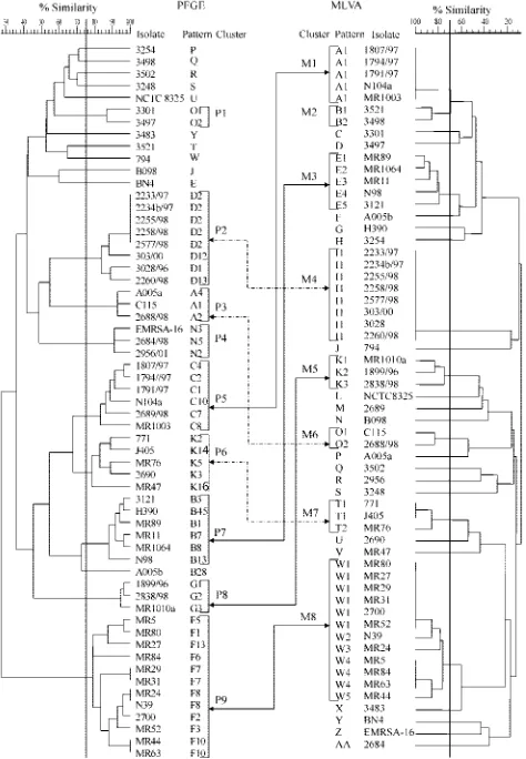
PFGE.Preparation of genomic DNA of the isolates, followed by SmaI (MBI Fermentas, Vilnius, Lithuania) digestion, and separation in a CHEF DR II apparatus (Bio-Rad, Hercules, Calif.) was performed as described previously (5). SmaI PFGE patterns were saved in the TIF format, exported to the Molecular Analyst database (Bio-Rad), and analyzed by using the Dice coefficient-UPGMA (unweighted pair-group method with arithmetic averages). A dendrogram was generated to examine relatedness of PFGE profiles for all study isolates, and cutoff levels of 75 and 92% were applied to this dendrogram (Fig. 1 and 2, respectively). With the 75% cutoff, isolates differing by up to six DNA fragments were clustered together, whereas the 92% cutoff corresponded to the difference of up to three bands within a cluster.
Preparation of total DNA for PCR.Total DNA of the isolates was purified by using the Genomic DNA Prep Plus kit (A&A Biotechnology, Gdynia, Poland) as previously described (25). The purified DNA concentration was estimated with a spectrophotometer (CE3021; Cecil Instruments, Cambridge, United Kingdom), and stock solutions were diluted to a concentration of 5 ng/l. A total of 5 ng of DNA were then used in each PCR.
MLVA typing.MLVA typing was performed as described previously (25). Gel images were exported as TIF files for further analysis by using the Molecular Analyst database. Construction of a dendrogram of banding patterns by using UPGMA was performed by using the Dice coefficient.
spatyping.Amplification of thespagene X region was performed as described previously (27), and amplicons were then sequenced by using an ABI 377 se-quencer (Applied Biosystems, Foster City, Calif.). Thespatypes were deter-mined with the Ridom SpaServer (12). Thespatypes with identical or similar repeat profiles were grouped into clusters according to the method of Koreen et al. (14). Any twospatypes that differed in the number of repeats but contained many identical repeats in common or showed a single deletion of the internalspa
sequence were classified into the same cluster.
MLST.MLST was performed according to protocol described by Enright et al. (8). Sequences of each locus were submitted to the Internet database (www.mlst .net), and resulting allelic profiles were assigned to particular sequence types (ST) for each isolate. START software (13) was used to classify different STs into clusters or clonal complexes (CCs) of phylogenetic relationships. Such clusters were composed of two or more isolates of the same STs or STs which differed at a single locus (single-locus variants) or two loci (double-locus variants) (10).
Calculation of concordance.Intermethod concordance was calculated as the maximum percentage proportion of isolates grouped together into unique pat-terns/profiles or clusters by two methods compared (24).
RESULTS
PFGE. The 59 S. aureus isolates produced 52 PFGE pat-terns. Using a cutoff similarity value of 75%, 48 of the isolates were classified into nine clusters designated from P1 to P9, while 11 isolates had separate positions in the dendrogram
(Fig. 1). The clusters almost perfectly matched PFGE types of the isolates that had been discerned in an earlier study aimed at description of the clonal structure of MRSA in Poland (original designations of the types O, D, A, N, C, K, B, G, and F, respectively), in which a much wider collection of isolates had been used (J. Krzyszton-Russjan, J. Empel, T. Leski, M. Gniadkowski, and W. Hryniewicz, Abstr. 11th International Symposium on Staphylococci and Staphylococcal Infections, abstr. ME-15, 2004). Isolates belonging to the same cluster or PFGE type fulfilled the criteria by Tenover et al. (30). The only exception was isolate A005b, which in the present study was excluded from cluster P7, corresponding to the original PFGE type B. The reason for the latter was that for the present study only a sample of isolates of this type were selected and did not include those that had linked this isolate by pattern similarity with the remaining isolates of the cluster.
[image:2.585.304.541.73.415.2]When the cutoff of 92% was applied, nine PFGE clusters comprising 28 isolates were distinguished and designated from P1⬘ to P9⬘ (Fig. 2). Of the clusters identified with the 75% cutoff, only two clusters, P3 and P8, remained unchanged as clusters P2⬘and P5⬘, respectively. Clusters P2, P5, and P7 were
FIG. 1. PFGE (left) and MLVA (right) dendrograms of the study isolates generated by the UPGMA algorithm. Isolate clusters were delineated with a 75 and 70% similarity cutoff values for PFGE and MLVA, respectively, as indicated by vertical lines. Double arrows connect the corresponding clusters discerned by both methods (differ-ent arrow forms are used only for clarity of visualization).
on May 15, 2020 by guest
http://jcm.asm.org/
reduced by three, four, and one isolate, respectively, into clus-ters P1⬘, P3⬘, and P4⬘. Cluster P9 was split into four smaller ones—P6⬘, P7⬘, P8⬘, and P9⬘—and two unique isolates. Finally, all isolates of clusters P1, P4, and P6 were classified as nonre-lated by PFGE with the raised cutoff value (Fig. 2).
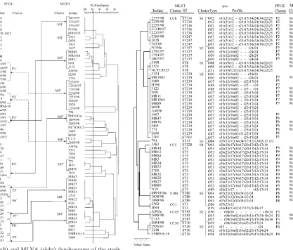
spa typing.Our analysis yielded 20 spatypes among 59S.
aureusisolates, including six new types (t386 to t391) (Fig. 3).
With the use of the criteria by Koreen et al. (14), isolates with similar spa repeat profiles were grouped into four clusters designated S1 to S4. Only a single isolate, isolate BN4 (t159), was classified outside any cluster.
MLST.MLST analysis identified 14 distinct allelic profiles or STs among the isolates, including one, designated ST501 (isolate 3301) (Fig. 3), that has not been described previously in the database at http://saureus.mlst.net/. Ten STs were rep-resented by more than one isolate. Six clusters of closely re-lated STs were distinguished, and these corresponded to S.
aureusclonal complexes CC1, CC5, CC8, CC30, and CC45,
with a cluster of three isolates belonging to ST80 (1, 9, 10). Only one isolate, isolate 794 of ST15, could not be assigned to any of the clusters.
[image:3.585.112.529.68.424.2]MLVA results and their comparison with the PFGE data. (i) Criteria for defining MLVA clusters.MLVA produced 40 dif-ferent DNA banding patterns among 59S. aureusisolates. In order to establish criteria for clustering the patterns into sim-ilarity groups, the MLVA typing results were compared to those obtained by PFGE. With the PFGE similarity cutoff of 75%, a comparable set of MLVA clusters was observed when the cutoff value between two MLVA patterns was set up at the level of 70% (Fig. 1). Isolates belonging to the same cluster differed by up to six bands, and isolates classified into different clusters differed by more than six bands. A total of eight MLVA clusters that comprised 40 isolates were distinguished. Almost all clusters delineated in the PFGE dendrogram had corresponding clusters in the MLVA dendrogram. PFGE clus-ters P2, P8, and P9 were identical to the MLVA clusclus-ters M4, M5, and M8, respectively (Fig. 1). A very good correlation was also observed between PFGE clusters P3, P5, P6, and P7 and MLVA clusters M6, M1, M7, and M3, respectively. The only difference was that MLVA groups were smaller by one or two
FIG. 2. PFGE (left) and MLVA (right) dendrograms of the study isolates. Isolate clusters were delineated with a 92 and 77% similarity cutoff values for PFGE and MLVA, respectively, as indicated by ver-tical lines. Double arrows connect the corresponding clusters discerned by both methods (different arrow forms are used only for clarity of visualization).
FIG. 3. Comparison of PFGE and MLVA results (cutoffs 75 and 70%, respectively) with those obtained by MLST andspatyping. The order in which isolates are listed from top to bottom is based on the MLST dendrogram (left side of the figure). Gaps withinspaprofiles indicate possible deletion/insertion events in thespalocus and were introduced in order to optimize the alignment of the profiles. In PFGE and MLST columns only cluster designations were used; the unique PFGE and MLVA types were not included for clarity.
on May 15, 2020 by guest
http://jcm.asm.org/
isolates than their PFGE counterparts; however, none of the “missing” isolates was clustered by MLVA together with any other isolates (isolate A005a, PFGE cluster P3; 2689/98, PFGE cluster P5; 2690 and MR47, PFGE cluster P6; and H390, PFGE cluster P7). Isolates in PFGE clusters P1 (isolates 3301 and 3497) and P4 (isolates EMRSA-16, 2684/98, and 2956/01) were all classified as unique isolates by MLVA analysis. The only isolates defined as similar by MLVA and nonrelated by PFGE were two isolates of the MLVA cluster M2 (isolates 3521 and 3498) (Fig. 1).
For the nine PFGE clusters defined by the 92% cutoff, a corresponding set of nine MLVA clusters, M1⬘ to M9⬘, was distinguished when the MLVA cutoff level was raised to 77% (Fig. 2). These clusters comprised 39 isolates, and isolates within a single cluster differed from each other by up to four bands. Clusters M1⬘, M2⬘, M3⬘, M4⬘, M6⬘, and M7⬘were iden-tical to those from the MLVA analysis with the 70% cutoff. The original cluster M5 was reduced by one isolate into M5⬘, and cluster M8 was divided into two smaller ones, M8⬘ and M9⬘. All but one PFGE clusters correlated with particular MLVA clusters; however, none of these were identical to its MLVA counterpart. PFGE clusters P1⬘, P2⬘, P3⬘, P4⬘, P5⬘, and P9⬘corresponded to MLVA clusters M4⬘, M6⬘, M1⬘, M3⬘, M5⬘, and M9⬘, respectively, and were usually smaller by one to three isolates than the MLVA ones. Isolates from clusters P7⬘and P8⬘were classified together by MLVA into cluster M8⬘. The only exception was PFGE cluster P6⬘, which had no a corre-sponding MLVA cluster, and its two isolates were split be-tween MLVA clusters M8⬘ and M9⬘ (Fig. 2). On the other hand, isolates of the MLVA clusters M2⬘ and M7⬘ were de-fined as nonrelated by PFGE with the 92% cutoff.
We also searched for a level of variability in PFGE patterns that would always allow classifying any two isolates to corre-sponding clusters by using MLVA and PFGE methods. Any two isolates with either indistinguishable PFGE patterns (the cutoff 92% clusters P1⬘, P7⬘, and P9⬘, and isolates MR24 and N39 of cluster P8⬘) or differing by a single band (cluster P3⬘) were always assigned to a single, separate MLVA cluster. These differed by no more than four bands within the MLVA clusters.
(ii) Comparison of MLVA withspaand MLST clusters.In all cases,S. aureusisolates of a given MLVA cluster, defined by the 70% cutoff value, were grouped together within the same
spacluster and MLST clonal complex or cluster (Fig. 3). More-over, such isolates were usually indistinguishable from each other byspatyping and MLST. Exceptions included isolates 3121 (cluster M3) and N39 (cluster M8) that differed inspa
types from other isolates in their clusters, and isolates MR1003 (cluster M1), and 2260/98, 3028 and 303/00 (cluster M4) that varied with respect to their STs.
Of the four clusters revealed byspatyping, almost each one was split by MLVA into several clusters and singular isolates. The one exception was thespacluster S4 with 13 isolates, 12 of which corresponded to the MLVA cluster M8 and one isolate (isolate 3483) was sorted as unique. Similar observations were made when the six MLST clusters were compared to MLVA clusters. The CC5 complex, corresponding to thespacluster S4, matched precisely the MLVA cluster M8 and the single isolate 3483. Isolate 3483 slightly differed from remaining iso-lates of the group inspa typing and MLST. The only other
MLST clusters that overlapped with MLVA (andspa) clusters were those occurring less often among isolates analyzed: clus-ter ST80 (corresponding to M5 and S3) and CC45 (M6 and the
spa subcluster S2⬘). CC30 was split completely into unique isolates by MLVA, as was the case of CC1 and all other typing methods used in the present study.
Concordance between methods.The concordance values be-tween the typing methods compared are listed in Table 1. The highest levels of correlation were found between the results produced on one hand by MLVA and PFGE (72.9%), and, on the other, byspatyping and MLST (72.9%). Of the remaining combinations of method pairs, a higher correlation level was observed only in the case of MLVA andspatyping (62.7%).
DISCUSSION
In this study we compared the newS. aureustyping method, MLVA (25), with PFGE, MLST, andspatyping on a group of 59 nosocomialS. aureus, mostly MRSA isolates. In contrast to other approaches it is rapid, inexpensive, and easy to use, which is a general characteristic of PCR-based typing tech-niques. DNA banding patterns generated by MLVA are simple and easy to interpret. Moreover, together withspatyping and MLST, MLVA offers the possibility of unambiguous interlabo-ratory comparisons of results.
[image:4.585.301.542.82.169.2]In general, typing techniques with high discriminatory power have a better level of concordance between themselves than with those of lower resolution potential. By analogy, methods with low discriminatory power correlate better with each other than with highly discriminatory techniques. According to this rule we have observed a high level of congruence between PFGE and MLVA on one hand andspatyping and MLST on the other. Comparability of the resolution potential between MLVA and PFGE has already been demonstrated in our ear-lier study (25). What is interesting, however, is that the level of concordance between typing techniques can vary and depends on the collection of isolates used for investigation. With a highly diverse collection of isolates, one may observe a good correlation even between typing techniques which differ greatly in discriminatory power. In this way we can explain the differ-ence in the concordance level between PFGE and MLST ob-tained in the present study (32.2%) and in the analysis per-formed by Grundmann et al. (67%) (11). In these two studies two different types ofS. aureuscollections were used. Whereas our collection included only nosocomial and mostly related
TABLE 1. Correlation between four typing methods forS. aureus
Typing method
% Correlation between patterns/profiles (correlation between clusters)
PFGE MLST MLVA spa
typing
PFGE – – – –
MLST 32.2 (49.1) – – –
MLVA 72.9 (64.4) (39.0)a 44.1 (42.4) – – spatyping 42.4 (49.1) 72.9 (66.1) 62.7 (47.5) –
a
–, Concordance value referring to the comparison of PFGE and MLVA clusters, in which similarity cutoff levels of 92% and 77% were used in the two methods, respectively. In all other cases, comparisons were done with a PFGE cutoff of 75% and an MLVA cutoff of 70%.
on May 15, 2020 by guest
http://jcm.asm.org/
MRSA isolates, Grundmann et al. (11) studied a collection of mainly MSSA isolates circulating in the community.
The concordance level between MLVA and PFGE was re-markably high. The comparison of the results obtained by both methods was performed at two similarity cutoff levels in the PFGE analysis: 75 and 92%. With the PFGE cutoff of 75%, which referred to the interpretation criteria by Tenover et al. (30), isolates that differed by no more than six bands in MLVA (MLVA cutoff of 70%) could be regarded as related in PFGE and therefore classified into the same MLVA cluster. MLVA differentiated all isolates of different PFGE clusters, and al-most all clusters distinguished with one of the methods had their counterparts in the second analysis. Most differences ob-served in PFGE and MLVA clustering were not accidental but could be explained by other observations. Sporadic isolates of PFGE clusters P1, P3, P4, P5, P6, and P7 that were segregated as unique by MLVA differed slightly from each other or from remaining isolates of their clusters also inspatypes (isolates 2689/98, 2690, and A005a), STs (isolates 3301 and 3497), or both (isolates 2684/98 and EMRSA-16). On the other hand, two isolates of the MLVA cluster M2 (isolates 3521 and 3498), which were separated from each other by PFGE, turned out to be the samespatype and ST. The PFGE interpretation criteria by Tenover et al. (30) allow for identifying broader isolate clusters that have been useful in larger-scale epidemiological or phylogenetic studies (6, 15, 16). However, short-term epi-demiological studies aimed mostly at outbreak investigations usually require more stringent criteria, especially in the case of highly clonal organisms, such asS. aureus.
Using the PFGE cutoff of 92%, which reflected up to a three-band difference between two PFGE patterns, it was pos-sible to raise the MLVA cutoff level to 77% in order to obtain the most stringent criteria in MLVA interpretation. Such MLVA clusters grouped DNA patterns that differed by up to four bands from each other. Under these conditions, almost each of the PFGE clusters had a corresponding MLVA cluster, with the only exception of the PFGE cluster P6⬘, the two isolates of which were split into MLVA clusters M8⬘and M9⬘. With the MLVA cutoff of 70%, these two clusters were com-bined into a single, broad M8, and it is possible that with more diverse isolates of this phylogenetic lineage included in the analysis, clusters M8⬘ and M9⬘ would form a single one also under the stringent conditions. The even better results were obtained when the MLVA cutoff 77% clusters were assigned to PFGE clusters that grouped isolates of up to one band differ-ence only. In this case, all PFGE clusters had their MLVA counterparts. All of the observations discussed above suggest that MLVA, with its resolution power and clustering capability comparable to PFGE, may be a good tool for the same appli-cations, especially in outbreak analyses and monitoring short-er-time interhospital spread ofS. aureusstrains.
Lower levels of correlation between MLVA (cutoff, 70%) andspatyping and MLST at the cluster level could be attrib-uted to obvious differences in discriminatory power between these methods; however, isolates of a specific MLVA type were usually of the same spa type and ST. Exceptional cases of variation (isolates 3121 and N39 inspaand isolates MR1003, 2260/98, 3028, and 303/00 in MLST) were still confined to the samespacluster (S2 or S4) or MLST clonal complex or cluster (CC8) and were either due to a single deletion/insertion event
within thespalocus or due to changes in only one or two of the loci analyzed by MLST. The correlation between MLVA and
spatyping at the DNA banding pattern level was especially noteworthy. In order to show dynamics of clone circulation, MLVA and spa typing may be combined, which would de-crease the time and cost of typing procedures and allow for interlaboratory comparisons of results. Since allspatypes cor-responded to specific MLVA patterns, it may be proposed that representatives of MLVA types byspagene may be routinely typed by using partial sequencing. On the other hand, because
spatyping results correlated very well with the MLST data, the representatives of spa types should be good candidates for MLST analysis.
The good agreement observed between MLVA and PFGE on one hand andspatyping and MLST on the other indicates a low level of genetic recombination inS. aureus. Its clonal population structure arises mostly from point mutations (10), which explains the presence of a phylogenetic signal within the
S. aureuspopulation (14). However, long-term evolution may
be also influenced by chromosomal replacements that compli-cate the view of the relationships between distantly related isolates. Clusters revealed byspatyping and MLST have not always corresponded in our study. The largest MLST clonal complex CC8 was also the most diverse with six different STs, including ST8 and ST239. This cluster was split into twospa
clusters, S1 and S2 (with ST8 and ST239 isolates, respectively), of which cluster S2 was also identified in another MLST group, CC30 (with ST30). Isolates of ST8 and ST239 were considered closely related because they were single-locus variants of each other, differing only in thearcC locus. However, analysis of Robinson and Enright, which includedspaand seven surface protein-encoding genes sas, revealed that a large (⬃557-kb) chromosomal fragment containing all of these genes andarcC
was in ST239 identical to that of ST30 and not that of ST8. Therefore, a chromosomal replacement between the ST30 par-ent and the ST8 parpar-ent which resulted in the ST239 mosaic chromosome (23) was most likely responsible for the observed incongruence. The second case was found for thespacluster S3 and the MLST complex CC1 and cluster ST80. The CC1 iso-late 3502 was indistinguishable from isoiso-late BN4 by MLST but different byspatype. On the other hand, itsspatype t386 was closely related to the type t044, which is characteristic for three ST80 isolates (cluster S3). There are no detailed data on ge-nome structure of these clones; therefore, we suggest that a chromosomal replacement encompassing the spa locus, fol-lowed by its mutational changes, could have also occurred in the evolution ofS. aureusCC1 (ST1) clone.
In summary, the main objective of the present study was to compare MLVA with other approaches of typing ofS. aureus
clinical isolates. Its discriminatory power was found to be lower but comparable with PFGE; moreover, both of these methods were highly concordant in terms of discerning clusters of re-lated and probably rere-lated isolates. Therefore, we conclude that MLVA may appear to be useful and reliable in shorter-term epidemiological investigations ofS. aureus. The obvious advantage of this method is that it is PCR based, which makes it available for many laboratories, including hospital-based ones. Its good correlation with methods of lower resolution, such asspatyping, suggests that MLVA may also be useful in indicating clonal representatives for larger-scale analyses
on May 15, 2020 by guest
http://jcm.asm.org/
formed at the Reference Center level. However, this method needs further validation against other typing methods, espe-cially PFGE, with use of other collections ofS. aureusisolates. Studies focused on MSSA isolates would also be of interest.
ACKNOWLEDGMENTS
This study was supported by two grants (grants 2 P04A 001 27 and 2P05D 081 26) from the Polish Committee for Scientific Research (KBN).
REFERENCES
1.Aires de Sousa, M., C. Batzavali, I. Spiliopoulou, I. Santos Sanches, M. I. Criso´stomo, and H. de Lencastre.2003. Two international methicillin-resis-tantStaphylococcus aureusclones endemic in a university hospital in Patras, Greece. J. Clin. Microbiol.41:2027–2032.
2.Aires de Sousa, M., and H. de Lencastre.2004. Bridges from hospitals to the laboratory: genetic portraits of methicillin-resistantStaphylococcus aureus
clones. FEMS Immunol. Med. Microbiol.40:101–111.
3.Bannerman, T. L., G. A. Hancock, F. C. Tenover, and J. M. Miller.1995. Pulsed-field gel electrophoresis as a replacement for bacteriophage typing of
Staphylococcus aureus. J. Clin. Microbiol.33:551–555.
4.Chiou, C. S., H. L. Wei, and L. C. Yang.2000. Comparison of pulsed-field gel electrophoresis and coagulase gene restriction profile analysis techniques in the molecular typing ofStaphylococcus aureus. J. Clin. Microbiol.38:2186– 2190.
5.Chung, M., H. de Lencastre, P. Matthews, A. Tomasz, I. Adamsson, M. Aires de Sousa, T. Camou, C. Cocuzza, A. Corso, I. Couto, A. Dominguez, M. Gniadkowski, R. Goering, A. Gomes, K. Kikuchi, A. Marchese, R. Mato, O. Melter, D. Oliveira, R. Palacio, R. Sa-Leao, I. Santos Sanches, J. H. Song, P. T. Tassios, and P. Villari.2000. Molecular typing of methicillin-resistant
Staphylococcus aureus by pulsed-field gel electrophoresis: comparison of results obtained in a multilaboratory effort using identical protocols and MRSA strains. Microb. Drug Resist.6:189–198.
6.Crisostomo, M. I., H. Westh, A. Tomasz, M. Chung, D. C. Oliveira, and H. de Lencastre.2001. The evolution of methicillin resistance inStaphylococcus aureus: similarity of genetic backgrounds in historically early methicillin-susceptible and -resistant isolates and contemporary epidemic clones. Proc. Natl. Acad. Sci. USA98:9865–9870.
7.Deplano, A., A. Schuermans, J. Van Eldere, W. Witte, H. Meugnier, J. Etienne, H. Grundmann, D. Jonas, G. T. Noordhoek, J. Dijkstra, A. van Belkum, W. van Leeuwen, P. T. Tassios, N. J. Legakis, A. van der Zee, A. Bergmans, D. S. Blanc, F. C. Tenover, B. C. Cookson, G. O’Neil, M. J. Struelens, et al.2000. Multicenter evaluation of epidemiological typing of methicillin-resistantStaphylococcus aureusstrains by repetitive-element PCR analysis. J. Clin. Microbiol.38:3527–3533.
8.Enright, M. C., N. P. Day, C. E. Davies, S. J. Peacock, and B. G. Spratt.2000. Multilocus sequence typing for characterization of methicillin-resistant and methicillin-susceptible clones ofStaphylococcus aureus. J. Clin. Microbiol. 38:1008–1015.
9.Enright, M. C., D. A. Robinson, R. Randle, E. J. Feil, G. Grundmann, and B. G. Spratt.2002. The evolutionary history of methicillin-resistant Staphy-lococcus aureus(MRSA). Proc. Natl. Acad. Sci. USA99:7687–7692. 10.Feil, E. J., J. E. Cooper, H. Grundmann, D. A. Robinson, M. C. Enright, T.
Berendt, S. J. Peacock, J. M. Smith, M. Murphy, B. G. Spratt, C. E. Moore, and N. P. Day.2003. How clonal isStaphylococcus aureus? J. Bacteriol. 185:3307–3316.
11.Grundmann, H., S. Hori, M. C. Enright, C. Webster, A. Tami, E. J. Feil, and T. Pitt.2002. Determining the genetic structure of the natural population of
Staphylococcus aureus: a comparison of multilocus sequence typing with pulsed-field gel electrophoresis, randomly amplified polymorphic DNA anal-ysis, and phage typing. J. Clin. Microbiol.40:4544–4546.
12.Harmsen, D., H. Claus, W. Witte, J. Rothganger, H. Claus, D. Turnwald, and U. Vogel.2003. Typing of methicillin-resistantStaphylococcus aureusin a university hospital setting by using novel software forsparepeat determina-tion and database management. J. Clin. Microbiol.41:5442–5448. 13.Jolley, K. A., E. J. Feil, M. S. Chan, and M. C. Maiden.2001. Sequence type
analysis and recombinational tests (START). Bioinformatics17:1230–1231. 14.Koreen, L., S. V. Ramaswamy, E. A. Graviss, S. Naidich, J. M. Musser, and B. N. Kreiswirth.2004.spatyping method for discriminating among Staph-ylococcus aureusisolates: implications for use of a single marker to detect genetic micro- and macrovariation. J. Clin. Microbiol.42:792–799. 15.Mato, R., F. Campanile, S. Stefani, M. I. Crisostomo, M. Santagati, S. I.
Sanches, and H. de Lencastre.2004. Clonal types and multidrug resistance patterns of methicillin-resistantStaphylococcus aureus(MRSA) recovered in Italy during the 1990s. Mikrob. Drug Resist.10:106–113.
16.McDougal, L. K., C. D. Steward, G. E. Killgore, J. M. Chaitram, S. K. McAllister, and F. C. Tenover.2003. Pulsed-field gel electrophoresis typing of oxacillin-resistantStaphylococcus aureusisolates from the United States: establishing a national database. J. Clin. Microbiol.41:5113–5120. 17.Mulvey, M. R., L. Chui, J. Ismail, L. Louie, C. Murphy, N. Chang, and M.
Alfa.2001. Development of a Canadian standardized protocol for subtyping methicillin-resistant Staphylococcus aureususing pulsed-field gel electro-phoresis. J. Clin. Microbiol.39:3481–3485.
18.Murchan, S., M. E. Kaufmann, A. Deplano, R. de Ryck, M. Struelens, C. E. Zinn, V. Fussing, S. Salmenlinna, J. Vuopio-Varkila, N. El Solh, C. Cuny, W. Witte, P. T. Tassios, N. Legakis, W. van Leeuwen, A. van Belkum, A. Vindel, I. Laconcha, J. Garaizar, S. Haeggman, B. Olsson-Liljequist, U. Ransjo, G. Coombes, and B. Cookson.2003. Harmonization of pulsed-field gel electro-phoresis protocols for epidemiological typing of strains of methicillin-resis-tantStaphylococcus aureus: a single approach developed by consensus in 10 European laboratories and its application for tracing the spread of related strains. J. Clin. Microbiol.41:1574–1585.
19.Oliveira, D. C., A. Tomasz, and H. de Lencastre.2001. The evolution of pandemic clones of methicillin-resistantStaphylococcus aureus: identification of two ancestral genetic backgrounds and the associated mecelements. Microb. Drug Resist.7:349–361.
20.Quelle, L. S., A. Corso, M. Galas, and D. O. Sordelli.2003. STAR gene restriction profile analysis in epidemiological typing of methicillin-resistant
Staphylococcus aureus: description of the new method and comparison with other polymerase chain reaction (PCR)-based methods. Diagn. Microbiol. Infect. Dis.47:455–464.
21.Roberts, R. B., A. de Lencastre, W. Eisner, E. P. Severina, B. Shopsin, B. N. Kreiswirth, A. Tomasz, et al.1998. Molecular epidemiology of methicillin-resistantStaphylococcus aureusin 12 New York hospitals. J. Infect. Dis. 178:164–171.
22.Roberts, R. B., A. M. Tennenberg, W. Eisner, J. Hargrave, L. M. Drusin, R. Yurt, and B. N. Kreiswirth.1998. Outbreak in a New York City teaching hospital burn center caused by the Iberian epidemic clone of MRSA. Mi-crob. Drug Resist.4:175–183.
23.Robinson, D. A., and M. C. Enright. 2004. Evolution ofStaphylococcus aureusby large chromosomal replacements. J. Bacteriol.186:1060–1064. 24.Robinson, D. A., S. K. Hollingshead, J. M. Musser, A. J. Parkinson, D. E.
Briles, and M. J. Crain.1998. The IS1167insertion sequence is a phyloge-netically informative marker among isolates of serotype 6BStreptococcus pneumoniae. J. Mol. Evol.47:222–229.
25.Sabat, A., J. Krzyszton-Russjan, W. Strzalka, R. Filipek, K. Kosowska, W. Hryniewicz, J. Travis, and J. Potempa.2003. New method for typing Staph-ylococcus aureusstrains: multiple-locus variable-number tandem repeat anal-ysis of polymorphism and genetic relationships of clinical isolates. J. Clin. Microbiol.41:1801–1804.
26.Schlichting, C., C. Branger, J. M. Fournier, W. Witte, A. Boutonnier, C. Wolz, P. Goullet, and G. Doring.1993. Typing ofStaphylococcus aureusby pulsed-field gel electrophoresis, zymotyping, capsular typing, and phage typ-ing: resolution of clonal relationships. J. Clin. Microbiol.31:227–232. 27.Shopsin, B., M. Gomez, S. O. Montgomery, D. H. Smith, M. Waddington,
D. E. Dodge, D. A. Bost, M. Riehman, S. Naidich, and B. N. Kreiswirth.1999. Evaluation of protein A gene polymorphic region DNA sequencing for typing ofStaphylococcus aureusstrains. J. Clin. Microbiol.37:3556–3563. 28.Stranden, A., R. Frei, and A. F. Widmer.2003. Molecular typing of
methi-cillin-resistantStaphylococcus aureus: can PCR replace pulsed-field gel elec-trophoresis? J. Clin. Microbiol.41:3181–3186.
29.Tang, Y. W., M. G. Waddington, D. H. Smith, J. M. Manahan, P. C. Kohner, L. M. Highsmith, H. Li, F. R. Cockerill III, R. L. Thompson, S. O. Mont-gomery, and D. H. Persing.2000. Comparison of protein A gene sequencing with pulsed-field gel electrophoresis and epidemiologic data for molecular typing of methicillin-resistantStaphylococcus aureus. J. Clin. Microbiol.38: 1347–1351.
30.Tenover, F. C., R. D. Arbeit, R. V. Goering, P. A. Mickelson, B. E. Murray, D. H. Persing, and B. Swaminathan.1995. Interpreting chromosomal DNA restriction patterns produced by pulsed-field gel electrophoresis: criteria for bacterial strain typing. J. Clin. Microbiol.33:2233–2239.
31.van Belkum, A., J. Kluytmans, W. van Leeuwen, R. Bax, W. Quint, E. Peters, A. Fluit, C. Vandenbroucke-Grauls, A. van den Brule, H. Koeleman, W. Melchers, J. Meis, A. Elaichouni, M. Vaneechoutte, F. Moenes, N. Maes, M. Struelens, F. Tenover, and H. Verbrugh.1995. Multicenter evaluation of arbitrarily primed PCR for typing ofStaphylococcus aureusstrains. J. Clin. Microbiol.33:1537–1547.
32.Wichelhaus, T. A., K. P. Hunfeld, B. Boddinghaus, P. Kraiczy, V. Schafer, and V. Brade.2001. Rapid molecular typing of methicillin-resistant Staphy-lococcus aureusby PCR-RFLP. Infect. Control Hosp. Epidemiol.22:294– 298.