JOURNAL OFCLINICALMICROBIOLOGY, Feb. 2005, p. 818–825 Vol. 43, No. 2 0095-1137/05/$08.00⫹0 doi:10.1128/JCM.43.2.818–825.2005
Copyright © 2005, American Society for Microbiology. All Rights Reserved.
In Vitro Method To Study Antifungal Perfusion in
Candida
Biofilms
Y. H. Samaranayake, J. Ye, J. Y. Y. Yau, B. P. K. Cheung, and L. P. Samaranayake*
Oral Biosciences, Faculty of Dentistry, The University of Hong Kong, Hong Kong, Special Administrative Region, People’s Republic of China
Received 20 October 2003/Returned for modification 11 February 2004/Accepted 5 August 2004
Antimycotic perfusion throughCandidabiofilms was demonstrated by a modification of a simple in vitro diffusion cell bioassay system. Using this model, the perfusion of three commonly used antifungal agents, amphotericin B, fluconazole, and flucytosine, was investigated in biofilms of three differentCandidaspecies (i.e.,Candida albicans,Candida parapsilosis, andCandida krusei) that were developed on microporous filters. Scanning electron microscopy revealed that C. albicans formed a contiguous biofilm with tightly packed blastospores and occasional hyphae compared withC. parapsilosisandC. krusei, which developed confluent biofilms displaying structural heterogeneity and a lesser cell density, after 48 h of incubation on nutrient agar. Minor structural changes were also perceptible on the superficial layers of the biofilm after antifungal perfusion. The transport of antifungals to the distal biofilm-substratum interface was most impeded byC.
albicans biofilms in comparison toC. parapsilosis and C. krusei. Fluconazole and flucytosine demonstrated
similar levels of perfusion, while amphotericin B was the least penetrant through all three biofilms, although the latter appeared to cause the most structural damage to the superficial cells of the biofilm compared with the other antifungals. These results suggest that the antifungal perfusion through biofilm mode of growth in
Candidais dependent both on the antimycotic and theCandidaspecies in question, and in clinical terms, these
phenomena could contribute to the failure ofCandidabiofilm-associated infections. Finally, the in vitro model we have described should serve as a useful system to investigate the complex interactions that appear to operate in vivo within the biofilm-antifungal interphase.
Candida species are opportunistic pathogens which cause superficial and systemic disease in compromised individuals (26).Candida albicansis the main causative agent of oropha-ryngeal candidiasis, while the clinical significance of other non-C.albicansspecies such asCandida parapsilosisand Can-dida kruseiare being increasingly recognized (16, 24, 28). The first step in the pathogenesis of candidal infection entails the adhesion of the yeast to the host surface or an associated prostheses, such as a denture or intravascular catheter material (18, 27, 34). It is thought that the presence of an indwelling denture or a catheter modifies the normal planktonic mode of growth of the yeasts in a yet unknown manner to a sessile biofilm mode of growth, leading to a structured community of cells encased in an extracellular polymer matrix. Once the biofilm mode of growth is established on prostheses such as heart valves and surgical implants (17, 19), the sessile yeasts begin to display unique characteristics, including resistance to antifungal agents (6). This in turn leads to failure of antifungal therapy and chronic infection, remediated by surgery and/or removal of these devices (33). Factors thought to contribute to antimycotic resistance inCandidabiofilms include the growth rate of the yeast (2) and the low diffusion gradient of the antimicrobial through the extracellular polymer matrix of the biofilm (2, 3).
Although there are many reports on the phenomenon of incomplete penetration of antimicrobial agents through bacte-rial biofilms (1, 31, 32, 35), none so far have investigated this
particular feature in Candida biofilms. Therefore, the main objective of the present study was to evaluate qualitatively the antifungal perfusion inCandidabiofilms with a modified, novel assay system. For this purpose, we used a representative isolate of each ofC. albicans,C. parapsilosis, andC. kruseiand three common antifungals: amphotericin B, fluconazole, and flucy-tosine. In addition, ultrastructural features were evaluated by scanning electron microscopy (SEM) to visualize and compare the cellular damage to the superficial layers of the Candida
biofilm before and after antifungal perfusion.
MATERIALS AND METHODS
Candidastrains.ThreeCandidastrains used in this study wereC. albicans
HK1Sa, from a human immunodeficiency virus-infected patient in Hong Kong, andC. parapsilosisATCC 22019 andC. kruseiATCC 6258, from the American Type Culture Collection. The identity of all three strains were reconfirmed by using the API20C AUX system, germ tube formation, and the CHROM agar technique prior to the experiments. All threeCandidastrains were maintained on Sabouraud’s dextrose agar slopes at⫺4°C.
To prepare the yeast inoculum for the development of biofilms, a loopful of the candidal strain under investigation was transferred into 10 ml of liquid yeast nitrogen base (YNB; Difco) supplemented with 50 mM galactose and incubated at 37°C overnight on a rotary shaker at 75 rpm. Cells were harvested and washed with phosphate-buffered saline (PBS; pH 7.2), and a cell suspension of approx-imately 107
CFU/ml was prepared by using a spectrophotometer (at 520 nm). Two types of agar media were used. First, Sabouraud’s dextrose agar supple-mented with 500 mM galactose was used for the development of biofilms, and second, Sabouraud’s dextrose agar supplemented with both 500 mM galactose and the specific antimycotic agent was used for the antifungal penetration studies (see below).
Antifungals.Three antifungals commonly used to treat oropharyngeal and
systemic candidiasis were selected for the study, viz., amphotericin B (Sigma), fluconazole (Pfizer), and flucytosine (Sigma). Three concentrations of the drugs, i.e., 150, 300, and 600g/ml were prepared for each antifungal. Stock solutions of the drugs in dimethyl sulfoxide (amphotericin B), dimethyl formamide (flu-conazole), and sterile distilled water (flucytosine) were prepared prior to use and
* Corresponding author. Mailing address: Dean’s Office, Faculty of Dentistry, University of Hong Kong, 34 Hospital Rd., Hong Kong, SAR, People’s Republic of China. Phone: 852 2859 0480. Fax: 852 2547 6133. E-mail: [email protected].
818
on May 16, 2020 by guest
http://jcm.asm.org/
added to a YNB–500 mM galactose agar solution at a temperature of 40°C for the preparation of the nutrient agar plates, which were then stored at 4°C and used within 1 day.
Development ofCandidabiofilms for antifungal penetration studies.Candida
biofilms were developed on Isopore membrane filters (diameter, 25 mm; pore size, 12m; Millipore) by using a modification of a method described by Anderl et al. (1). A schematic presentation of this method is shown in Fig. 1. At first the filter membranes were sterilized by exposure to UV light (15 min per side), and each membrane was aseptically placed on a Sabouraud’s dextrose agar (500 mM galactose) plate. Afterwards, 50 l of an overnight Candidainoculum (107 CFU/ml) was carefully deposited on the membrane with a pipette. The nutrient agar plates with the inoculum were incubated at 37°C for 1 h (to dry the deposit), and then the plate was inverted and reincubated at 37°C for a period of 48 h. During this incubation period, each filter with the growing biofilm was manually lifted and repositioned on a fresh location on the plate every 10 to 12 h.
After 48 h, the biofilms on the membrane filters were placed on an antibiotic-incorporated agar medium prepared as described above with the biofilm facing outwards. Black microporous membrane filters (diameter, 13 mm; pore size, 0.2
m; Millipore) were then placed on top of 48-h-oldCandidabiofilms. A blank antibiotic (AB) disk (diameter, 9 mm; Difco) moistened with 10l of PBS (pH 7.2) was then placed on top of the 13-mm-diameter membrane (wetting the disk obviates passive capillary perfusion of the antifungal through the biofilm). The whole assembly was then incubated at 37°C for 4 h (Fig. 1).
A semiquantitative evaluation of the antifungals that diffused into an AB disk through the biofilm was performed as follows, withC. parapsilosisas an indicator organism. The disk was removed after the specified incubation time and placed on a fresh RPMI agar plate spread plated with 100l of (planktonic)C. parap-silosis. For this assay,C. parapsilosiswas grown on Sabouraud’s dextrose agar at 37°C, and the growth was diluted with PBS to an optical density of 0.05 (at 520 nm) prior to plating. The control used for the experiment was an AB disk placed on the two-membrane system devoid of a biofilm. Then the RPMI plates were incubated at 37°C for 24 h, and the diameters of the growth inhibition zones (with and without biofilms) were measured by a computerized image analysis system (Quantimet 500 Qwin; Leica, Cambridge, United Kingdom). The assay
was conducted in duplicate on three separate occasions for eachCandidastrain tested.
Standard curves for antifungal concentration versus zone of inhibition.
Stan-dard curves of the drug concentration versus the radius of growth inhibition of the indicator organism for amphotericin B, fluconazole, and flucytosine were constructed by using the methodology described above.
[image:2.585.109.469.73.321.2]FIG. 1. (A) Schematic representation of the experimental system used to monitor antifungal penetration throughCandidabiofilms. The biofilm (a) was developed on a 25-mm-diameter microporous polycarbonate membrane (b) resting on the agar medium. A 13-mm-diameter microporous polycarbonate membrane (c) was placed on top of the biofilm, and then a moistened antibiotic disk (d) was placed on top of the polycarbonate membrane. The entire unit (components a through d) was then transferred to antifungal-laced agar (e). (B) Transverse view of the Isopore filter membrane placed on YNB–500 mM galactose agar and a surface view of the Isopore membrane inoculated with the yeast suspension (method adapted from Anderl et al. [1]).
FIG. 2. Standard curves for the three antifungal agents, showing the relationship between drug concentration and the radius of growth inhibition of a lawn ofC. parapsilosison RPMI agar. 5FC, flucytosine; FL, fluconazole; AmB, amphotericin B.
on May 16, 2020 by guest
http://jcm.asm.org/
[image:2.585.302.544.507.683.2]SEM.For SEM, biofilms were developed on microporous filters for 4, 9, 19, and 24 h, as described earlier. The biofilms were then rinsed in (4% [vol/vol] formaldehyde, 1% [vol/vol] phosphate buffer) and placed in 1% osmium tetrox-ide for 1 h. Samples were subsequently washed in distilled water, dehydrated in a series of ethanol washes (70% for 10 min, 95% for 10 min, and 100% for 20 min), and air dried in a desiccator prior to sputter coating with gold. Afterwards, the surface topographies of biofilms were visualized with a scanning electron microscope (Philips XL30CP) in high-vacuum mode at 10 kV, and the images were processed for display with Photoshop software (Adobe Systems, Inc., Mountain View, Calif.).
Statistics.The Kruskal-Wallis test was used to determine (i) significant
dif-ferences in diffusion of the three concentrations of each of the drugs with each of the threeCandidaspecies and (ii) significant differences between a single
Candidaspecies and a similar concentration of the three respective drugs. The Mann-Whitney U test was used to compare significant differences between the control and each single drug concentration for each of theCandidaspecies.
RESULTS
Methodology. All three Candida species evaluated devel-oped satisfactory biofilms on membrane filters, and the whole assembly could be easily detached from the substrate surface for the diffusion studies. The use of the AB disk with the antifungal and the antifungal sensitivity assay withC. parapsi-losisas the indicator organism yielded reproducible results on repeat experiments conducted on separate occasions, indicat-ing satisfactory sensitivity of the assay system.
Standard curves for antifungal concentration versus zone of inhibition.Standard curves of the drug concentration versus the radius of growth inhibition of the indicator organism for the three antifungals were constructed as shown in Fig. 2. However, these standard curves were curvilinear and hyper-bolic, indicating that the radius of growth inhibition could be used to derive the perfusion of only the very low drug concen-trations. For instance, amphotericin B and flucytosine showed a concentration-dependent difference in the range of 10 to 100
g/ml, while fluconazole exhibited a slightly higher range of 10 to 220g/ml. Therefore, the percent inhibition of drug pene-tration for each experiment was expressed in a semiquantita-TABLE 1. Percent drug penetration of the three antifungals through the three differentCandidabiofilms compared with
biofilm-free controlsa
Candidaspecies Drug
% Penetration (SD) at concn (g/ml):
Pvalueb
150 300 600
C. albicans Amphotericin B 0.00 (0.00) 0.35 (0.05) 2.02 (0.72) 0.027
Fluconazole 55.98 (4.57) 60.80 (6.12) 65.36 (4.42) 0.275
Flucytosine 51.62 (6.94) 59.14 (7.91) 67.86 (6.66) 0.045
C. parapsilosis Amphotericin B 0.00 (0.00) 1.28 (0.27) 1.68 (0.45) 0.027
Fluconazole 63.81 (7.40) 74.64 (2.45) 86.87 (3.86) 0.037
Fluocytosine 67.53 (12.50) 79.27 (12.04) 87.99 (5.59) 0.037
C. krusei Amphotericin B 0.00 (0.00) 1.14 (0.32) 2.02 (0.72) 0.027
Fluconazole 95.99 (12.27) 92.31 (3.08) 91.92 (3.33) 0.346
Flucytosine 87.43 (12.89) 89.92 (12.63) 89.30 (7.59) 0.346
a
Calculations were made, using the radii of growth inhibition zones, of lawns ofC. parapsilosisgrown of RPMI agar. b
[image:3.585.43.543.89.207.2]Kruskel-Wallis H test.
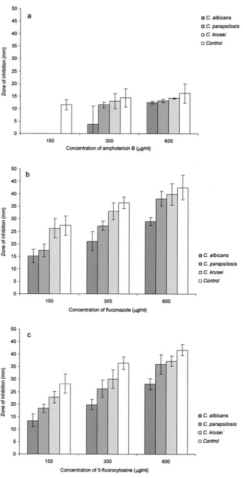
FIG. 3. Penetration of various concentrations (i.e., 150, 300, and 600 g/ml) of the three antifungals, amphotericin B (a), fluconazole (b), and flucytosine (c), through 48-h-oldC. albicans,C. parapsilosis, andC. kruseibiofilms shown in terms of the radii of growth inhibition zones on a lawn ofC. parapsilosison RPMI agar.
820 SAMARANAYAKE ET AL. J. CLIN. MICROBIOL.
on May 16, 2020 by guest
http://jcm.asm.org/
[image:3.585.47.285.259.726.2]tive manner by using data obtained with a corresponding con-trol AB disk without a biofilm (Table 1).
Antifungal perfusion.The variations in antifungal perfusion in the three differentCandidabiofilms for the three antimy-cotics studied were quantified in terms of the zone of inhibition of an indicator organism (C. parapsilosis) on RPMI agar. Sta-tistical analysis revealed significant differences in drug pene-tration through the three differentCandidabiofilms for each of the drug concentrations tested (P⬍0.05) (Fig. 3).
Amphotericin B. When the permeation of amphotericin B through the three differentCandidabiofilms was compared, a statistically significant reduction in drug diffusion was noted for all threeCandida species for the 150-g/ml concentration of the drug (P⬍0.037). However, 300-g/ml amphotericin B was inhibited significantly only byC. albicans(P⬍0.046), while at a higher concentration of this drug, its penetration was signif-icantly inhibited byC. parapsilosis(P⬍0.05) andC. albicans(P
⬍0.046) but not byC. krusei(P⫽0.346) (Fig. 3a).
Fluconazole.The profile of fluconazole penetration through the biofilms was noted to be different than that of either am-photericin B or flucytosine. Thus, fluconazole permeation throughC. albicansbiofilms was significantly lower for all of the tested drug concentrations (P⬍0.05), whileC. parapsilosis
exhibited a significant difference only for 150 and 300g/ml (P
⬍0.05) andC. kruseiexhibited a significant difference only for 150g/ml (P⬎0.05). This implied that theC. albicansbiofilm was the most resistant to fluconazole perfusion. However, with
increasing concentrations of fluconazole, a corresponding sig-nificant increase in drug perfusion (P⬍0.05) was seen with all threeCandidabiofilms (Fig. 3b).
Flucytosine.ForC.albicans, flucytosine did not demonstrate a significant difference in drug perfusion with either a 150-, 300-, or 600-g/ml concentration compared with the control biofilm (P ⫽ 0.05). A similar trend in drug perfusion was observed for bothC.parapsilosisandC.kruseibiofilms. How-ever, as observed for fluconazole, all threeCandida biofilms demonstrated a significant increase in drug perfusion (P ⬍
0.05) with increasing flucytosine concentrations (Fig. 3c). Ultrastructural features.The ultrastructure of biofilm ma-trix observed through SEM varied depending on theCandida
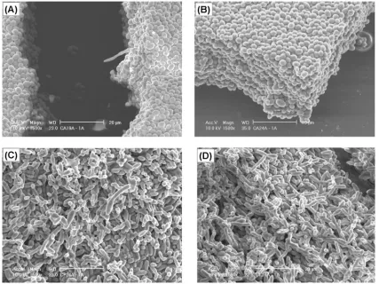
species investigated. Thus,C. albicansproduced a more pro-fuse biofilm relative to the two less commonly pathogenicC. parapsilosisandC. krusei. The latter two species first developed a basal blastospore cell layer sparsely dispersed on the filter surface devoid of either pseudohyphae or hyphae, whereasC. albicansessentially developed into a more contiguous biofilm intimately packed with blastospores and occasional pseudohy-phae after 4 h of growth (Fig. 4A). Also, during the early phase of biofilm growth, C. parapsilosis cells adhered in relatively large numbers compared toC. krusei. These cells then gradu-ally grew and developed a denser structure (at 7 h) mainly composed of noncontiguous small blastospore aggregates and channels in between (Fig. 4B and C). BothC. parapsilosisand
[image:4.585.84.499.73.383.2]C. kruseibiofilms at this stage were less densely packed thanC.
FIG. 4. SEM images of theCandidabiofilms on polycarbonate microporous filters (diameter, 25 mm; pore size, 12m; Millipore). (A)C. albicansat 4 h; (B)C. parapsilosisat 7 h; (C)C. kruseiat 7 h; (D)C. albicansat 7 h.
on May 16, 2020 by guest
http://jcm.asm.org/
albicans(Fig. 4D) and were devoid of extracellular polymeric material.
After 19 h of growth, C. albicans exhibited a multilayer biofilm structure with a few hyphae penetrating the matrix (Fig. 5A). However, even after 24 h, extracellular polymeric material could not be visualized inC. albicansbiofilms, which at this stage was 12 to 15 cell layers thick (Fig. 5B).C. parap-silosis and C. krusei also developed confluent biofilms after approximately 24 h. Characteristically, the latter two species displayed structural heterogeneity with a lesser cell density (Fig. 5C and D) and channels traversing the cell mass thanC. albicans.
Visualization of the ultrastructure in general revealed that the most damage to the biofilm constituents was caused by the highest concentrations of the three antifungals. However, am-photericin B caused the most extensive damage, especially to the most superficial cell layers of all threeCandida biofilms, compared to fluconazole and flucytosine (Fig. 6, 7, and 8). Amphotericin B-treated sessileC.albicanscells demonstrated a range of changes, such as shrinkage of cells, ballooning blas-tospores, ruptured cell walls, and shrunken, wrinkled, or fused
Candida blastospores. This suggests that despite the relative minimal diffusion, amphotericin B, compared with other drugs, may be exerting a fairly severe metabolic interference in Can-dida biofilms. Amphotericin B-perfused C. parapsilosis cells also demonstrated a high degree of destruction with ruptured
and shrunken cells, whileC.kruseiblastospores exhibited the least damage.
The effect of fluconazole perfusion was more subtle. Effects of this drug were barely noticeable withC. albicansbiofilms, whereas a small number of distorted cells were seen in theC. parapsilosisandC. kruseibiofilms, especially on the superficial layers.
DISCUSSION
The development and characterization ofCandidabiofilms on bioprosthetic surfaces that are components of indwelling devices are well documented (5, 13, 14, 23). These studies have shown thatCandidabiofilm development is closely associated with the generation of an extracellular matrix and that mature biofilms show a highly heterogeneous structure and grow vari-ably depending on the topography of the substrate. It has also been shown thatC. albicansforms larger and more complex biofilms on silicone elastomer catheter material thanC. parap-silosis,Candida pseudotropicalisandCandida glabrataand that differences occur between invasive and noninvasive Candida
isolates from infective foci (12, 14).
It is now recognized that antibiotic resistance is a general trait associated with biofilm organisms, including bacteria (21, 22) and fungi (15, 23). Several theories have been proposed to explain this phenomenon, including the growth rate differen-FIG. 5. SEM images showing the biofilm architecture of differentCandidabiofilms on polycarbonate microporous filters (diameter, 25 mm; pore size, 12m; Millipore). (A)C. albicansat 19 h; (B)C. albicansat 24 h; (C)C. parapsilosisat 24 h; (D)C. kruseiat 24 h. Note the lesser cell density of the latter two species than that ofC. albicans.
822 SAMARANAYAKE ET AL. J. CLIN. MICROBIOL.
on May 16, 2020 by guest
http://jcm.asm.org/
[image:5.585.79.506.69.389.2]FIG. 6. SEM images of 48-hC. albicans HK1Sa. (A) ControlC. albicansHK1Sa; (B)C. albicansHK1Sa biofilm exposed to 600-g/ml amphotericin B for 4 h; (C)C. albicans HK1Sa biofilm exposed to 600-g/ml fluconazole for 4 h. Note the wrinkled, shrunk, ruptured, and ballooning effect of the drug on yeast cells.
FIG. 7. SEM images of 48-hC. parapsilosisATCC 22019. (A) Con-trol; (B)C. parapsilosis ATCC 22019 biofilm exposed to 600-g/ml amphotericin B for 4 h; (C)C. parapsilosisATCC 22019 biofilm ex-posed to 600-g/ml fluconazole for 4 h. Note the ruptured and shrunken effect of the drug on yeast cells.
on May 16, 2020 by guest
http://jcm.asm.org/
[image:6.585.43.284.88.658.2]tials (9, 10), a recalcitrant phenotypic state (4), the production of antibiotic-degrading enzymes (11), and the extracellular polymeric material that may act as an adsorbent or a reactant with the antimicrobial (30). Most of the foregoing studies, up to now, have been conducted with bacterial biofilms, and sparse information is available on the diffusion parameters of antimy-cotics inCandidabiofilms. For instance, Baillie and Douglas ob-served that 20 times the MIC of commonly used antifungals such as amphotericin B, fluconazole, or flucytosine is required to cause a significant reduction in cell numbers (2). Further Chandra et al. (6) reported thatC. albicansrequired low MICs of polyenes and fluconazole during the early biofilm development phase. How-ever, during biofilm maturation, they became highly resistant to these drugs. These studies are hampered by the lack of an appro-priately standardized model system to evaluate drug diffusion in candidal biofilms, and the present model was an attempt to ad-dress this issue. The advantages of such an in vitro membrane-supported biofilm system are (i) flexibility to investigate antifun-gal resistance by using simultaneous and parallel biofilm samples, (ii) the accessibility to both sides of the biofilm, after its removal from the membrane surface, and (iii) the possibility of using these biofilms as primitive models of pseudomembranous candidal in-fections. In our hands, we found this system to be relatively sim-ple, inexpensive, and reliable for measurement of the qualitative differences in antifungal perfusion throughCandidabiofilms.
Our SEM studies revealed that the biofilms on micropore filters were distinct from those on polymethyl methacrylate strips or silicone elastomer disks, as reported by previous work-ers (5, 14). For instance, we observed large numbwork-ers of C. albicansmicrocolonies on the filter surface after only 4 h of incubation, unlike the smaller numbers that appeared during a similar incubation period with acrylic strips (5). There were only extremely sparse hyphal forms ofC. albicans in mature filter surface biofilms, in comparison to profuse filamentation seen on polymethyl methacrylate strips (5), silicone elastomers (14), or polyvinyl chloride surfaces (13). This anomaly may be due to strain variation in germ tube and hyphal development (8, 25), and further studies with multipleCandidastrains with the present system are required to confirm our findings.
Upon prolonged incubation,C.albicansbiofilms developed into a dense mass stacked in a palisadic manner devoid of intra-cellular spaces. It is tempting to speculate that this exuberant growth mode may easily retard antifungal penetration into the basal layers of the biofilms, thus protecting them from the drug action. We also noted thatC. albicansbiofilms developed barely discernible matrix material under static incubation on nutrient agar plates, an observation similar to that of Hawser et al. (13).
In addition toC. albicans, we also usedC. parapsilosisandC. kruseistrains for the present pilot studies. The latter, in par-ticular, exhibits intrinsic resistance to azoles, and to our knowl-edge, no one has evaluated its biofilms. Interestingly, theC. parapsilosis biofilms that were developed resembled their counterparts on polymethyl methacrylate disks cultured in YNB medium, where the yeast form was predominant (5). Compared withC. albicans, sparse numbers ofC. parapsilosis
[image:7.585.44.284.94.667.2]and C. krusei microcolonies were seen on the filter surface after 4 h, and these slowly developed into less-dense biofilms, mostly of blastospores, during the ensuing 48-h period. When compared with C. albicans, these biofilms were of reduced thickness, less profuse, and devoid of hyphal elements. FIG. 8. SEM images of 48-hC. kruseiATCC 6258. (A) ControlC.
kruseiATCC 6258; (B)C. kruseiATCC 6258 biofilm exposed to 600-g/ml amphotericin B for 4 h; (C) C. krusei ATCC 6258 biofilm exposed to 600-g/ml fluconazole for 4 h.
824 SAMARANAYAKE ET AL. J. CLIN. MICROBIOL.
on May 16, 2020 by guest
http://jcm.asm.org/
When we evaluated antifungal perfusion in 48-h biofilms, it was noted that amphotericin B was likely to be the least pen-etrant through the biofilms of all threeCandidaspecies com-pared with fluconazole and flucytosine, both of which demon-strated almost a similar degree of drug penetration. However, further kinetic analyses have to be performed to confirm this data.One reason for the foregoing observation could be the large size of the amphotericin B molecule compared with flucy-tosine and fluconazole (20), which may hinder its diffusion. Another could be the hydrophobicity of amphotericin B, which lowers its solubility and, hence, its biofilm perfusion (7). Al-though, of the tested antifungals, amphotericin B was the least penetrant through the threeCandidabiofilms, SEM observa-tions revealed that it caused the most damage to the biofilm surface layer in comparison to fluconazole and flucytosine.
In general, biofilms are encased within an exopolymer matrix which may restrict the diffusion of solutes and also bind anti-microbials. For example, extracellular polysaccharide matrices of bacterial biofilms are known to differentially regulate the diffusion of antibiotics (29). Stewart et al. (30) observed that chlorosulfamate penetrated bacterial biofilms more rapidly than hypochlorite due to a slower reaction rate with biofilm constituents. On the other hand, the ability of an antibiotic to penetrate the biofilm can be severely retarded if the antimi-crobial agent is neutralized by its reactivity with the biofilm (30). These observations suggest that the extent of antibiotic penetration through bacterial biofilms is agent and organism specific, and it is likely that similar conditions may operate in candidal biofilms.
In conclusion, the results of our study indicate that the ultrastructure of differentCandidabiofilms is species specific, varies considerably, and may be affected by the substrate con-tents. The major differences in the spatial configurations of the three differentCandidabiofilms seen in our SEM images tend to suggest that genes encoding biofilm formation may be dif-ferentially regulated during this mode of growth. Finally, the present method appears to be simple and versatile for the study of differential perfusion of antifungals or other solutes through the biofilms of various Candida species. In clinical terms, it is difficult to determine if this type of in vitro biofilm morphology we observed resembles in vivo candidiasis on mu-cosal surfaces, inert catheters, or denture surfaces. Further studies with a large number of clinicalCandidastrains with the proposed method are required to demystify the complexities associated with the biofilm-solute interphase.
REFERENCES
1.Anderl, J. N., M. J. Franklin, and P. S. Stewart.2000. Role of antibiotic
penetration limitation inKlebsiella pneumoniaebiofilm resistance to ampi-cillin and ciprofloxacin. Antimicrob. Agents Chemother.44:1818–1824.
2.Baillie, G. S., and L. J. Douglas.1998. Effect of growth rate on resistance of
Candida albicansbiofilms to antifungal agents. Antimicrob. Agents Che-mother.42:1900–1905.
3.Baillie, G. S., and L. J. Douglas.1999.Candidabiofilms and their
suscepti-bility to antifungal agents. Methods Enzymol.310:644–656.
4.Brown, M. R. W., and P. Gilbert.1993. Sensitivity of biofilms to antimicrobial
agents. J. Appl. Bacteriol.74:87S–97S.
5.Chandra, J., D. M. Kuhn, P. K. Mukherjee, L. L. Hoyer, and M. A.
Ghan-noum.2001. Biofilm formation by the pathogenC. albicans: development,
architecture, and drug resistance. J. Bacteriol.183:5385–5394.
6.Chandra, J., P. K. Mukherjee, S. D. Leidich, F. F. Faddoul, L. L. Hoyer, L. J.
Douglas, and M. A Ghannoum.2001. Antifungal resistance ofCandida
biofilms formed on denture acrylicin vitro.J. Dent. Res.80:903–908.
7.Costerton, J. W.1999. Introduction to biofilm. Int. J. Antimicrob. Agents
11:217–221.
8.Cutler, J. E.1991. Putative virulence factors ofCandida albicans.Annu. Rev.
Microbiol.45:187–218.
9.Evans, D. J., D. G. Allison, M. R. W. Brown, and P. Gilbert.1991.
Suscep-tibility of Pseudomonas aeruginosa andEscherichia colibiofilms towards ciprofloxacin: effect of specific growth rate. J. Antimicrob. Chemother.27:
177–184.
10.Gilbert, P., P. J. Collier, and M. R. W. Brown.1990. Influence of growth rate
on susceptibility to antimicrobial agents: biofilms, cell cycle, dormancy, and stringent response. Antimicrob. Agents Chemother.34:1865–1868.
11.Giwercman, B., E. T. Jensen, N. Hoiby, A. Kharazmi, and J. W. Costerton.
1991. Induction of-lactamase production inPseudomonas aeruginosa bio-films. Antimicrob. Agents Chemother.35:1008–1010.
12.Hawser, S. P., and L. J. Douglas.1994. Biofilm formation byCandidaspecies
on the surface of catheter materials in vitro. Infect. Immun.62:915–921.
13.Hawser, S. P., G. S. Baillie, and L. J. Douglas.1998. Production of
extra-cellular matrix byCandida albicansbiofilms. J. Med. Microbiol.47:253–256.
14.Kuhn, D. M., T. George, J. Chandra, P. K. Mukherjee, and M. A.
Ghan-noum.2002. Comparison of biofilms formed byCandida albicansand
Can-dida parapsilosison bioprosthetic surfaces. Infect. Immun.70:878–888.
15.Kuhn, D. M., T. George, J. Chandra, P. K. Mukherjee, and M. A.
Ghan-noum.2002. Antifungal susceptibility ofCandidabiofilms: unique efficacy of
amphotericin B lipid formulations and echinocandins. Antimicrob. Agents Chemother.46:1773–1780.
16.Levin, A. S., S. F. Costa, N. S. Mussi, M. Basso, S. I. Sinto, C. Machado,
D. C. Geiger, M. C. B. Villares, A. Z. Schreiber, A. A. Barone, and M. L. M.
Branchini.1998.Candida parapsilosisfungemia associated with implantable
and semi-implantable central venous catheters and the hands of healthcare workers. Diagn. Microbiol. Infect. Dis.30:243–249.
17.Mermel, L. A., B. M. Farr, R. J. Sheretz, I. I. Raad, N. O’Grady, J. S. Harris,
and D. E. Craven.2001. Guidelines for the management of intravascular
catheter-related infections. Clin. Infect. Dis.32:1249–1272.
18.Nguyen, M. H., J. E. Peacock, D. C. Tanner, A. J. Morris, M. L. Nguyen,
D. R. Snydman, M. M. Wagener, and V. L. Yu.1995. Therapeutic approaches
in patients with candidemia. Evaluation in a multicenter, prospective, obser-vational study. Arch. Intern. Med.155:2429–2435.
19.Nucci, M., A. L. Colombo, F. Silveira, R. Richtmann, R. Salomao, M. L.
Branchini, and N. Spector.1998. Risk factors for death in patients with
candidemia. Infect. Control Hosp. Epidemiol.19:846–850.
20.Odds, F. C.1988. Candida and candidosis: a review and bibliography, 2nd ed,
p. 288–313. Bailliere Tindall, London, England.
21.Olsen, M. E., H. Ceri, D. W. Morck, A. G. Buret, and R. R. Read.2002.
Biofilm bacteria: formation and comparative susceptibility to antibiotics. Can. J. Vet. Res.66:86–92.
22.Passerini, L., K. Lam, J. W. Costerton, and E. G. King.1992. Biofilms on
indwelling vascular catheters. Crit. Care Med.20:665–673.
23.Ramage, G., K. VandeWalle, S. P. Bachmann, B. L. Wickes, and J. L.
Lopez-Ribot.2002. In vitro pharmacodynamic properties of three antifungal
agents against preformedCandida albicansbiofilms determined by time-kill studies. Antimicrob. Agents Chemother.46:3634–3636.
24.Reichart, P., L. P. Samaranayake, Y. H. Samaranayake, M. Grote, E. Pow,
and B. Cheung.2002. High oral prevalence ofCandidaspecies, especially
Candida krusei, in burnt-out leprosy patients in northern Thailand. J. Clin. Microbiol.40:4479–4485.
25.Richardson, M. D., and H. Smith.1981. Production of germ tubes by virulent
and attenuated strains ofCandida albicans.J. Infect. Dis.144:565–568.
26.Samaranayake, L. P., L. K. Cheung, and Y. H. Samaranayake.2002.
Can-didiasis and other fungal diseases of the mouth. Dermatol. Ther.15:252–270.
27.Samaranayake, L. P., and T. W. MacFarlane.1980. Anin vitrostudy of the
adherence ofCandida albicansto acrylic surfaces. Arch. Oral Biol.25:603– 609.
28.Samaranayake, Y. H., and L. P. Samaranayake.1994.Candida krusei:
biol-ogy, epidemiolbiol-ogy, pathogenicity and clinical manifestations of an emerging pathogen. J. Med. Microbiol.41:295–310.
29.Spoering, A. L., and K. Lewis.2001. Biofilms and planktonic cells of
Pseudo-monas aeruginosa have similar resistance to killing by antimicrobials. J. Bacteriol.183:6746–6751.
30.Stewart, P. S., J. Rayner, F. Roe, and W. M. Rees.2001. Biofilm penetration
and disinfection efficacy of alkaline hypochlorite and chlorosulfamates. J. Appl. Microbiol.91:525–532.
31.Stewart, P. S., and J. W. Costerton.2001. Antibiotic resistance of bacteria in
biofilms. Lancet358:135–138.
32.Suci, P. A., M. W. Mittelman, F. P. Yu, and G. G. Geesey.1994. Investigation
of ciprofloxacin penetration intoPseudomonas aeruginosabiofilms. Antimi-crob. Agents Chemother.38:125–2133.
33.Tunney, M. M., S. P. Gorman, and S. Patrick.1996. Infection associated with
medical devices. Rev. Med. Microbiol.7:195–205.
34.Weems, J. J., Jr.1992.Candida parapsilosis: epidemiology, pathogenecity,
clinical manifestations and antimicrobial susceptibility. Clin. Infect. Dis.14:
757–766.
35.Zheng, Z., and P. S. Stewart.2002. Penetration of rifampin through
Staph-ylococcus epidermidisbiofilms. Antimicrob. Agents Chemother.46:900–903.