Open Access
Research
Comparison of the effect of pressure loading on left ventricular size,
systolic and diastolic function in canines with left ventricular
dysfunction with preserved and reduced ejection fraction
Steven J Lavine*
1,2,3and Donald A Conetta
1,2Address: 1Wayne State University, Detroit, MI 48202, USA, 2University of Florida, Jacksonville, USA and 3Cardiovascular Center, 655 West 8th Street, Jacksonville, FL 32209, USA
Email: Steven J Lavine* - [email protected]; Donald A Conetta - [email protected] * Corresponding author
Abstract
Background: Decompensated heart failure may present with severe hypertension in patients with preserved (PreEF) or reduced left ventricular (LV) ejection fraction (RedEF) and is clinically indistinguishable. Previously, we demonstrated that arterial pressure elevation increases LV filling pressures in a canine model of chronic LV dysfunction with PreEF or RedEF. It is not clear whether any differences in hemodynamics, LV volume or performance, or diastolic function can be demonstrated between canines with PreEF or RedEF in response to arterial pressure elevation. We hypothesized that the LV systolic, diastolic, and hemodynamic response to pressure loading would be similar in RedEF or PreEF.
Methods: We studied 25 dogs with chronic LV dysfunction due to coronary microsphere embolization with RedEF (35 ± 4%) and 20 dogs with PreEF (50 ± 3%). Arterial pressure was increased with methoxamine infusion and hemodynamics and echo-Doppler parameters of LV size, function, transaortic and transmitral pulsed Doppler prior to and with methoxamine infusion was obtained.
Results: Though LV filling pressures were similar at baseline, LV size was larger (p < 0.01) and ejection fraction lower in dogs with RedEF (p < 0.001). With methoxamine, there were similar increases in LV size, LV pressures, and index of myocardial performance with the ejection fraction reduced similarly. Diastolic parameters demonstrated similar tau increases, E/A reduction, and diastolic filling shortening in RedEF and PreEF dogs. A similar extent of isovolumic contraction and relaxation times and index of myocardial performance prolongation occurred with pressure loading.
Conclusion: Pressure loading in a canine model of LV dysfunction with PreEF and RedEF resulted in similar degrees of LV dilatation, increased filling pressures, and increased index of myocardial performance.
Published: 18 November 2008
Cardiovascular Ultrasound 2008, 6:57 doi:10.1186/1476-7120-6-57
Received: 26 July 2008 Accepted: 18 November 2008
This article is available from: http://www.cardiovascularultrasound.com/content/6/1/57
© 2008 Lavine and Conetta; licensee BioMed Central Ltd.
Background
Decompensated heart failure is clinically indistinguisha-ble in patients with either preserved or reduced left ven-tricular (LV) ejection fraction and may be accompanied by elevated arterial pressures. Although systolic dysfunction is often suspected, it is only after noninvasive imaging that the clinician discovers that the ejection fraction may be in the normal range. This may occur in up to 40–50% of patients depending on age and sex and is more common in elderly females with diabetes [1,2]. In addition, in patients with heart failure and LV systolic dysfunction, a significant though lower percentage of patients also have hypertension. Using a chronic canine model of LV dys-function with either preserved or reduced ejection fraction induced by coronary microsphere embolization, we previ-ously demonstrated that acute arterial pressure elevation results in marked elevation of LV filling pressures associ-ated with prolonged relaxation and shortening of the diastolic filling period [3,4].
It is not entirely clear whether any differences in hemody-namics, LV volume or performance, or LV diastolic func-tion can be demonstrated between canines with reduced or preserved LV ejection fraction in response to a stressor (e.g arterial pressure elevation). Although there have been some information in the literature regarding multiple lev-els of LV dysfunction, the model employed has been "tachypacing" induced heart failure, and the heart has been unloaded or contractile performance improved with dobutamine [5,6]. Little data has been generated using the coronary microsphere embolization model which pro-duces a dilated, scarred left ventricle with reduced LV systolic function which has relevance to chronic coronary disease and LV dysfunction [7,8].
We hypothesized that the hemodynamic, LV volume and functional responses to the stress of pressure loading would be similar in a model of chronic LV dysfunction with reduced or preserved LV ejection fraction and would demonstrate similar elevations of LV filling pressures and volumes associated with similar direction and extent of abnormalities in indices describing diastolic function.
Methods
The animals used in this study were maintained in accord-ance with the guidelines of the Committee on Animal Studies at Wayne State University School of Medicine and with the position of the American Heart Association on research animal use. The study was approved by the Wayne State University Animal Investigation Committee. Anesthesia was induced in 45 conditioned mongrel dogs (16–24 kg) with intramuscular morphine sulfate (1.5 mg/ kg) and acepromazine (1.1 mg/kg) followed in 15 min-utes by 30 mg/kg of intravenous ketamine hydrochloride. Maintenance anesthesia was produced by intravenous
morphine sulfate (1.5 mg/kg/hr) and pentobarbital (3 mg/kg/hr). The dogs were intubated and artificially venti-lated with a Harvard respirator using room air. Using fluoroscopic guidance, two 7 F high fidelity catheters (Millar Instruments) were introduced via the right carotid artery and advanced to the left ventricle and ascending aorta. A #8 multipurpose Judkins catheter was introduced through a sheath (Cordis) into the right femoral artery and advanced into the left coronary ostium. Continuous electrocardiographic monitoring was performed using lead II. At held end expiration, ECG, LV pressures, dP/dt, and central aortic pressures were obtained at 100 mm/s using an 8 channel physiologic recorder (Gould). Simul-taneous 2 dimensional echocardiograms and Doppler were obtained from with the use of a phased array echocardiograph (Aloka). Transesophageal 4 and 2 cham-ber view with color flow were obtained from a 5 MHz biplane probe placed in the mid-esophagus with both transaortic and transmitral pulsed Doppler recordings were obtained from the LV outflow tract and from beyond the tips of the mitral leaflets in the left ventricle at 100 mm/s.
Model of LV dysfunction with preserved LV ejection fraction
To test our hypothesis we employed a previously described canine model of LV dysfunction using coronary microsphere embolization [7,8]. In the development of this model, we discovered that the degree of LV dysfunc-tion produced could be titrated based on microsphere number per injection, number of injections, and micro-sphere size. Re-embolization had been required to create models of moderate LV dysfunction when only minimal dysfunction had been previously created. Despite mini-mal systolic dysfunction, there had been remodeling with increased LV volumes, increased LV mass, and mild LV filling pressure elevation despite LV ejection fractions >50% [3,7,9].
instrumented as above. Additional embolizations were performed reducing peak +dP/dT an additional 10–20% or until the LV ejection fraction was <50% (for PreEF) or <40% (for RedEF) following 45 minutes of stabilization. At this time, hemodynamics and Echo-Doppler imaging were repeated as above. This limited embolization approach ultimately leads to a chronic model of LV dys-function and increased LVEDP characterized by patchy myocardial interstitial and replacement fibrosis (7–9) with an ejection fraction in the normal range (>50%) for PreEF or ejection fraction in the moderate dysfunction range (35–40%) for RedEF. Following 45 minutes of sta-ble hemodynamics, the above parameters were repeated. The right carotid and femoral arteries were repaired and the dogs were allowed to recover without any dog suc-cumbing in the PreEF group and 2 dogs in the RedEF group in the 1st 48 hours.
At 8 weeks post coronary microsphere embolization, the animals in both groups were anaesthetized, intubated, ventilated, instrumented, and imaged as above. Atrial pac-ing was instituted at least 5 beats above the baseline rate with a PR interval <160 msec. The above hemodynamics, cardiac outputs, echocardiographic imaging and Doppler recordings were obtained after 10 minutes of steady state pacing. Arterial pressure was increased with a methoxam-ine infusion to increase LV systolic pressure >40 mm Hg. Methoxamine was chosen as it increases arterial pressure without significant change in peak + dP/dT in this model. The above parameters were again obtained. The pacer was turned off and the dogs were permitted to return to their baseline hemodynamic state of chronic LV dysfunction.
Hemodynamic, echocardiographic, and transmitral doppler measurements
For all stages and time periods, LV pressures, dP/dt, car-diac outputs, and aortic pressures were measured from the average of 3 consecutive cycles at held end expiration. Peak LV systolic pressure, LV minimal pressure, and LVEDP were measured. The time constant of LV pressure decline (tau) was calculated using the Weiss method (10). A frame-by-frame assessment of LV volumes using trans-esophageal apical views throughout the cardiac cycle was calculated using the biplane Simpson's rule from the aver-age of 3 determinations. LV foreshortening rarely occurs in the canine as compared to humans. LV end diastolic volume was defined as the largest volume and end systolic volume as the smallest volume. LV ejection fraction was calculated as the difference between end diastolic volume and end systolic volume (stroke volume) divided by end diastolic volume. LV mass was calculated by the area length method. Effective arterial elastance was calculated as LV end systolic pressure/LV end systolic volume.
For all stages and time periods in both groups of dogs, all
Doppler indices were measured from the average of 3 con-secutive cycles at held end expiration. From transmitral Doppler indices, peak rapid filling velocity (E) and peak atrial filling velocity (A) were measured. The rapid filling deceleration time was calculated as the time interval from the peak rapid filling velocity to the time mitral flow decelerated to the zero baseline. The tracing was extrapo-lated to the zero baseline if atrial filling commenced prior to mitral flow fully decelerating to zero. The length of the diastolic filling period was obtained as the interval from beginning to the end of transmitral spectral tracing. When rapid and atrial filling velocity spectra demonstrated any degree of merging, the onset of atrial filling was defined at the point of the end of the p wave on the ECG. The time from the R wave to the end of the mitral time velocity spectrum and to the onset of the mitral time velocity spec-trum (onset of flow) was obtained. The severity of mitral regurgitation was assessed as the ratio of maximal left atrial color flow jet area during systole divided by the simultaneous left atrial area.
Index of myocardial performance
Index of myocardial performance (IMP) is defined as the sum of isovolumic contraction time and isovolumic relax-ation time divided by the LV ejection time [3,4,11]. Using sequential pulse-wave Doppler tracing of the mitral inflow and transaortic outflow, IMP was calculated:
IMP = (a-b)/b
a = The period of time from the end of transmitral velocity spectrum of 1 beat to the onset of the transmitral velocity spectrum of the next beat. b = LVET is time interval from the onset of the pulsed Doppler transaortic spectrum to the end of the transaortic spectrum.
The isovolumic relaxation time (IRT) was measured as R wave to the onset of transmitral velocity spectrum minus the time from the R wave to the end of the aortic spectral tracing. The isovolumic contraction time (ICT) was meas-ured as "a" minus the sum of LV ejection time and IRT.
LV pressure-volume composite plots were constructed for each stage and time period from mean LV pressures and LV volumes obtained throughout the cardiac cycle. An estimate of the operational LV chamber stiffness constant at end diastole was calculated using the approach of Marino, et al [12]. Essentially, the difference between LV pressure minimum and LVEDP was divided by the change in LV volume from the time of LV pressure minimum to end diastole.
blinded to the dates of the studies, name of the dog, and experimental conditions. Intra-observer and inter-observer variability for LV volume was determined by selecting end diastolic and end systolic frames from the echocardiogram of 10 previously studied dogs. Each frame was analyzed 3 weeks apart by 2 observers (see acknowledgement). The average difference for end diasto-lic or end systodiasto-lic volume was 2.2 cc's for intraobserver variability and 3.4 cc's for inter-observer variability.
Statistics
All data was expressed as mean ± standard deviation. Dif-ferences between a variable among stages was assessed using analysis of variance for repeated measures. If the F statistic (p < 0.05) indicated a significant difference, then Tukey's test was utilized to determine where the signifi-cant differences existed. A p < 0.05 was considered signif-icant.
Results
Table 1 1summarizes parameters of LV size, systolic and diastolic function, and LV pressures at baseline and fol-lowing induction of either chronic LV dysfunction with PreEF or RedEF in canines. LV size and mass increased, LV ejection fraction declined, LV filling pressures increased, and tau was prolonged in both groups following the induction of LV dysfunction. LV end diastolic and end systolic volumes were greater (p < 0.01) and the LV ejec-tion fracejec-tion was further reduced in the RedEF group as compared to the PreEF group as per the design (p < 0.001). Despite, the lower ejection fraction and larger LV volumes, LVEDP and LV minimal pressures were similar in both the PreEF and RedEF groups. Transmitral Doppler parameters describing diastolic function revealed a reduc-tion in decelerareduc-tion time, and increases in IRT and ICT in both groups. IMP demonstrated an increase in both groups with a greater increase in the RedEF group (p < 0.05). This was due to a non-significantly greater IRT and ICT in the RedEF group. Mitral regurgitation was noted in 3 dogs with PreEF (minimal or mild in all 3 dogs; jet area/ left atrial area = 4%, 5%, and 9%) and 5 dogs with RedEF (minimal or mild in 5 dogs; jet area/left atrial area = 2%, 5%, 7%, 8%, and 11%)
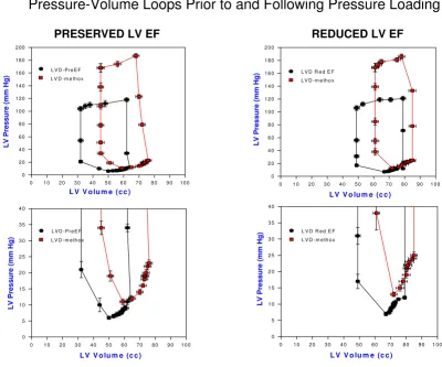
Table 2 1summarizes the results of arterial pressure incre-mentation with methoxamine in both groups. LV vol-umes increased with a reduction of LV ejection fraction in both groups. Stroke volume declined only in the RedEF group. LVEDP and LV minimal pressures, effective arterial elastance and chamber stiffness increased in both groups. Effective arterial elastance was lower in the RedEF group at baseline LV dysfunction (p < 0.05) and with pressure loading (p < 0.05) than in the PreEF group. Figure 1 dis-plays composite LV pressure-volume plots (with mean ± standard error of the mean) prior to and following
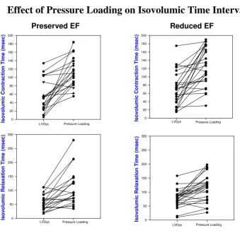
meth-oxamine infusions for PreEF (left) and RedEF (right) groups. Both groups demonstrate a similar rightward and upper shift of the pressure-volume curve from the base-line LV dysfunction plot. LV volumes (p < 0.01) were greater and LV ejection fraction (p < 0.001) were further reduced in the RedEF group than the PreEF group with arterial pressure elevation. Table 3 1summarizes the results of arterial pressure elevation in both groups with regard to diastolic filling parameters, IMP and its compo-nents. For both groups, E and E/A declined, the time to onset of mitral velocity was delayed, and was associated with shortening of diastolic filling, prolongation of IRT and ICT with marked increases in IMP. The IMP value with pressure loading was significantly more elevated in the RedEF group. Figure 2 and 3 summarizes the individ-ual canine response in each group to pressure loading with regard to IMP and diastolic filling period (figure 2) and IRT and ICT (figure 3). The responses of each of these parameters to pressure loading were similar in both groups of dogs. Mitral regurgitation was noted in 5 dogs with PreEF with methoxamine infusion (mild in all; jet area/left atrial area = 6%, 6%, 8%, 10%, and 11%) and 8 dogs with ReEF (mild in all; 6%, 7%, 9%, 11%, 12%, 14%, 15% and 17%)
Table 4 1summarizes the changes in parameters with pres-sure loading in both the PreEF and RedEF groups. The direction, quantity, and percentage (data not shown) change in each parameter with arterial pressure elevation was similar in both canine LV dysfunction groups.
Discussion
In this study we used a chronic canine model of LV dys-function with elevated LVEDP's induced by coronary microsphere embolization with either a PreEF or RedEF to determine whether a stressor to LV performance would produce differences in hemodynamics, LV volume and systolic performance, and diastolic function. Methoxam-ine infusion was administered to increase arterial pressure >40 mm Hg above baseline. The effect of arterial pressure elevation on LV volumes, LV filling pressures, parameters of diastolic function, and IMP were determined.
IMP being larger and ejection fraction lower in the RedEF group prior to pressure loading. Changes in effective arte-rial elastance and operational LV chamber stiffness were similar as were qualitative changes in the composite LV pressure-volume plots. In summary, hemodynamic, LV volume, LV pressures, and diastolic changes were indistin-guishable.
Previous Literature
Using pressure-volume plots, the effect of pressure load-ing on LV dysfunction has been well described both clini-cally and experimentally [4,13]. However, only a modest amount of information is available for various levels of LV dysfunction and only in the model produced by rapid ventricular pacing [5,6]. Rahko [5] demonstrated that the
position of the pressure-volume plot changed with vary-ing levels of LV dysfunction as did the slope of the rela-tion. However, he used inferior vena caval occlusion and the extent of LV dysfunction was not stable for an extended period of time as occurs with coronary micro-sphere embolization [7,8]. Pressure loading in the pacing model might have resulted in similar rightward shifts of the pressure-volume plot (with varying levels of LV dys-function) with similar changes in effective chamber com-pliance as compared to the coronary microsphere model. The expected increases in LV volumes with both levels of LV dysfunction may induce pericardial constraint result-ing in a similar rightward and upward shift in the pres-sure-volume plot [14]. Moe [6] studied the recovery from pacing induced LV dysfunction and demonstrated a
Composite LV pressure-volume plots (mean ± standard error of the mean) at paced LV dysfunction and with peak methoxam-ine (LVD-methox) are shown for canmethoxam-ines with LV dysfunction and preserved LV ejection fraction (LVD-PreEF) on the left and LV dysfunction with reduced ejection fraction (LVD Red EF) on the right
Figure 1
Composite LV pressure-volume plots (mean ± standard error of the mean) at paced LV dysfunction and with peak methoxamine (LVD-methox) are shown for canines with LV dysfunction and preserved LV ejection frac-tion (LVD-PreEF) on the left and LV dysfunction with reduced ejection fraction (LVD Red EF) on the right.
Below are the expanded LV pressure-volume plots truncated at 40 mm Hg to demonstrate diastolic pressure differences more clearly. The pressure-volume curves are shifted rightward and upward for both groups of dogs. LV volumes are greater at both baseline LV dysfunction and with methoxamine in the group with reduced ejection fraction.
PRESERVED LV EF
L V V o lu m e (c c )
0 1 0 2 0 3 0 4 0 5 0 6 0 7 0 8 0 9 0 1 0 0
L
V
Pressu
re (
m
m
Hg
)
0 2 0 4 0 6 0 8 0 1 0 0 1 2 0 1 4 0 1 6 0 1 8 0 2 0 0
L V D -P re E F L V D -m e th o x
L V V o lu m e (c c )
0 1 0 2 0 3 0 4 0 5 0 6 0 7 0 8 0 9 0 1 0 0
LV Pr
es
sure
(
m
m Hg)
0 5 1 0 1 5 2 0 2 5 3 0 3 5 4 0
L V D -P re E F L V D -m e th o x
REDUCED LV EF
Pressure-Volume Loops Prior to and Following Pressure Loading
L V V o lu m e (c c )
0 1 0 2 0 3 0 4 0 5 0 6 0 7 0 8 0 9 0 1 0 0
LV
P
res
s
u
re
(mm Hg)
0 2 0 4 0 6 0 8 0 1 0 0 1 2 0 1 4 0 1 6 0 1 8 0 2 0 0
L V D R e d E F L V D -m e th o x
L V V o lu m e (c c )
0 1 0 2 0 3 0 4 0 5 0 6 0 7 0 8 0 9 0 1 0 0
LV Pressur
e
(mm Hg)
0 5 1 0 1 5 2 0 2 5 3 0 3 5 4 0
downward shift in the velocity of circumferential shorten-ing-end systolic stress plot with recovery. However, this study did not stress the left ventricle. Pressure-volume studies in pacing induced heart failure using afterload augmentation with phenylephrine and dobutamine aug-mentation also demonstrated a reduction in ventricular arterial coupling [15], but the effect of multiple levels of stable LV dysfunction were not addressed. Unfortunately, phenylephrine is also a positive inotrope initially unless beta blockade is used ([16] and personal observations). Using an isolated heart cross-perfused model of LV dys-function previously produced by coronary microemboli-zation, Todaka [17] demonstrated that the end diastolic pressure-volume relation was shifted rightward. However,
multiple levels of LV dysfunction and afterload stress were not employed. Consequently, there is a paucity of studies examining the effects of afterload stress on LV systolic and diastolic performance with more than 1 level of LV dys-function. This study served to help fill this void and points out that though the resting diastolic pressure-volume plots may differ in position in that more severe LV dys-function is shifted rightward, the response to pressure loading is hemodynamically similar. The coronary micro-sphere model of LV dysfunction is uniquely suited to explore this issue as both varying levels of LV dysfunction can be produced for prolonged periods of time [7,8] and responses to pharmacologic interventions can be assessed [18]. Furthermore, this model can also be used to assess
Index of myocardial performance at baseline LV dysfunction (LVDys) and following pressure loading in dogs with LV dysfunc-tion and preserved LV ejecdysfunc-tion fracdysfunc-tion (left upper) and heart failure with reduced ejecdysfunc-tion fracdysfunc-tion (right upper) demonstrates similar extent of increases with pressure loading
Figure 2
Index of myocardial performance at baseline LV dysfunction (LVDys) and following pressure loading in dogs with LV dysfunction and preserved LV ejection fraction (left upper) and heart failure with reduced ejection fraction (right upper) demonstrates similar extent of increases with pressure loading. Also, the diastolic filling period at baseline LV dysfunction and following pressure loading in dogs with LV dysfunction and preserved ejection fraction (left lower) and LV dysfunction with reduced ejection fraction (right lower) demonstrates a similar degree of shortening with pressure loading.
Preserved EF Reduced EF
Effect of Pressure Loading on Index of Myocardial Performance and the Diastolic Filling Period
In
de
x of
My
oc
ard
ial
P
e
rf
o
rman
c
e
0.0 0.5 1.0 1.5 2.0 2.5 3.0
LVDys Pressure Loading
Di
a
s
to
li
c Fil
li
ng Pe
ri
od
(m
s
e
c
)
0 50 100 150 200 250 300 350 400 450 500 550 600
LVDys Pressure Loading
In
de
x of My
oc
a
rd
ia
l Per
fo
rm
a
nc
e
0.0 0.5 1.0 1.5 2.0 2.5 3.0
LVDys Pressure Loading
Di
as
tol
ic
Fil
lin
g Pe
ri
od (m
s
e
c
)
0 50 100 150 200 250 300 350 400 450 500 550 600
the force frequency relationship which may trend down-ward at higher rates with various causes of LV dysfunction [19].
Patients with decompensated heart failure commonly present in the emergency room with marked increases in arterial pressure. It is only after noninvasive evaluation of the LV ejection fraction that clinicians discover that the ejection fraction may be preserved or in the normal range. Heart failure with normal or PreEF has an increased pro-portion of elderly females and associated hypertension with rates of morbidity and mortality that have been described as either lower or equivalent to patients with RedEF [20-23]. It has been difficult to distinguish between PreEF and RedEF clinically in the presentation of acute
decompensated heart failure. Acute decompensated heart failure may present as 1 of 3 presentations: cardiogenic shock, decompensation of chronic heart failure, and pul-monary edema with hypertension [24]. The ADHERE reg-istry demonstrated at least 50% of patients and the Euro Heart Survey II described 34% of patients with decompen-sated heart failure presented with PreEF with lower mor-tality than patients with reduced ejection fraction though subsets of patients (reduced systolic blood pressure and renal insufficiency) had comparable mortality [22,25].
Studies attempting to differentiate PreEF from RedEF at the time of acute decompensation have been limited to surveys and registries. As the pathophysiology of this pres-entation is still not well understood, clinical or
experi-Isovolumic contraction time at baseline LV dysfunction (LVDys) and following pressure loading in dogs with LV dysfunction and preserved ejection fraction (left upper) and LV dysfunction with reduced ejection fraction (right upper) demonstrates similar extent of increases with pressure loading
Figure 3
Isovolumic contraction time at baseline LV dysfunction (LVDys) and following pressure loading in dogs with LV dysfunction and preserved ejection fraction (left upper) and LV dysfunction with reduced ejection fraction (right upper) demonstrates similar extent of increases with pressure loading. Similarly, the isovolumic relaxation period at baseline LV dysfunction (LVDys) and following pressure loading in dogs with LV dysfunction and preserved ejection fraction (left lower) and LV dysfunction with reduced ejection fraction (right lower) demonstrates a similar degree of lengthen-ing with pressure loadlengthen-ing.
Preserved EF Reduced EF
Effect of Pressure Loading on Isovolumic Time Intervals
Is
ov
olu
m
ic
Co
nt
ra
c
tion
Time
(
m
s
e
c
)
0 20 40 60 80 100 120 140 160 180 200
LVDys Pressure Loading
Is
ov
olu
m
ic
C
o
n
tra
c
tion
Tim
e
(ms
e
c
)
0 20 40 60 80 100 120 140 160 180 200
LVDys Pressure Loading
Is
o
v
o
lum
ic
R
e
la
xa
tio
n
Ti
me
(m
s
e
c
)
0 50 100 150 200 250 300
LVDys Pressure Loading Is
ov
ol
u
m
ic
Rel
a
x
a
ti
on
Ti
me (
m
s
ec)
0 50 100 150 200 250 300
mental trials addressing this issue have not yet been performed. As hypertension appears as an important co-morbidity, it stands to reason that it may participate in its pathogenesis. Reviews of therapy for decompensated heart failure have suggested normalizing blood pressure, diuresis, use of either angiotensin converting enzyme inhibitors or angiotensin receptor blockers, beta blockers, aldosterone inhibitors, and possibly nitrates for therapy [26-28]. Many of the above agents are useful in both heart failure with RedEF and PreEF especially agents that pro-mote regression of LV hypertrophy, avoidance of tachycar-dia [29], reduction in interstitial collagen and reduction in the renin angiotensin system activity [30].
Limitations
First, the appropriateness of the coronary microemboliza-tion model for the producmicroemboliza-tion of LV dysfuncmicroemboliza-tion is open to question. As the microsphere model of LV dysfunction results in diffuse fibrotic changes throughout the myocar-dium, its histopathology bears great similarity to the his-topathology seen in hypertensive and diabetic cardiomyopathy [31,32], ischemic cardiomyopathy, and hypertensive heart disease with and without LV dysfunc-tion. The applicability to all patients with heart failure and PreEF (ejection fraction >40%) is open to question. It has applicability to the group of patients with hypertensive heart disease as the etiology of heart failure. Alternatively, the use of a renovascular model of hypertension in aged dogs produces an excellent model (increased myocardial fibrosis) of LV dysfunction with PreEF for testing where hypertensive heart disease is the etiology of heart failure [33]. This study does not address the issue of the appropri-ateness of whether the etiology and pathogenesis of heart failure with PreEF or RedEF should be different. This study is simply assessing the hemodynamic and LV volume and function response to pressure loading in experimental groups based on LV ejection fraction. Certainly, the etiol-ogy may be the same or different for both heart failure with PreEF and RedEF. However, there is similarity of hemodynamic, LV volume, systolic and diastolic responses with PreEF vs RedEF.
Second, one must raise the question whether pressure loading is the appropriate stressor. Earlier studies using volume loading increased LV size and LV filling pressures but lengthened LV ejection time [34]. Patients with LV dysfunction often have increased IMP's due to lengthen-ing of the isovolumic indices and shortenlengthen-ing of LV ejec-tion time [11,35]. Also, pressure loading increases IMP by lengthening the isovolumic indices and insignificantly shortening LV ejection time [3,4], a finding that mirrors the expected findings in a patient with severe LV systolic function. Finally, a substantial number of patients present with pulmonary edema and hypertension who rapidly respond to lowering of their arterial pressure and have
PreEF or RedEF [20,22]. However, methoxamine may not be a pure afterload stressor as 1 study has suggested that it may increase preload to a greater extent than angiotensin II [36]. In addition, an alpha adrenergic agonist may also increase contractility [37] though peak positive dP/dT was unchanged in this study with methoxamine infusion.
Third, as this is an experimental study, the applicability of these findings and relation to humans is always a limita-tion. The general anesthesia utilized may influence the results, and the results may differ with conscious canines. However, this anesthesia regimen has been used in numerous studies in our laboratory and results in lower arterial pressures and LV filling pressures, which may lessen the impact of our intervention. Nevertheless, the experimental data cited here clearly indicates that there is little hemodynamic difference to pressure loading based on LV ejection fraction. This is clearly paralleled clinically by our experience in the emergency room when patients who are severely hypertensive and present with acute decompensated heart failure. There are few clinical cues as to whether the LV ejection fraction is normal or reduced. The importance of blood pressure control may assume even greater importance in patients with hypertensive heart disease and symptoms of heart failure.
Conclusion
Pressure loading in a canine model of LV dysfunction with PreEF or RedEF resulted in similar degrees of LV dilata-tion, increased filling pressures, and increased IMP associ-ated with similar delays in isovolumic relaxation and contraction despite a larger left ventricle and a lower ejec-tion fracejec-tion in the RedEF group.
Abbreviations
LV: left ventricular; PreEF: preserved ejection fraction; LVEDP: left ventricular end diastolic pressure; RedEF: reduced ejection fraction; E: peak rapid filling velocity; A: peak atrial filling velocity; IMP: index of myocardial per-formance; IRT: isovolumic relaxation time; ICT: isovolu-mic contraction time.
Competing interests
The authors declare that they have no competing interests.
Authors' contributions
Additional material
Acknowledgements
We would like to thank the American Heart Association of Michigan for their support. We would like to thank Petar Prcevski D.V.M. and Vicki Johnson for both their invaluable technical assistance and the echocardio-graphic analysis in this investigation
References
1. Kitzman DW: Diastolic heart failure in the elderly. Heart fail Rev
2002, 7:17-27.
2. Vasan RS, Larson MG, Benjamin EJ, Evans JC, Reiss CK, Levy DV:
Congestive heart failure in subjects with normal versus reduced ejection fraction: prevalence and mortality in a pop-ulation based cohort. J Am Coll Cardiology 1999, 33:1948-1955. 3. Lavine SJ, Lavine JA: The effect of acute hypertension on left
ventricular diastolic pressures in a canine model of left ven-tricular dysfunction with preserved ejection fraction and
ele-vated LV filling pressures. J Am Soc Echocardiogr 2006,
19(11):1350-8.
4. Lavine SJ: Index of myocardial performance is afterload
dependent in the normal and abnormal left ventricle. J Am
Soc Echocardiogr 2005, 18:342-50.
5. Rahko PS: Comparative efficacy of three indexes of left ven-tricular performance derived from pressure-volume loops in heart failure induced by tachypacing. J Am Coll Cardiol 1994,
23:209-18.
6. Moe GW, Grima EA, Howard RJ, Seth R, Armstrong PW: Left ven-tricular remodeling and disparate changes in contractility and relaxation during the development of and recovery from experimental heart failure. Cardiovasc Res 1994, 28:66-71. 7. Lavine SJ, Held AC, Prcevski P, Johnson V: Experimental model of
global left ventricular dysfunction secondary to left coronary microembolization. J Am Coll Cardiol 1991, 18(7):1794-1803. 8. Sabbah HN, Stein PD, Kono T, Gheorghiade M, Levine TB, Jafri S,
Hawkins ET, Goldstein S: A canine model of chronic heart fail-ure produced by multiple sequential coronary microembol-izations. Am J Physiol (United States) 1991, 260(4 Pt 2):pH1379. 9. He KL, Dickstein M, Sabbah HN, Yi GH, Maurer M, Wei CM, Wang
J, Burkhoff D: Mechanisms of heart failure with well preserved ejection fraction in dogs following limited coronary micro-embolization. Cardiovasc Res 2004, 64:72-83.
10. Weiss JL, Fredrickson JW, Weisfeldt ML: Hemodynamic determi-nants of the time course of fall in canine left ventricular pres-sure. J Clin Invest 1976, 58:751-760.
11. Tei C, Ling LH, Hodge DO, Bailey KR, Oh JK, Rodeheffer RJ, Tajik AJ, Seward JB: New index of combined systolic and diastolic myo-cardial performance: A simple and reproducible measure of cardiac function-a study in normals and dilated cardiomyop-athy. J Cardiol 1995, 26:357-366.
12. Marino P, Little WC, Rossi A, Barbieri E, Anselmi M, Destro G, Prioli A, Lanzoni L, Zardini P: Can left ventricular diastolic stiffness be measured noninvasively? J Am Soc Echocardiogr 2002:935-943. 13. Grossman W, Braunwald E, Mann T, McLaurin LP, Green LH:
Con-tractile state of the left ventricle in man as evaluated from end-systolic pressure-volume relations. Circulation 1977,
56:845-52.
14. Lavine SJ: Genesis of the restrictive filling pattern: pericardial constraint or myocardial restraint. J Am Soc Echocardiogr 2004,
17:152-60.
15. Prabhu SD: Altered left ventricular-arterial coupling precedes pump dysfunction in early heart failure. Heart Vessels 2007,
22:170-7.
16. Kronenberg MW, McCain RW, Boucek RJ Jr, Grambow DW, Sagawa K, Friesinger GC: Effects of methoxamine and phenylephrine on left ventricular contractility in rabbits. J Am Coll Cardiol
1989, 14:1350-8.
17. Todaka K, Leibowitz D, Homma S, Fisher PE, Derosa C, Stennett R, Packer M, Burkhoff D: Characterizing ventricular mechanics and energetics following repeated coronary microemboliza-tion. Am J Physiol 1997, 272(1 Pt 2):H186-94.
18. Lavine SJ: Effect of changes in contractility on the index of myocardial performance in the dysfunctional left ventricle. Cardiovasc Ultrasound 2006, 4:45.
19. Bombardini T, Gemignani V, Bianchini E, Venneri L, Petersen C, Pas-anisi E, Pratali L, Pianelli M, Faita F, Giannoni M, Picano E: Cardiac reflections and natural vibrations: Force-frequency relation recording system in the stress echo lab. Cardiovasc Ultrasound
2007, 5:42.
20. Maurer KS, King DL, Rumbarger EK, Packer M, Burkhoff D: Left heart failure with a normal ejection fraction: identification of different pathophysiologic mechanisms. J Card Fail 2005,
3:177-187.
21. Redfield MM, Jacobsen SJ, Burnett JC, Mahoney DW, Bailey KR, Rode-heffer RJ: Burden of systolic and diastolic ventricular dysfunc-tion in the community: appreciating the scope of the heart failure epidemic. JAMA 2003, 289:194-202.
22. Yancy CW, Lopatin M, Stevenson LW, De Marco T, Fonarow GC:
ADHERE Scientific Advisory Committee and Investigators. Clinical presentation, management, and in-hospital out-comes of patients admitted with acute decompensated heart failure with preserved systolic function: a report from the Acute Decompensated Heart Failure National Registry (ADHERE) Database. J Am Coll Cardiol 2006, 47:76-84.
23. Danciu SC, Gonzalez J, Ghandi N, Sadhu S, Herrera CJ, Kehoe R:
Comparison of six-month outcomes and hospitalization rates in heart failure patients with and without preserved left ventricular ejection fraction and with and without intraven-tricular conduction defects. Am J Cardiol 2006, 15:256-9. 24. Zannad F, Mebazza A, Juilliere Y, Cohen-Solal A, Guize L, Alla F,
Rouge P, Blin P, Bartlet MH, Palozzi L, Vincent C, Desnos M, Samii K:
EFICA Investigators. Clinical profile, contemporary man-agement and one-year mortality in patients with acute
severe heart failure syndromes: The EFFICA study. Eur J
Heart Fail 2006, 8:697-705.
25. Nieminen MS, Brutsaert D, Dickstein K, Drexler H, Follath F, Harjola VP, Hochadel M, Komajda M, Lassus J, Lopez-Sendon JL, Ponikowski P, Tavazzi L: EuroHeart Survey Investigators; Heart Failure Association, European Society of Cardiology. EuroHeart Failure Survey II (EHFS II): a survey on hospitalized acute heart failure patients: description of population. Eur Heart J
2006, 27:2725-36.
26. Fonarow GC, Heywood JT, Heidenreich PA, Lopatin M, Yancy CW:
ADHERE Scientific Advisory Committee and Investigators. Temporal trends in clinical characteristics, treatments, and outcomes for heart failure hospitalizations, 2002 to 2004: findings from Acute Decompensated Heart Failure National Registry (ADHERE). Am Heart J 2007, 153:1021-8.
27. Dec GW: Management of acute decompensated heart failure. Curr Probl Cardiol 2007, 32(6):321-66.
28. Friedewald VE, Gheorghiade M, Yancy CW, Young JB, Roberts WC:
The editor's roundtable: Acute decompensated heart fail-ure. Am J Cardiol 2007, 99(11):1560-7.
29. Gutierrez C, Blanchard DG: Diastolic heart failure: challenges of diagnosis and treatment. Am Fam Physician 2004, 69:2609-16. 30. Burlew B: Diastolic dysfunction in the elderly. Am J Geriatr
Car-diol 2004, 13:29-38.
31. Fein FS, Capasso JM, Aronson RS, Cho S, Nordin C, Miller-Green B, Sonnenblick EH, Factor SM: Combined renovascular hyperten-sion and diabetes in rats: a new preparation of congestive cardiomyopathy. Circulation 1984, 70:318-20.
32. Factor SM, Bhan R, Minase T, Wolinsky H, Sonnenblick EH, Factor SM, Bhan R, Minase T, et al.: Hypertensive diabetic ardiomyopa-thy in the rat: an experimental model of human disease. Am J Pathol 1981, 102:219-228.
Additional file 1
Click here for file
Publish with BioMed Central and every scientist can read your work free of charge
"BioMed Central will be the most significant development for disseminating the results of biomedical researc h in our lifetime."
Sir Paul Nurse, Cancer Research UK
Your research papers will be:
available free of charge to the entire biomedical community
peer reviewed and published immediately upon acceptance
cited in PubMed and archived on PubMed Central
yours — you keep the copyright
Submit your manuscript here:
http://www.biomedcentral.com/info/publishing_adv.asp
BioMedcentral 33. Munagala VK, Hart CYT, Burnett JC, Meyer DM, Redfield MM:
Ven-tricular structure and function in aged dogs with renal hyper-tension: A model of experimental diastolic heart failure. Circulation 2005, 111:1128-1135.
34. Lavine SJ: Effect of heart rate and preload on the index of myo-cardial performance in the normal and abnormal left ventri-cle. J Am Soc Echocardiogr 2005, 18:133-41.
35. Poulsen SH, Jensen SE, Nielsen JC, Møller JE, Egstrup K: Serial changes and prognostic implications of a Doppler-derived index of combined left ventricular systolic and diastolic myo-cardial performance in acute myomyo-cardial infarction. Am J Car-diol 2000, 85:19-25.
36. Hausdorf G, Gluth NJ, Nienaber CA: Non-invasive assessment of end systolic pressure-length and stress shortening relation-ships in normal individuals: significance of different loading
conditions induced by methoxamine and angiotensin II. Eur
Heart J 1987, 8:1099-1108.
37. Scholz H, Brucker R, Mugge A, Reupcke C: Myocardial alpha-adrenoreceptors and positive inotropy. J Moll Cell Cardiol 1986,