RESEARCH ARTICLE
Two-step analytical procedure
for the characterization and quantification
of metal soaps and resinates in paint samples
Jacopo La Nasa, Anna Lluveras‑Tenorio, Francesca Modugno and Ilaria Bonaduce
*Abstract
Metal soaps and resinates are known to be spontaneously formed in artistic paintings, as a product of the reac‑ tion between aliphatic and terpenoid acids released by hydrolysis and oxidation of the organic media and certain cations contained in some inorganic pigments. In this paper we present an optimization and the validation of a GC/MS method for the qualitative and quantitative analysis of mixtures of terpenoid acids and aliphatic mono and dicarboxylic acids and metal carboxylates of terpenoid and aliphatic mono and dicarboxylic acids in the same paint microsample. This is based on a two‑step analytical approach entailing the subsequent use of two silylating agents, N,O‑bis(trimethylsilyl)trifluoroacetamide for the analysis of free acids and metal carboxylates, and 1,1,1,3,3,3‑hexam‑ ethyldisilazane for the analysis of free acids. The application of this approach is possible because of the good stability at room temperature of the TMS derivatives of aliphatic and terpenoid species, characterized by high boiling points and relatively low vapour pressures. The method was then applied to the characterization of samples collected from two reference paint layers aged for 20 years, a paint sample taken from a pulpit, and sample of the varnish coating of a wooden writing desk, both from the second half of the seventeenth century.
Keywords: Metal soaps, Metal resinates, Free fatty acids, GC/MS, TMS derivatives
© The Author(s) 2018. This article is distributed under the terms of the Creative Commons Attribution 4.0 International License (http://creat iveco mmons .org/licen ses/by/4.0/), which permits unrestricted use, distribution, and reproduction in any medium, provided you give appropriate credit to the original author(s) and the source, provide a link to the Creative Commons license, and indicate if changes were made. The Creative Commons Public Domain Dedication waiver (http://creat iveco mmons .org/ publi cdoma in/zero/1.0/) applies to the data made available in this article, unless otherwise stated.
Introduction
Metal soaps are known to be spontaneously formed in artistic oil paintings, as a product of reaction between the excess of carboxylic acid moieties released by the hydrol-ysis of triglycerides as well as the oxidative cleavage of unsaturated fatty acids, and the metal ions contained in certain pigments [1–8]. Moreover, in manufactured oil paint formulations, since the 19th century, metal soaps (most commonly Zn and Al stearates) have commonly been added to facilitate the dispersion of pigments into the medium [9–11].
Metal carboxylates are also formed between terpenoid acids-deriving from plant resins used in coatings, var-nishes and as ingredients of paint binders, and certain cations, as it has been discussed in several publications [12–17].
Finally, a special class of carboxylates, which are ubiq-uitous to paintings (and other artistic objects), is that of oxalates. These are considered to be the products of reac-tion between oxalic acid, formed upon oxidative deg-radation of the organic medium of a paint, and cations present in the paint film [18–21].
Formation of metal soaps, fatty acid efflorescences, drips, ooze and melting impastoes, and development of solvent sensitivity are important conservation issues in modern oil paintings [22–25]. Their molecular causes, the kinetics of their development, and mechanical impli-cations are still object of open debate among the scien-tific community. The idenscien-tification and quanscien-tification of mobile unbound fractions in paint layers may be impor-tant tools to be used to try to understand these phenom-ena, in the analysis of model samples and paintings.
The identification and quantification of free fatty and dicarboxylic acids, and free carboxylates of fatty and dicarboxylic acids (not bound to the polymeric network,
Open Access
*Correspondence: [email protected]
nor to glycerides) in samples from paintings and other artistic objects can be achieved using a new GC/MS ana-lytical approach, based on the use of two different silylat-ing agents: hexamethyldisilazane (HMDS), which reacts only with free fatty and dicarboxylic acids and N,O -bis(trimethylsilyl)acetamide (BSTFA), which reacts both with free fatty and dicarboxylic acids and of carboxylates of fatty and dicarboxylic acids [26, 27]. The drawback of this analytical approach is related to the sample size: the method entails the separation of the sample into two ali-quots, one to be derivatized with HMDS and the other with BSTFA. This analytical approach can be considered exhaustive if applied on a sample with a good homogene-ity. However, paint samples are often highly heterogene-ous, and the amount of sample available is generally very low, making this approach unpractical in some cases.
To address this limitation, we improved this analytical procedure [26], in order to perform the analysis on one single sample aliquot, in two subsequent steps: after deri-vatisation with HMDS the sample is dried under nitrogen flow, and thus derivatised with BSTFA. In order to eval-uate if this approach is feasible and reliable, as the pro-cedure is aimed at performing quantitative analyses, we performed statistical tests to verify that no sample losses occur.
As the original analytical procedure [26] was only tested on fatty and dicarboxylic acids, we also evaluated the possibility of using this quantitative approach also on terpenoid acids which have different chemical structures than fatty and dicarboxylic acids, and may, thus, exhibit different reactivity towards silylating agents. To this aim, we synthetized lead abietate and we performed a full val-idation of the procedure. Abietic acid and lead abietate exhibit the same reactivity towards HMDS and BSTFA than fatty and dicarboxylic acids: the lead carboxylate is only derivatised with BSTFA and the free terpenoid acid is derivatised by both HMDS and BSTFA. By performing calibration curves, quantitative analyses are thus possi-ble. In general it is possible that soaps between the same carboxylate and a different cation might have a different reactivity. A lower reactivity may be expected when the bond between the carboxylate and the metal is covalent with respect to when it is ionic. As it has been shown that lead carboxylates are characterized by bonds with a substantial covalent character [26, 28], we thus decided investigate into the reactivity of lead soaps.
The procedure was also tested for the derivatisation of oxalates and oxalic acids. Results indicate that oxalates are not derivatised by either derivatising agents, and thus their analysis by GC–MS requires a previous hydrolysis step [29].
This new analytical approach allows to characterize and quantify in one single micro-sample the free acids
and metal carboxylates deriving from the lipid portion and to obtain for the first time a full e quantitative pic-ture of the metal resinates present in the paint sample.
The method was applied to characterise two model paint layers containing a drying oil and a pigment natu-rally aged since 1999, and two historical samples from Norway of the second half of the 17th century: a paint sample taken from a polychrome pulpit and sample of the varnish coating of a wooden writing desk.
Materials and methods Chemicals
Bidistilled water, absolute ethanol, acetone, diethyl ether, n-hexane and iso-octane (HPLC grade, Sigma Aldrich, USA). Lead acetate, manganese chloride, cop-per acetate, cobalt acetate, lead nitrate, and stearic acid (purity > 99%, Sigma-Aldrich).
Abietic acid, hexadecane, tridecanoic, hexade-canoic and octadehexade-canoic acid, (purity 99%), (N,O
-bis(trimethylsilyl)trifluoroacetamide (BSTFA) containing 1% trimethylchlorosilane (TMCS) and
1,1,1,3,3,3-hexamethyldisilazane (HMDS) were all pur-chased from Sigma-Aldrich.
Fatty acid solutions were prepared in acetone, and contained lauric (4.10 µg/g), suberic (4.27 µg/g), azelaic (3.95 µg/g), myristic (4.11 µg/g), sebacic (3.85 µg/g), palmitic acid (4.39 µg/g), oleic acid (6.32 µg/g) and stearic (6.62 µg/g) acids. All standard solutions were used to derive calibration curves. The acids were pur-chased from Sigma-Aldrich, purity > 99%.
Tridecanoic acid (purity 99%; Sigma-Aldrich) solu-tion in iso-octane, 139.91 µg/g, was used as internal standard for derivatization; hexadecane (purity 99%; Sigma-Aldrich), solution in iso-octane,142.00 µg/g, was used as internal standard for injection. Lead stearate and palmitate were synthesized according to the reac-tion condireac-tions reported in [26].
Oxalic acid (purity > 99%, Sigma-Aldrich) solu-tion was prepared in acetone with a concentrasolu-tion of 10 µg/g while calcium oxalate was purchased from Sigma-Aldrich (purity 99.9%).
Lead abietate synthesis
Samples
The analytical procedure was tested on three types of samples (weights ranging between 150 and 650 μg):
• Two reference paint layers provided by Prof Leslie Carlyle, made in 1999 as part of the MOLART Pro-gram [30]. These contained linseed oil as binder and two different pigments, lead white and vine black. Samples were naturally aged and preserved in labora-tory at room temperature and RH%.
• Two historical samples: a paint sample taken from a polychrome pulpit from Norway (sample B53) and sample of the varnish coating of a wooden writing desk (DHU), both from the second half of the seven-teenth century. The presence of pine resin and resin-ates in both samples was previously ascertained by means of GC/MS and FTIR.
Attenuated total reflectance infrared spectroscopy (ATR‑FTIR)
ATR-FTIR spectra were collected using a Perkin Elmer Spectrum 100 spectrometer coupled with a MIRacle TM ATR accessory equipped with a SeZn crystal. Sixteen scans were collected from 4000 to 650 cm−1 with a
reso-lution of 4 cm−1. The analyses were performed at room
temperature. Jasco Spectra Manager software was used for data analysis.
GC/MS analysis of saccharide, proteinaceous and lipid‑resinous materials
The GC–MS system was made up of a 6890 N GC cou-pled with a 5975 Mass Selective Detector single quadru-pole mass spectrometer, equipped with a PTV injector (Agilent Technologies). The mass spectrometer was oper-ated in the electron impact (EI) positive mode (70 eV). The MS transfer line temperature was 280 °C, the MS ion source temperature was kept at 230 °C, and the MS quadruple temperature was at 150 °C. A combined pro-cedure that allows the separation and characterization in the same microsample of three different fractions corre-sponding to saccharide, proteinaceous and lipid-resinous materials was used for the GC/MS analysis 31.
Derivatization of free fatty acids with HMDS
To the samples 5 µL of tridecanoic acid solution was added, and then dried under nitrogen flow at room tem-perature to remove the solvent. The residual was sub-jected to derivatization for the GC/MS analysis with 20 µL of HMDS, 150 µL of iso-octane at 60 °C for 30 min. 5 µL of hexadecane solution were added just before injec-tion as injecinjec-tion internal standard. 2 µL of the resulting solution were injected into the GC/MS.
GC/MS instrumentation consisted of an Agilent Tech-nologies 6890 N Gas Chromatograph coupled with a 5973 Mass Selective Detector single-quadrupole mass spectrometer.
Samples were injected in splitless mode at 280 °C. GC separation was performed on a fused silica capillary col-umn HP-5MS (J&W Scientific, Agilent Technologies, stationary phase 5% diphenyl-95% dimethyl-polysilox-ane, 30 m length, 0.25 mm i.d., 0.25 μm film thickness). Chromatographic conditions were: initial temperature 80 °C, 2 min isothermal, 20 °C/min up to 280 °C, 10 min isothermal.
MS parameters: electron impact ionization (EI, 70 eV) in positive mode; ion source temperature 230 °C; scan range 50–700 m/z; interface temperature 280 °C. The injection volume was 2 µL.
Derivatization of metal soaps and free fatty acids with BSTFA containing 1% TMCS
The mixture obtained from the derivatization with HMDS was dried under nitrogen flow at room tempera-ture, and subsequently added with 20 µL of BSTFA and 150 µL of iso-octane. The reaction time and temperature were set at 80 min and 80 °C. 2 µL of the resulting solu-tion were injected into the GC/MS.
The GC/MS set up is reported in “Derivatization of free fatty acids with HMDS” section.
Quantitative analysis
The GC/MS quantitative analysis of trimethysilyl esters of lauric, suberic, azelaic, myristic, sebacic, palmitic, oleic, stearic, abietic, and oxalic acids obtained after derivatisation with both HMDS and BSTFA was per-formed using calibration curves obtained operating the MS in Select Ion Monitoring (SIM) mode: lauric acid m/z 117–257, suberic acid m/z 149–303, azelaic acid m/z 149–317, myristic acid m/z 117–285, sebacic acid m/Z 149–331, palmitic acid m/z 117–313, oleic acid m/z 117–339, stearic acid m/z 117–341, abietic acid m/z 256– 374, dehydroabietic acid m/z 239–372, oxalic acid m/z 73–147.
Results and discussion
Two‑step derivatization procedure
that it is possible to evaporate under nitrogen flow at room temperature the solution containing HMDS to remove the excess derivatizing agent and the solvent, without affecting trimethylsilyl esters of the analytes of interest.
To confirm this hypothesis, a solution containing tri-decanoic acid trimethylsilyl ester in iso-octane (8.0 µg/g) (obtained after derivatization with HMDS [23]) was ana-lysed in triplicates before and after drying the solution under nitrogen flow and reconstituting the solution with iso-octane. The areas of the chromatographic peaks were compared using a t-test (CI 99%) and resulted to belong to the same statistical population. This proves that dry-ing a solution of trimethylsilyl esters (TMS) of fatty acids under nitrogen flow at room temperature doesn’t cause a loss of TMS esters of fatty acids. This suggests that it is possible to discriminate and quantify free fatty and dicar-boxylic acids and carboxylates of free fatty and dicarbox-ylic acids on the same sample aliquot, by performing:
• A first derivatization step with HMDS to derivatise free carboxylic and dicarboxylic acids;
• GC/MS analysis of the TMS esters produced from free fatty and dicarboxylic acids;
• Drying the residual sample solution under nitrogen flow to remove the excess of HMDS and the solvent; • A second derivatization step with BSTFA to
deriva-tise metal carboxylates;
• GC/MS analysis of the TMS esters produced from both free acids and carboxylates.
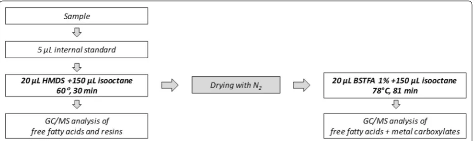
The scheme of the proposed analytical procedure is shown in Fig. 1.
Mixtures of a standard solution of fatty and dicarbox-ylic acids and known amounts (10–40 µg) of lead palmi-tate and stearate were analysed in triplicate according to the scheme described before. Derivatization yields were
calculated based on the content of fatty acid carboxylates in the synthesized metal soaps evaluated as described in paragraph 2.1.
The analytical results are reported in Table 1 and show that the proposed approach can successfully discriminate and quantify mixtures of free fatty and dicarboxylic acids and carboxylates of free fatty and dicarboxylic acids in the same sample aliquot.
Derivatization of abietic acid and lead abietate
The new analytical protocol was then tested in tripli-cate experiments on a solution containing abietic acid (10 µg/g) mixed with lead abietate (10–40 µg), in order to explore the possibility of discriminating and quantifying mixtures of terpenoid carboxylates and acids. Quantita-tive analyses were performed on the basis of calibration curves. Results are shown in Table 1, showing that abietic acid and lead abietate can be quantitatively determined with the described procedure. Limit of detection and quantification were calculated for all the species, obtain-ing values in the range 0.1–0.3 µg/g for the LOD and
Fig. 1 Analytical approach for the characterization of free aliphatic and terpenoid acids, and metal carboxylates by GC/MS analysis
Table 1 Derivatization yields of acids and carboxylates with the two derivatising agents HMDS and BSTFA and relative coefficients of variation (CV %)
Analytes HMDS BSTFA
Derivatization
yield (%) CV% Derivatization yield (%) CV%
Acids Palmitic acid 97 15 100 10
Stearic acid 102 11 100 9
Abietic acid 100 6 100 9
Oxalic acid – – 98 11
Soaps Lead palmitate – – 98 18
Lead stearate – – 100 16
Lead abietate – – 96 6
0.2–0.5 µg/g for the LOQ. Moreover, intra and inter day reproducibility were evaluated, obtaining values lower than 6.0%.
Derivatization oxalic acid and calcium oxalate
The new analytical protocol was tested in triplicate exper-iments on a solution containing oxalic acid (10 µg/g) and samples of calcium oxalate (10–40 µg). The results are reported in Table 1. Oxalic acid is derivatised by BSTFA but not by HMDS, and calcium oxalate is not derivatised by either silylating agents.
Paint samples
Preliminary characterization of the historical samples
The historical samples analyzed are a pictorial sample from Norwegian pulpit and a coating of a writing desk both from the second half of the seventeenth century.
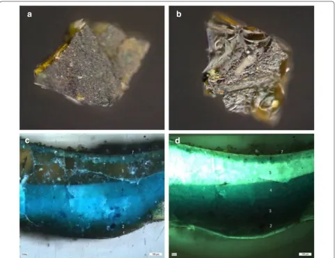
The writing desk belongs to the Museu del Disseny de Barcelona (MADB64160 reference number) and it has been previously analyzed by means of FTIR spectros-copy [33]. The sample showed a dark coating layer of around 50 μm applied on a silver foil on a red bole and a gypsum preparation. The coating layer consisted of a heterogeneous brownish material showing the presence of green particles (Fig. 2a, b). The infrared spectrum of a green area showed the characteristic absorption bands of verdigris (1602, 1444, 692 cm−1), copper II acetate
monohydrate (Cu(CH3COO)2·H2O), while the brownish
matrix showed the characteristic features of a Pinaceae resin (2933, 2872, 1706, 1460, 1386, 1240 cm−1) and the
characteristic COO− antisymmetric stretching band at
1612 cm−1 together with a band at 1400 cm−1 associated
to the symmetric stretching of COO− group of copper
abietate. Copper carboxylates resulted from the reaction
between the abietic acid and abietane skeleton acids from the resin and the copper pigment to produce copper salts.
However, in the 1600 cm−1 regions, absorption bands
of copper carboxylates (both copper acetate from the pig-ment and the salts formed as a result of the paint layer long term chemistry) and the resin C=C stretching bands overlap. A new band formed at 1400 cm−1 has been
asso-ciated to the symmetric stretching of COO− group of
copper abietate and used for identification and imaging of these compounds. The distribution map of verdigris (690 cm−1), Pinaceae resin (1700 cm−1) and copper
abi-etate (1400 cm−1) showed the formation of carboxylates
close to the pigment particles [33].
The sample from the Norwegian pulpit was taken from the blue/green dress from one of the decorative figures. The sample shows a complex build-up (Fig. 2) made of seven pictorial layers from the transparent foundation layer (1) applied on the wood support to a superficial dark layer (7) passing by different green–blue paint layers (2,3,4 and 6) and a thick transparent layer (5). Both the foundation layer (1) and the transparent layer (5) show a white light blue fluorescence under UV light.
A bulk sample of less than 1 mg was analysed by GC/ MS by means of an analytical procedure that allows the analysis of the lipid, resinous and proteinaceous content of the sample [31]. The analysis of the lipid resinous frac-tion of the samples permitted to assess the absence of a lipid material but the presence of abietane and pima-rane acids and their oxidation products indicative of the presence of a diterpenoid resin. The main diterpenoid compounds identified are dehydroabietic acid (DHA) and their oxidation products dehydro-dehydroabietic acid (Di-DHA) and 7-oxo-dehydroabietic acid (7-oxo-DHA) together with other abietane acids. The presence of animal glue was also established from the analy-sis of the proteinaceous fraction due to the presence of hydroxyproline, marker of collagen, though the amount of proteinaceous material in the sample was between the detection and the quantitation limit.
A sample taken from the backside of another figure showing only the thick transparent layer on the wooden support showed the presence of abietane and pimarane acids and their oxidation products (being dehydroabietic acid the most abundant) pointing to the use of a diterpe-noid resin in this layer.
Characterization of free aliphatic and abietane acids and free metal soaps of aliphatic and abietane acids
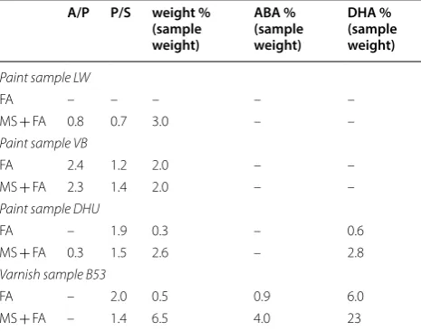
In order to evaluate the performances of the new analyti-cal method when analysing real paint matrices, including pigments and the cross-linked network of the polymer-ised paint, samples of increasing complexity were ana-lysed. Table 2 reports the relative amounts of free acids
(FA weight%), determined by derivatization with HMDS as the sum of the content of the quantified silyl esters of aliphatic mono and dicarboxylic acids normalized for the sample weight, and the relative content of free acids and their relative carboxylates (MS + FA weight%), determined by derivatization with BSTFA, as the sum of the content of the quantified silyl esters of aliphatic mono and dicarboxylic acids normalised for the sample weight. The relative content of the silyl esters of abietic acid (ABA%) and dehydroabietic acid (DHA%), as deter-mined after derivatization with HMDS (FA) and BSTFA (MS + FA) are reported as well.
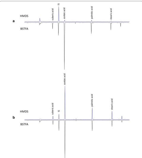
The chromatograms obtained in the analysis of the two model paint layers, one containing linseed oil and lead white (LW) and another containing linseed oil and vine black (VB), are reported in Fig. 3.
These model paints were previously analysed with the original analytical procedure [26], entailing the derivati-zation of two sample aliquots. The data obtained with the new approach, reported in Table 2, are in accordance with the previously published results [26] (t-test, CI 99%).
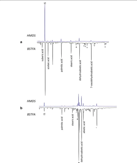
The two historical samples previously described were thus analysed: the paint sample from a Norwegian pulpit (B53) and the coating of a writing desk (DHU), both from the second half of the seventeenth century.
The chromatograms obtained for the two samples are reported in Fig. 4.
The main compounds identified in the chromatograms relative to the FA fractions of both samples correspond to diterpenoid compounds deriving from Pinaceae resin (pimaric, abietic, dehydroabietic and didehydroabietic acid).
Both samples show significant differences between the FA and MS + FA fractions, highlighted by the higher rela-tive amount of silyl esters of aliphatic and of terpenoid
Table 2 GC/MS data relative to the paint samples analysed
FFA free acid, MS metal salt, A/P azelaic acid/palmitic acid, P/S palmitic acid/ stearic acid, ABA abietic acid, DHA dehydroabietic acid
A/P P/S weight % (sample weight) ABA % (sample weight) DHA % (sample weight)
Paint sample LW
FA – – – – –
MS + FA 0.8 0.7 3.0 – –
Paint sample VB
FA 2.4 1.2 2.0 – –
MS + FA 2.3 1.4 2.0 – –
Paint sample DHU
FA – 1.9 0.3 – 0.6
MS + FA 0.3 1.5 2.6 – 2.8
Varnish sample B53
FA – 2.0 0.5 0.9 6.0
acids in the MS + FA fraction. This indicates the pres-ence, in both samples, of metal carboxylates of fatty acids (palmitic and stearic mainly) and of diacids (suberic, azelaic and sebacic acid), and also of abietic and pimaric
The Pinaceae resin shows different molecular pat-terns in the two samples. The sample from the desk var-nish (DHU) contains mostly dehydroabietic acid and its oxidation product 7-oxo-dehydroabietic acid. The pres-ence of copper carboxylates in this sample was evident based on the FTIR spectra [33]. Copper carboxylates result in this case from the reaction between the abie-tane acids from the Pinaceae resin present and the cop-per pigment, verdigris, a copcop-per acetate [33].
The paint sample from the pulpit (B53) shows instead several peaks corresponding to pimarane and abietane compounds, among which the most abun-dant are pimaric and dehydroabietic acid (DHA). The relative amount of DHA with respect to its oxidation products, as well as the presence of pimaradiene com-pounds such as pimaric and isopimaric acids, point to a well-preserved Pinaceae resin, which had undergone to relatively little oxidation. This suggests the use of the resin in a non-superficial finishing layer of the complex sample stratigraphy: e.g. the foundation layer directly applied onto the wood support below the pigmented paint layers, or the thick brownish layer among paint layers. The presence of a consistent amount of resinates in the sample from the pulpit (B53) points to the for-mation of carboxylates due to the reaction of the abi-etane and pimarane acids from the resin from one or more unpigmented layer with the metal ions present in the adjacent pigmented paint layers, indicating the abil-ity of the paint constituents to migrate through the dif-ferent strata.
Conclusions
In this work we presented an optimisation of a recently developed analytical approach, based on the sequential use of two different silylating reactions for the GC/MS analysis of mixtures of free aliphatic and terpenoid acids (transformed in the correspondent TMS ester by reaction with HMDS) and their free metal soaps (transformed in the correspondent TMS ester by reaction with BSTFA).
This new analytical protocol can be applied on a single sample aliquot, which is an important requirement for a technique used to characterise precious and unique sam-ples from works of art. This analytical approach proved also capable of characterising the molecular components of diterpenoid resins, and their products of reaction with metal cations present in the same or adjacent layers.
The analytical results showed that this procedure is a useful tool to obtain a comprehensive picture of the molecular composition of the non-polymerised/esteri-fied components of a paint sample, and to contribute to the study of the molecular changes undergone by the organic constituents of a paint layer upon ageing.
Authors’ contributions
JLN and ALT performed the analyses. JLN, ALT and IB performed the data elaboration. JLN, FM, ALT and IB planned the experimental set‑up and wrote the manuscript. All authors read and approved the final manuscript..
Acknowledgements
Authors with to thank Donatella Banti, Aviva Burnstock, Klaas Jan van den Berg, Bronwyn Ormsby and all CMOP project members for the useful discussions.
Authors wish to thank Catharina Blaensdorf and Barbara Wapler (Technical University of Munich) and Nati Salvado (Universitat Politècnica de Catalunya) for kindly providing the samples from the from the polychrome Norwegian pulpit and the coating from a 17th century a writing desk, respectively. The
“Museu del Disseny de Barcelona” is acknowledged for allowing the access to the desk. Prof. Leslie Carlyle (REQUIMTE, Lisbon, Portugal) is thanked for pro‑ viding the lead white and vine black paint layer samples analyzed in this work.
Moreover, this work has received funding from University of Pisa project “Analytical chemistry for the knowledge of materials and techniques in mod‑ ern and contemporary art” (PRA_2016_13).
Competing interests
The authors declare that they have no competing interests.
Availability of data and materials
On request, the authors will gladly share other additional files and data not published in the article.
Funding
This work was performed within the context of the JPI CMOP project: “Clean‑ ing of modern oil paints” (Heritage Plus Joint Call Project 2015e2018).
Publisher’s Note
Springer Nature remains neutral with regard to jurisdictional claims in pub‑ lished maps and institutional affiliations.
Received: 7 July 2018 Accepted: 21 September 2018
References
1. Boon, JJ, Hoogland F, Keune K. Chemical processes in aged oil paints affecting metal soap migration and aggregation. In: AIC paintings spe‑ cialty group postprints; american institute for conservation of historic & artistic works: providence, Rhode Island, vol. 19; 2007. p. 16–23. 2. Boon JJ, van der Weerd J, Keune K, Noble P, Wadum J. Mechanical and
chemical changes in Old Master paintings: dissolution, metal soap formation and remineralization processes in lead pigmented ground/ intermediate paint layers of 17th century paintings. In: Vontobel R, eds. 13th triennial meeting of the ICOM committee for conservation in Rio De Janeiro Preprints, vol. 406. London: James & James; 2002. p. 401. 3. Cotte M, Checroun E, De Nolf W, Taniguchi Y, De Viguerie L, Burghammer
M, Walter P, Rivard C, Salomé M, Janssens K, Susini J. Lead soaps in paint‑ ings: friends or foes? Stud Conserv. 2017;62:2–23.
4. Cotte M, Checroun E, Susini J, Dumas P, Tchoreloff P, Besnard M, Walter P. Kinetics of oil saponification by lead salts in ancient preparations of pharmaceutical lead plasters and painting lead mediums. Talanta. 2006;70:1136–42.
5. Keune K, van Loon A, Boon JJ. SEM backscattered‑electron images of paint cross sections as information source for the presence of the lead white pigment and lead‑related degradation and migration phenomena in oil paintings. Microsc Microanal. 2011;17:696–701.
6. Robinet L, Corbeil MC. The characterization of metal soaps. Stud Conserv. 2003;48:23–40.
8. Osmond G, Keune K, Boon J. A study of zinc soap aggregates in a late 19th century painting by RG Rivers at the Queensland Art Gallery. AICCM Bull. 2005;29:37–46.
9. Osmond G. Zinc white: a review of zinc oxide pigment properties and implications for stability in oil‑based paintings. AICCM Bull. 2012;33:20–9. 10. Tumosa CS. A brief history of aluminum stearate as a component of paint,
vol. 23. New York: Waac newsletter; 2001.
11. Izzo FC, van den Berg KJ, van Keulen H, Ferriani B, Zendri E. Modern oil paints—formulations, organic additives and degradation: some case studies. In: van den Berg KJ, Burnstock A, de Keijzer M, Krueger J, Learner T, de Tagle A, Heydenreich G, editors. Issues in contemporary oil paint. Cham: Springer; 2014. p. 75–104.
12. Poli T, Piccirillo A, Nervo M, Chiantore O. Interactions of natural resins and pigments in works of art. J Colloid Interface Sci. 2017;503:1–9.
13. Doménech‑Carbó MT, Kuckova S, de la Cruz‑Cañizares J, Osete‑Cortina L. Study of the influencing effect of pigments on the photoageing of terpe‑ noid resins used as pictorial media. J Chromatogr A. 2006;1121:248–58. 14. Arbizzani R, Casellato U, Fiorin E, Nodari L, Russo U, Vigato PA. Decay
markers for the preventative conservation and maintenance of paintings. J Cultu Herit. 2004;5:167–82.
15. Altavilla C, Ciliberto E. Copper resinate: an XPS study of degradation. Appl Phys A. 2006;83:699–703.
16. Gunn M, Chottard G, Rivière E, Girerd J‑J, Chottard J‑C. Chemical reac‑ tions between copper pigments and oleoresinous media. Stud Conserv. 2002;47:12–23.
17. Poli T, Piccirillo A, Zoccali A, Conti C, Nervo M, Chiantore O. The role of zinc white pigment on the degradation of shellac resin in artworks. Polym Degrad Stab. 2014;102:138–44.
18. Zoppi A, Lofrumento C, Mendes NFC, Castellucci EM. Metal oxalates in paints: a Raman investigation on the relative reactivities of different pig‑ ments to oxalic acid solutions. Anal Bioanal Chem. 2010;397:841–9. 19. Alessandrini G. The oxalate films: origin and significance in the conserva‑
tion of works of art. Milan: Centro Congressi Cariplo; 1989.
20. Otero V, Vilarigues M, Carlyle L, Cotte M, De Nolf W, Melo MJ. A little key to oxalate formation in oil paints: protective patina or chemical reactor? Photochem Photobiol Sci. 2018;17:266–70.
21. Sotiropoulou S, Sciutto G, Tenorio AL, Mazurek J, Bonaduce I, Prati S, Mazzeo R, Schilling M, Colombini MP. Advanced analytical investigation on degradation markers in wall paintings. Microchem J. 2018;139:278–94. 22. Van den Berg KJ, Burnstock A, De Keijzer M, Krueger J, Learner T, De Tagle A, Heydenreich G. Issues in contemporary oil paint. New York: Springer; 2014.
23. Lee J, Bonaduce I, Modugno F, La Nasa J, Ormsby B, van den Berg KJ. Scientific investigation into the water sensitivity of twentieth century oil paints. Microchem J. 2018;138:282–95.
24. Lee J, Ormsby B, Burnstock A, van der Berg KJ. The chemical characterisa‑ tion of water sensitive modern oil paint swatches by Winsor & Newton. In: ICOM CC Copenhagen Congress, 2017.
25. Baij L, Hermans JJ, Keune K, Iedema P. Time‑Dependent ATR‑FTIR Spectro‑ scopic Studies on Fatty Acid Diffusion and the Formation of Metal Soaps in Oil Paint Model Systems. Angewandte Chemie. 2018;57:7351–4. 26. La Nasa J, Modugno F, Aloisi M, Lluveras‑Tenorio A, Bonaduce I. Develop‑
ment of a GC/MS method for the qualitative and quantitative analysis of mixtures of free fatty acids and metal soaps in paint samples. Anal Chim Acta. 2018;1001:51–8.
27. Banti D, La Nasa J, Tenorio AL, Modugno F, Jan van den Berg K, Lee J, Ormsby B, Burnstock A, Bonaduce I. A molecular study of modern oil paintings: investigating the role of dicarboxylic acids in the water sensi‑ tivity of modern oil paints. RSC Adv. 2018;8:6001–12.
28. Burrows HD, Lobo VM. 3 potentiometric study on the interactions between divalent cations and sodium carboxylates in aqueous solution. Progress in organic and physical chemistry: structures and mechanisms, vol. 23. New York: Academic Press; 2013.
29. Rampazzi L, Andreotti A, Bonaduce I, Colombini MP, Colombo C, Toniolo L. Analytical investigation of calcium oxalate films on marble monu‑ ments. Talanta. 2004;63:967–77.
30. Carlyle L. Molart Fellowship Report: historical reconstructions of artists’s oil paint: an investigation of oil processing methods and the use of medium‑modifiers. Report no 72894; Canadian Conservation Institute, 2000; vol. Report no 72894.
31. Lluveras A, Bonaduce I, Andreotti A, Colombini MP. GC/MS analytical pro‑ cedure for the characterization of glycerolipids, natural waxes, terpenoid resins, proteinaceous and polysaccharide materials in the same paint microsample avoiding interferences from inorganic media. Anal Chem. 2010;82:376–86.
32. Sigma‑Aldrich: Derivatization reagents: for selective response and detec‑ tion in complex matrice. 2011. https ://www.sigma aldri ch.com/conte nt/ dam/sigma ‑aldri ch/migra tionr esour ce4/Deriv atiza tion%20Rgt s%20bro chure .pdf. Accessed Sept 2018.
33. Beltran V, Salvadó N, Butí S, Cinque G. Micro infrared spectroscopy dis‑ crimination capability of compounds in complex matrices of thin layers in real sample coatings from artworks. Microchem J. 2015;118:115–23. 34. Degano I, La Nasa J. Trends in high performance liquid chromatography
for cultural heritage. In: Mazzeo R, editor. Analytical chemistry for cultural heritage. Cham: Springer International Publishing; 2017. p. 263–90. 35. Blanco‑Zubiaguirre L, Ribechini E, Degano I, La Nasa J, Carrero JA, Iñañez
J, Olivares M, Castro K. GC–MS and HPLC–ESI–QToF characterization of organic lipid residues from ceramic vessels used by Basque whalers from 16th to 17th centuries. Microchem J. 2018;137:190–203.
36. Degano I, La Nasa J, Ghelardi E, Modugno F, Colombini MP. Model study of modern oil‑based paint media by triacylglycerol profiling in positive and negative ionization modes. Talanta. 2016;161:62–70.
37. La Nasa J, Zanaboni M, Uldanck D, Degano I, Modugno F, Kutzke H, Tveit ES, Topalova‑Casadiego B, Colombini MP. Novel application of liquid chromatography/mass spectrometry for the characterization of drying oils in art: elucidation on the composition of original paint materials used by Edvard Munch (1863–1944). Anal Chim Acta. 2015;896:177–89. 38. Carlesi S, Ricci M, Cucci C, Nasa JL, Lofrumento C, Picollo M, Becucci
M. Multivariate analysis of combined fourier transform near‑infrared spectrometry (FT‑NIR) and raman datasets for improved discrimination of drying oils. Appl Spectrosc. 2015;69:865–76.
39. La Nasa J, Degano I, Modugno F, Colombini MP. Industrial alkyd resins: characterization of pentaerythritol and phthalic acid esters using integrated mass spectrometry. Rapid Commun Mass Spectrom. 2014;29:225–37.