R E V I E W
Open Access
Spinal cord stimulation in chronic pain:
evidence and theory for mechanisms
of action
Jacob Caylor
1, Rajiv Reddy
1, Sopyda Yin
1, Christina Cui
1, Mingxiong Huang
3,4, Charles Huang
4,5, Ramesh Rao
6,
Dewleen G. Baker
2,7, Alan Simmons
2,7, Dmitri Souza
8, Samer Narouze
8, Ricardo Vallejo
9,10,11and
Imanuel Lerman
1,2,4,6,12*Abstract
Well-established in the field of bioelectronic medicine, Spinal Cord Stimulation (SCS) offers an implantable, non-pharmacologic treatment for patients with intractable chronic pain conditions. Chronic pain is a widely heterogenous syndrome with regard to both pathophysiology and the resultant phenotype. Despite advances in our understanding of SCS-mediated antinociception, there still exists limited evidence clarifying the pathways recruited when patterned electric pulses are applied to the epidural space. The rapid clinical implementation of novel SCS methods including burst, high frequency and dorsal root ganglion SCS has provided the clinician with multiple options to treat refractory chronic pain. While compelling evidence for safety and efficacy exists in support of these novel paradigms, our understanding of their mechanisms of action (MOA) dramatically lags behind clinical data. In this review, we reconstruct the available basic science and clinical literature that offers support for mechanisms of both paresthesia spinal cord stimulation (P-SCS) and paresthesia-free spinal cord stimulation (PF-SCS). While P-SCS has been heavily examined since its inception, PF-SCS paradigms have recently been clinically approved with the support of limited preclinical research. Thus, wide knowledge gaps exist between their clinical efficacy and MOA. To close this gap, many rich investigative avenues for both P-SCS and PF-SCS are underway, which will further open the door for paradigm optimization, adjunctive therapies and new indications for SCS. As our understanding of these mechanisms evolves, clinicians will be empowered with the possibility of improving patient care using SCS to selectively target specific pathophysiological processes in chronic pain.
Keywords:Spinal cord stimulation, Biomarker, Neurophysiology, Chronic pain, Complex regional pain syndrome,
Failed back surgery syndrome, Mechanisms of action, Neuropathic pain, Objective measures, Neuroinflammation
Background
Chronic Pain is a heterogenous, complex syndrome with significant burden for both the patient and the healthcare system. While the advent of multi-modal and multidiscip-linary treatment approaches has improved strategies for chronic pain management, the push for opiate-free therapies has inspired development of novel approaches to both nociceptive and neuropathic pain syndromes. The
gate-control theory of pain proposed by Melzack and Wall in 1965 spurred development of conventional Spinal Cord Stimulation (SCS), first surgically implanted in 1967 by Shealy, who noted that paresthesia elicited by electrical stimulation of the dorsal columns (DC) inhibited deep pain due to metastatic lung cancer (Melzack & Wall,
1965; Shealy et al.,1967). This groundbreaking work into
the field of bioelectronic medicine ultimately opened the door for SCS and the rise of targeted neuromodulation.
SCS has been successfully utilized over the last half century in the adjunctive treatment of refractory pain syndromes not amenable to conservative therapy. Though the gate-control theory initially postulated a re-duction in nociceptive pain in response to SCS, clinical
© The Author(s). 2019Open AccessThis article is distributed under the terms of the Creative Commons Attribution 4.0 International License (http://creativecommons.org/licenses/by/4.0/), which permits unrestricted use, distribution, and reproduction in any medium, provided you give appropriate credit to the original author(s) and the source, provide a link to the Creative Commons license, and indicate if changes were made. The Creative Commons Public Domain Dedication waiver
(http://creativecommons.org/publicdomain/zero/1.0/) applies to the data made available in this article, unless otherwise stated.
* Correspondence:[email protected]
1
Department of Anesthesiology, Center for Pain Medicine, University of California San Diego School of Medicine, La Jolla, CA, USA
2VA Center of Excellence for Stress and Mental Health, VA San Diego
Healthcare System, La Jolla, CA, USA
experience demonstrated that patients with neuropathic and other rarer pain syndromes also received benefit. Current indications for SCS include Failed Back Surgery Syndrome (FBSS), Chronic Regional Pain Syndrome (CRPS), neuropathic pain, visceral abdominal pain and intractable angina pectoris (Lindblom &
Meyer-son, 1975; Linderoth & Foreman, 2017). While
paresthesia-based SCS (P-SCS) now represents the traditional approach to neuromodulation of these dysregulated pain pathways, novel SCS paradigms and new anatomical targets have rapidly entered clinical use in the field of bioelectronic medicine. These include Dorsal Root Ganglion stimulation (DRG-S) as well as the paresthesia-free SCS (PF-SCS) paradigms: Burst SCS (B-SCS) and High Frequency SCS (HF-SCS). An additional PF-SCS paradigm, Evoked Compound Action Potential SCS (ECAP-SCS) has recently been developed, utilizing closed-loop monitoring to improve charge delivery to spinal targets. Together, the rapid clinical implementation of these novel paradigms has outpaced the bandwidth of the preclinical sciences to decipher the mechanisms and recruited pathways responsible for their efficacy. While some work has been completed, the trajectory of this ascension has left a sizeable knowledge gap between the basic sciences and clinical research,
necessitating a review of evidence and theory for the mechanisms of action of these devices. In this review, we will 1) elucidate pertinent dysregulated pathways respon-sible for chronic pain syndromes, 2) identify the neuro-anatomical targets thought to be modulated by SCS paradigms, 3) discuss proposed mechanisms of P-SCS, PF-SCS, DRG-S, and ECAP-PF-SCS, and 4) present and evaluate the preclinical and clinical evidence in the context of these proposed mechanisms. Collectively, this review serves to recognize gaps in current knowledge and identify poten-tially rich avenues for future investigation.
Parameters for spinal cord stimulation
Prescribed by the device generator, the implanted SCS
leads deliver a charge “dose” to the target tissue. This
generates a local electric field (EF). Neural bodies, synapses and axons which project through the EF
may be modulated (Miller et al., 2016). While leads
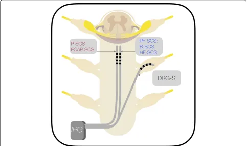
from P-SCS and PF-SCS enter the epidural space and deliver charge to the midline structures, DRG-S leads enter the epidural space, exit the neuroforamina and
deliver charge to the adjacent DRG (Fig. 1) (Miller et
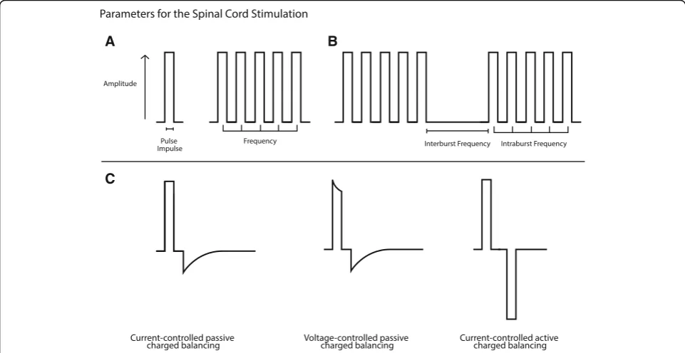
al.,2016). The fundamental unit of a SCS program is the
pulse, which is defined by the amplitude, pulse width (PW)
and delivery waveform (Fig. 2, Panel a). By convention,
frequency describes the number of pulses within one
sec-ond (Miller et al., 2016). B-SCS utilizes a novel firing
pat-tern, wherein stacked pulses are delivered followed by a period of quiescence which is then repeated. Thus, B-SCS can also be defined by intra-burst frequency and inter-burst
frequency (Fig. 2, Panel b) (Chakravarthy et al., 2018a).
Parameters for SCS charge delivery are reviewed in Fig.2.
Lead impedance represents the inherent resistance of tissue to changes in charge. In SCS, the impedance is determined by the interaction between the lead contact and target, which may vary in an acute or chronic man-ner. Acute alterations in impedance reflect alterations in the anatomic relationship of the lead to the DC. These include lead migration, spine position such as lumbo-thoracic extension or abrupt changes in cerebrospinal fluid (CSF). Also caused by CSF volume expansion, local tissue fibrosis and scarring are thought to contribute more significantly to chronic elevations in impedance
(Abejon & Feler, 2007). Interestingly, Kramer et al.
(2015) noted these impedance changes are mitigated
with close lead contact and use of the relatively
immo-bile DRG-S (Kramer et al., 2015). The charge delivery
strategy determines whether voltage or current will be
fixed in relation to the system’s impedance (Fig.2, Panel
c). In current-controlled (CC) systems, the current is set; variations in impedance cause a change in voltage. In voltage-controlled (VC) systems, the voltage is set; varia-tions in impedance cause a change in current (Miller et
al.,2016; Schade et al.,2010). These systems will deliver
a pulse with either constant voltage or constant current for a specified period of time (PW). The repolarization waveform is determined by the charge-balancing (CB)
strategy (Fig.2, Panel c) (Miller et al.,2016). Active CB
creates a symmetric, biphasic pulse. Passive CB yields an asymmetric pulse. In either CB strategy, the initial pulse is followed by equal and opposite current movement to return the net charge to baseline; this avoids buildup of charge in the tissue, which may lead to injury if allowed
to accumulate (Miller et al.,2016).
In response to a single pulse, axons and cell bodies within the EF exhibit a spectrum of modulation including no change, sub-threshold depolarization or generation of a mature action potential (AP). Intermittent, repeated pulses can induce summative APs. The susceptibility of fibers to depolarization is dependent on fiber thickness, myelination and distance from the lead. This susceptibility
A B
C
is summarized by each fiber’s strength-duration curve. As the total charge delivery of each pulse is the product of amplitude and PW, this curve represents total charge
delivery necessary for AP generation (Abejón et al.,2015;
West & Wolstencroft,1983). Central to the perception of
paresthesia in P-SCS, threshold depolarization of a DC Aβ
fiber generates an AP, which may travel in an orthodromic
and antidromic manner (West & Wolstencroft, 1983;
Holsheimer, 2002). The coverage and intensity of
paresthesia produced by P-SCS are determined by the amplitude and PW. Higher amplitude currents or voltages provide stronger paresthesia. Increasing the PW leads to greater fiber recruitment and increases the distribution of
paresthesia (Holsheimer,2002; Lee et al.,2011; Hershey et
al.,2010). Frequency also plays a role in determining PW,
as they are inversely related: as frequency increases, the
available PW decreases. Additionally, as neurons
intrinsically have maximal firing frequencies due to refrac-tory periods, providing an overdriving stimulus would not allow every threshold pulse to generate an AP. Neuromo-dulation can occur without generating a paresthesia when the generated pulse is below the AP threshold of the fiber, as in PF-SCS paradigms that utilize properties of the strength-duration curve. The shape of this inverse hy-perbolic curve can be exploited such that very large quan-tities of charge can be delivered without generating a paresthesia-evoking AP, especially at the extremes of PW
and amplitude (Miller et al., 2016; Hershey et al., 2010).
Subthreshold depolarizations by a weak EF are postulated to modulate neural networks by causing firing desyn-chrony, AP inhibition and changes in the resting
mem-brane potential (Miller et al.,2016). Novel PF-SCS such as
HF-SCS utilizes a charge delivery strategy such that a single, limited DC AP is generated only when PF-SCS is initiated, though no paresthesia is reported by the patient. These paradigms likely provide analgesia through novel mechanisms which remain to be elucidated and will be
reviewed in their respective sections (Crosby et al.,2016).
Despite advances in P-SCS and PF-SCS program settings, the common basis for classification of SCS remains frequency, with the exception of DRG-S which is an anatomic specification, and ECAP-SCS, which is a novel
program (Miller et al., 2016). While this makes device
categorization simple in colloquial contexts, in reality all pulse parameters must be considered to influence the
physiologic response to SCS (Miller et al., 2016). Below,
we will discuss the dysregulated pain pathways thought to be modulated by the charge delivery of SCS.
Mechanisms of hyperalgesia and allodynia
Pain is a context-dependent sensory or emotional experience associated with actual or potential damage to tissue. A peripheral nociceptive stimulus is perceived via pathways projecting to cortical and subcortical regions,
resulting in the conscious and affective pain experience. Chronic pain is a product of the dysregulation in pain processing in response to peripheral or central injury. Serving to guide clinical therapies, preclinical models attempt to recreate these dysregulated pain pathways, further our understanding of antinociceptive mecha-nisms and have spurred the development of novel SCS paradigms. Animal models of hyperalgesia and allodynia include chronic constriction injury (CCI), spinal nerve ligation (SNL) and spared nerve injury (SNI); these models generate an incomplete nerve injury represen-tative of typical human pathology, in opposition to
complete transection models (Todd, 2010). SNI and
SNL animals develop tactile allodynia within the first day, whereas CCI animals develop allodynia over the course of one week. The target of the CCI and SNI models are the sciatic nerves whereas SNL targets the
L4/L5 spinal nerves (Todd, 2010). Measuring paw
withdrawal thresholds (PWT) to evaluate pain behavior before and after intervention, animal models are routinely employed in the assessment of SCS paradigms and elucida-tion of their respective mechanisms. At the crux of chronic pain pathogenesis is central sensitization (CS), which the
IASP Task Force defines as “increased responsiveness of
nociceptive neurons in the central nervous system (CNS)
to normal or subthreshold afferent input” (Sandkuhler,
2010; Taxonomy ITFo, 1994). Peripheral injury leads to
pathologic activation of post-synaptic nociceptive projec-tion neurons (PN) and has become the focus of
re-search in animal models (Sandkuhler, 2009).
SCS-mediated pain relief is thought to act through modulation of the maladaptive aggregate response to local injury, neu-roinflammation and CS at both the segmental and supraspinal levels, processes which are hallmarks of hyperalgesia and allodynia. The ligand-receptor pairs of interest to the process of CS include: 1) Glutamate, which
activates N-methyl-D-aspartate (NMDA),
amino-3-hydroxy-5-methyl-4-isoxazole propionate (AMPA), and
metabotropic glutamate (mGLU) receptors (Todd, 2010;
Latremoliere & Woolf,2009), 2) Substance P (SP), which
activates Neurokinin-1(NK1) Receptors (Latremoliere &
Woolf, 2009), 3) Brain-derived neurotrophic factor
(BDNF) which activates Tropomyosin receptor kinase B
(TrkB) receptor (Latremoliere & Woolf, 2009), 4)
Cal-citonin Gene-related Peptide (CGRP) which activates
CGRP Receptor, 5) Gamma-aminobutyric Acid
(GABA) which activates GABA Receptors, including
the GABA A (GABAA) Receptor and GABA B
(GABAB) Receptor and 6) Glycine (Gly) which activates
the Gly Receptor (GlyR).
inhibition, neural plasticity and phenotypic
transform-ation of low-threshold Aβ afferents (Todd, 2010).
Windup represents the short-term, reversible temporal summation of slow C-fiber activation of NK-1 and CGRP receptors, leading further to NMDA receptor ac-tivation and prolongation of progressive membrane
depolarization (Latremoliere & Woolf, 2009). Though
windup is commonly ascribed to the pathogenic process of CS, LTP produces durable activation changes of the NK1-positive PN. Repetitive stimulation of NK1 positive
neurons leads to synaptic amplification through a Ca2+
-dependent mechanism (Latremoliere & Woolf, 2009;
Ikeda et al., 2006). Implicated in CS, repetitive
stimula-tion leads to elevastimula-tions in intracellular calcium.
Activa-tion of Ca2+-dependent protein kinases leads to receptor
ionophore phosphorylation and decreased potassium currents, resulting in increased receptor efficacy and po-tentiation of AMPA, NMDA and NK1 signaling
(Sand-kuhler, 2009; Latremoliere & Woolf, 2009). Similar to
excitatory LTP, nerve injury can also result in dysregula-tion of inhibidysregula-tion, primarily through a funcdysregula-tional loss of GABA signaling. Moreover, after nerve injury, PN may
be-come altered so that GABAAand glycine receptor
signal-ing becomes excitatory. As chloride ionophores, GABAA
and glycine receptors facilitate Cl− movement across its
transmembrane gradient, which is maintained by active
Cl−transporters. Noted after nerve injury, a reduction in
Cl− exporter function yields an increase in intracellular
Cl− concentration (Benzon et al.,2014). Transmembrane
gradient reversal may lead to paradoxical AP generation
with the activation of GABAA and glycine receptors,
whereby an initially inhibitory or non-noxious stimulus
now generates a nociceptive AP (Benzon et al.,2014). This
mechanism, in part, also explains the paradoxical
nocicep-tive response to Aβdrive, while pathological ectopic Aβ
sprouting also contributes (Benzon et al.,2014). While the
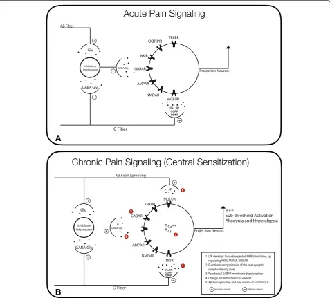
pathogenesis of CS is a heterogenous process, changes in receptor kinetics leading to sub-threshold and paradoxical AP generation are one explanation for hyperalgesia and
allodynia (Fig. 3). Paramount, modulation of CS at the
segmental level remains the mechanistic cornerstone of analgesia from SCS. In the non-pain state, inhibitory interneurons release GABA and glycine to suppress
post-synaptic depolarization through Cl− dependent
hyperpo-larization (Benzon et al., 2014). In nerve-injury models,
loss of GABAergic positivity in the dorsal horn (DH) has been observed with CS, due to cell death or the depletion
of GABA in terminals (Moore et al., 2002; Polgar et al.,
2003). GABA depletion is known to play a significant role
in CS, as allodynia is reduced with intrathecal GABAA
re-ceptor agonism and induced by nonspecific GABA
recep-tor antagonism (Hwang & Yaksh,1997; Malan et al.,2002).
Whereas GABA depletion, receptor potentiation and iono-phore kinetics offer competing hypothesized mechanisms
of hyperalgesia and allodynia, the CS phenotype is likely
oversimplified in clinical practice (Fig.3).
Reorganization and phenotypic transformation of
mechanoreceptive Aβ afferents also plays a role in the
pathogenesis of CS. Woolf et al. (1992) demonstrated
that the central terminals of Aβafferents, normally
pro-jecting to laminae III and IV, sprouted to lamina II after
axotomy (Woolf et al.,1992). This arborization of
non-nociceptive, low-threshold myelinated inputs to lamina II primarily populated by nociceptive terminals likely also contributes to tactile allodynia after nerve injury
(Woolf et al., 1992). Moreover, Aβ afferents began
ex-pressing SP after nerve injury, thought to contribute to
allodynia (Fig.3, Panel b) (Hughes et al.,2007; Neumann
et al., 1996). Lastly, abnormal polysynaptic circuits may
lead to the miscoding of a mechanoreceptive input as
nociceptive (Schoffnegger et al., 2008). While initially
it was thought that SCS activation of Aβ afferents
would inhibit ascending nociceptive inputs through the simplified gate-control mechanism, it is now clear that additional mechanisms at the segmental and
supraspinal levels play a role in SCS-mediated
analgesia. Offering an explanation for the variable response to SCS, seemingly similar clinical phenotypes may be generated by vastly different pathologic mech-anisms, explained by the heterogenous, maladaptive process of central sensitization.
Neuroanatomical targets of spinal cord neuromodulation
Spinothalamic tract
A functional understanding of the spinothalamic tract (STT) is critical to evaluating mechanisms of SCS-mediated anal-gesia. AP originating from the stimulation of peripheral nociceptors are transmitted to the CNS via the thinly
myelinated Aδand unmyelinated C fibers (Benzon et al.,
2011). Aδfibers, conducting at a moderate velocity of 5–
10 m/s, synapse in Rexed laminae I and III-V of the spinal
cord DH (Benzon et al.,2011). As Aδfibers conduct more
quickly than C fibers, they are considered the first pain signal, carrying acute pain, temperature and pressure
(McMahon et al., 2013). C fibers, conducting at a slower
velocity of < 2 m/s, synapse superficially in Rexed laminae I-II. AP to C fibers are initiated by multiple receptor types, including thermoreceptors, mechanoreceptors and che-moreceptors; thus these fibers are classified as polymodal
(Todd, 2010). C fibers carry the delayed pain response,
often characterized as poorly-localized burning or aching.
By synapse quantity, Aδ and C fibers primarily project to
interneuron circuitry, which serves as a conduit to con-duct inhibitory or excitatory signals to ascending PN
(Pol-gar et al., 2008). However, primary afferents also synapse
Specific (NS) neurons (Todd, 2010; McMahon et al.,
2013). WDR neurons, concentrated in laminae III-V,
re-ceive input from interneurons of Aβ, Aδand C fiber origin
(Todd,2010; McMahon et al.,2013). Because they receive
inputs from both noxious and non-noxious stimuli, it is fitting that WDR neurons have a graded increase in firing frequency and amplitude with repetitive polymodal stimuli
(Mendell,1966). NS neurons, concentrated superficially in
laminae I-II, only receive input from fibers carrying
noxious stimuli and do not exhibit a graded response to pain stimuli. Moreover, stimulation of independent sym-pathetic neurons shows significant activation of WDR neurons while NS neurons are not as impacted (Roberts
& Foglesong,1988). Axons of WDR and NS neurons
carry-ing crude touch, pain and temperature decussate and as-cend in the anterolateral STT, synapsing on nuclei in the posterior, medial and lateral thalamus. Collateral axons as-cend and project onto centers for autonomic regulation
A
B
and somatosensory modulation within the brainstem and midbrain. A functional understanding of this tract is critical to evaluating SCS mechanisms as it serves as the primary modulatory target via both segmental spinal and supraspinal mechanisms.
Dorsal column–medial lemniscus pathway
Though responsible for the paresthesia induced by direct stimulation in P-SCS, the DC normally carry fine touch, vibration and proprioceptive afferent inputs
from peripheral mechanoreceptors (Todd, 2010). These
peripheral APs enter the spinal cord via axons of pseudou-nipolar neurons at the DRG and ascend in the DC via the dorsal column medial lemniscus (DCML) pathway. As
discussed previously, Aβfibers also send inputs to circuits
of the DH, affecting afferent nociceptive AP and contrib-ute to CS. First order fibers ascending in the DCML then synapse on their respective nuclei within the ipsilateral medulla: the upper extremities synapse in nucleus cunea-tus laterally, whereas the lower extremities synapse on the
nucleus gracilis medially (McMahon et al.,2013). Second
order axons decussate in the brainstem forming the in-ternal arcuate fibers, then ascend cephalad through the brainstem and midbrain in the medial lemniscus, acquir-ing trigeminal inputs, ultimately synapsacquir-ing on the Ventral Posterior Lateral (VPL) and Ventral Posterior Medial (VPM) nuclei of the thalamus. Third and higher order neurons carrying inputs from the DCML and STT project
to the somatosensory cortex (McMahon et al., 2013).
While the stimulation of DC Aβ fibers activates DH
in-hibitory circuits in an antidromic manner (Fig. 4), other
important mechanisms contribute to SCS-mediated anal-gesia (Fig.5).
Native signal modulation: beyond gate control
The origins of Melzack and Wall’s gate-control theory of
pain developed from the hypothesis that cells of the substantia gelatinosa (SG) played a role in presynaptic
inhibition of cutaneous sensory input (Mendell, 2014).
Based on their input mapping and the discovery of
axo-axonal synapses, Szentagothai (1964) postulated that
spinal interneurons played a role in modulating
ascend-ing nociceptive signals (Mendell, 2014; Szentagothai,
1964). Specifically, they hypothesized the existence of a
single interneuronwith inhibitory input to the small and large fiber terminals; this interneuron itself received
stimulatory input from large fibers (Aβ) and inhibitory
input from small fibers (Aδ and C) (Melzack & Wall,
1965; Mendell,2014). Central to this theory, they
postu-lated that an imbalance between small and large fiber in-put could lead to disinhibition of the WDR neuron, thus leading to transmission of the ascending pain signal. Support for this model was provided by experiments
demonstrating presynaptic control in the SG by
stimulating small or large fibers and measuring evoked
dorsal root potentials (Mendell, 2014). Though the gate
control theory of pain continues to serve as a simplified model for framing pain signaling, it is current cannon that many additional factors contribute to spinal noci-ceptive modulation. Relevant to SCS-mediated analgesia, local interneurons, descending projections, glia and neu-roinflammation comprise the contributing SG
architec-ture to nociceptive processing (Fig.4).
Spinal interneurons
Inhibitory and excitatory interneurons play an important role in the sensory signaling cascade, modulating the activity of WDR and NS neurons in the DH. Adding to
Melzack and Wall’s gate-control theory, interneurons
form an adaptive neural circuitry critical in modulation of afferent nociceptive signaling that establishes a ba-lance between excitatory and inhibitory inputs. Laminae I-III of the DH are populated with a plexus of
interneu-rons (Todd, 2010). Excitatory interneurons secreting
glutamate comprise the non-GABAergic interneurons of laminae I-III, identified by immunostaining at the
synap-tic bouton (Todd, 2010; Todd et al., 2003). GABA and
glycine immunostaining in the rat DH exposed a dense plexus of inhibitory axons that largely arise from local
interneurons in laminae I-III (Polgar et al., 2003; Todd
& Sullivan, 1990). As evidence for circuit plasticity,
Keller et al. (2001) has demonstrated that inhibitory
synapses undergo maturation and tuning through
refine-ment of neurotransmitter release (Keller et al., 2001).
Potentially, SCS-mediated analgesia contributes to res-toration of non-pathologic equilibrium in this inherently
plastic circuitry (Saadé et al., 2006; Lind et al., 2016).
Attenuation of WDR neuron hyperexcitability through
Aβ-mediated inhibitory control is one proposed
mech-anism of P-SCS analgesia (Simone et al., 1991; Willis et
al., 1974; Chung et al., 1979). Careful neuroanatomical
studies show that both SG interneurons and WDR PN
are involved in GABAergic synapses (Todd, 2010;
Zeilhofer et al., 2012; Lekan & Carlton, 1995). Single
unit recording studies show dorsal column stimulation inhibits WDR neuron hyperexcitation within the DH
deep laminae (Hillman & Wall, 1969; Lindblom &
Meyerson, 1975; Foreman et al., 1976; Linderoth et al.,
2009; Zhang et al.,2014). Critically, P-SCS applied to the
DC prevented WDR sensitization secondary to C
Fiber-induced LTP and wind-up (Wallin et al., 2003; Guan et
al.,2010). Moreover, it has been observed that P-SCS
in-hibits WDR neuron activation in animal models of
neuropathic pain (Yakhnitsa et al.,1999). Numerous
pre-clinical studies have confirmed that Aβfiber suppression
of neuropathic pain is mediated through a GABAergic
mechanism (Sivilotti & Woolf, 1994; Duggan & Foong,
on GABAergic inhibitory interneurons are reviewed in depth in the 'gamma-aminobutyric acid' section, it should be noted that suppression of PN firing by P-SCS is dependent on intact cord architecture and lead
pro-ximity (Hillman & Wall, 1969; Foreman et al., 1976;
Smits et al.,2012). Taken together, there is strong evidence
for segmentally-mediated DH regulation of nociception, either through potentiation or inhibition of PN by local interneurons. The relative contribution of segmental and supraspinal antinociceptive mechanisms mediated by ei-ther P-SCS or PF-SCS remains an intense area of research.
Descending Antinociceptive systems
Paresthesia based spinal cord stimulation provided the first clear evidence that SCS activates the descending anti-nociceptive system (DAS), thus modulating the DH and
PN. First, Nashold et al. (1972) suggested that SCS
masked pain at the supraspinal level based on data showing that DC stimulation produced measurable EEG potentials consistent with known somatosensory evoked
potentials (Nashold et al.,1972). While investigating local
mechanisms of SCS, Foreman et al. (1976) further
suggested presence of a supraspinal mechanism given that mid-thoracic and cervical stimulation depressed spinotha-lamic tract activity measured at the lumbosacral
enlarge-ment (Foreman et al., 1976). Clarifying supraspinal
control over DH PN firing, Saade et al. (1985) utilized
DC-transected decorticate-decerebrate cats, showing that DC stimulation rostral to the transection as well as direct stimulation of nucleus raphe magnus inhibited firing in
DH neurons (Saade et al.,1985). Expanding on this
obser-vation, their group used carefully-designed brainstem lesioning experiments to elucidate the connections be-tween the DC, periaqueductal gray (PAG) and nuclei of
the reticular formations (Fig.5) (Saade et al.,1982; Saade
et al., 1983). In subsequent investigations in which they
stimulated only the DC nuclei, their group confirmed a supraspinal inhibitory loop in an awake rat model (Saade
et al., 1986). These studies accelerated the interest into
DAS as exploitable mechanisms of SCS (Fig.5).
Colloquially termed the serotonergic DAS, inputs to the PAG relay through the rostral ventromedial medulla (RVM), descend in the dorsal lateral funiculus (DLF)
and project to interneurons of the DH (Fig. 5). While
the serotonergic DAS largely utilizes the neurotransmit-ter serotonin (5-HT), opioidergic and GABAergic
mechanisms contribute at the supraspinal and segmental
levels (Cui et al., 1999). The PAG, a well-known
opioidergic pain center, does not have direct projections to the DH, and instead relays its descending signals
through the RVM (Behbehani & Fields, 1979). A spinal
relay for all descending non-noradrenergic pain
inhibition, the RVM is comprised of the nucleus raphe magnus and nuclei of the reticular formations (Newman,
1985). Both the PAG and RVM receive ascending inputs
from DH PN, creating a feedback loop (Dong et al.,
1999; Hardy,1986; Helmstetter et al.,1998; Sakata et al.,
1989). The PAG and RVM are highly innervated by pain
modulating centers, receiving projections from cortical
and subcortical structures, thus contributing the
conscious, stress and emotional responses to pain (Dong
et al.,1999; Hardy,1986; Helmstetter et al.,1998; Sakata
et al., 1989). Together, these supraspinal cortical and
subcortical communications comprise the neural sig-nature of pain, which will be evaluated in the context of P-SCS and PF-SCS in the 'SCS affects Cortical and subcortical pain processing' section.
Fibers from the RVM descend via the DLF to widely
innervate the DH (Todd, 2010; Basbaum et al., 1976).
RVM cells projecting to the DH have multiple classifica-tions: ON, OFF, 5-HT like or neutral. While neutral cells and 5-HT like cells have only been partially character-ized, more is understood about the firing patterns of ON
and OFF cells (Fields, 2004). Normally quiescent, ON
cells are activated by a nociceptive stimulus, enhancing
DH nociceptive transmission (Mendell,2014). Pathologic
ON cell activity is thought to play a role in CS and
opioid-induced hyperalgesia (Mendell, 2014). Tonically
suppressing DH pain transmission, pathological OFF cell deactivation leads to increased pain transmission in the DH. Their activity enhanced by opioids, RVM OFF cell regulation of the nociceptor-PN synapse creates an opioid-dependent supraspinal pain gate (McMahon et
al., 2013; Mendell, 2014). In the RVM, exogenously
ad-ministered opioids activate OFF cells and suppress ON cells through a GABAergic mechanism (McMahon et al.,
2013). Sharing a partial pathway with opioids, Song et al.
(2013) has shown that P-SCS results in DH
antinocicep-tion through activaantinocicep-tion of RVM OFF cells and 5-HT like
cells, but has no effect on ON cells (Fig. 5) (Song et al.,
2013a). Moreover, P-SCS mediated analgesia was attenu-ated with GABA receptor agonism but independent of opioidergic mechanisms within the RVM (Song et al.,
2013a). Because opioids and P-SCS share a partially re-dundant mechanism, opioidergic mechanisms may not contribute to P-SCS mediated analgesia, discussed in the 'Endogenous Opioids' section.
The descending noradrenergic pathway modulates ascending pain signals through the release of norepi-nephrine (NE) in the DH, specifically laminae I-III of the SG. Axons from the locus coeruleus (LC) and associated cell bodies provide descending spinal NE, synapsing on nociceptive afferents and lamina II spinal interneurons
(Kwiat & Basbaum, 1992). While the LC, A5 and A7 all
receive inputs from the PAG, the LC receives additional notable cortical and sub-cortical relays (Bajic & Proudfit,
1999; Bernard et al., 1996; Cedarbaum & Aghajanian,
1978). Ascending signals originating in the DH send
projections to the LC while redundant pathways relay from the insular cortices, amygdala and hypothalamus. This creates a neural circuit responsive to emotion and
stress (Bernard et al.,1996; Gauriau & Bernard,2002). It
is likely that part of the analgesic effect from activation of the PAG is due to recruitment of the noradrenergic
DAS (Cui et al.,1999; Bajic & Proudfit, 1999). Evidence
for SCS modulation of the noradrenergic and serotoner-gic DAS is discussed below.
Segmental and Supraspinal neurotransmitters
Preclinical pain models have served to clarify me-chanisms contributing to SCS-mediated analgesia. From careful lesioning experiments to direct DH sampling using microdialysis catheters, changes in neurotransmit-ter content, concentration and synthetic enzyme func-tion have molded our understanding of these pathways. However, differences in implementation, methodology and inability to isolate cell-specific contributions to neurotransmitter release makes it critical to constantly reassess our understanding of spinal and supraspinal pain mechanisms. Specifically, it is unclear the extent to which an observed neurotransmitter change can be as-cribed to a particular mechanism, as the pathways re-main incompletely elucidated. For example, the relative contributions of interneurons, glia, non-nociceptive fi-bers and DAS to GABA release in response to SCS re-mains unclear. While we recognize the limitations of experimental models, progress has been made with re-gard to clarifying these pathways. We therefore discuss current constructs of pain signaling in SCS as they relate to the specific neurotransmitter, site of action and pro-posed origin.
Serotonin
Modulated by SCS and released in the DH, serotonin (5-hydroxytrypamine, 5-HT) is a monoamine neuro-transmitter involved in the in the serotonergic DAS. A component of the RVM and a known serotonergic center, electrical stimulation of the nucleus raphe magnus led to GABA release in the DH, indicating that spinal 5-HT acts through a GABAergic
inter-mediary (Kato et al., 2006; Tazawa et al., 2015).
Inter-estingly, electrical stimulation of the PAG attenuated nociceptive inputs through a serotonergic mechanism, supporting that opioidergic mechanisms of analgesia
also utilize this 5-HT DAS (Akil & Liebeskind, 1975;
Liu et al., 1988). Preclinical pain models consistently
demonstrate that descending 5-HT originates from the RVM and P-SCS models confirm this relationship
(Vera-Portocarrero et al., 2006; Suzuki et al., 2004;
Peng et al. (1996) showed that analgesia from PAG
stimulation was blocked by 5-HT3 receptor
antago-nists, Li et al. (2014) demonstrated that DH 5-HT
resulting from P-SCS was exclusively synthesized in supraspinal nuclei and transported anterograde to
nerve terminals (Peng et al., 1996; Li et al., 2014).
Localizing the source of supraspinal 5-HT, Maeda et
al. (2009) showed that P-SCS induced expression of
c-Fos in the RVM (Maeda et al., 2009). Tazawa et al.
(2015) further showed that the supraspinal nuclei and
not the local spinal cord were responsible for pro-duction of 5-HT after P-SCS therapy. Moreover, they noted increased activation and number of seroto-nergic neurons in the dorsal raphe nucleus, a center adjacent to the ventral PAG with communications to
the RVM and LC (Tazawa et al., 2015). Lastly, they
noted that while both the serotonergic and noradre-nergic DAS contributed to P-SCS-mediated analgesia, the 5-HT pathway played a greater role in
antino-ciception (Tazawa et al., 2015). Together, these studies
demonstrate activation of the serotonergic DAS in response to P-SCS therapy, which in turn innervates the DH, providing antinociceptive drive. Linderoth et
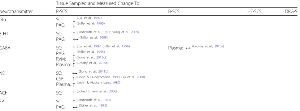
al. (1992) first showed that DC stimulation increased
5-HT levels in the DH (Table 1) (Linderoth et al.,
1992). Clarifying this observation, their group further
demonstrated the presence of 5-HT staining nerve termi-nals in the SG and noted that intrathecal 5-HT potenti-ated the antinociceptive effects of P-SCS. Moreover, by showing that antinociception from P-SCS was attenuated
with GABAB receptor antagonism and unchanged with
muscarinic 4 acetylcholine receptor (M4 mAChR) antag-onism, they demonstrated the presence of a GABAergic link and accelerated interest into 5-HT receptor subtyping
(Table 2) (Song et al., 2009). Through agonist-antagonist
studies, Song et al. (2011) utilized a rat model of
mono-neuropathy to clarify the role of 5-HT and GABA receptor subtypes on analgesia from P-SCS therapy (Song et al.,
2011). Specifically, they noted that agonists of 5-HT2 and
5-HT3 receptors enhanced P-SCS analgesia while the
benefits from 5-HT3 receptor agonists were inhibited
more with antagonism of GABABthan GABAA
recep-tors. While they did not discover any difference with
5-HT1, 5-HT6and 5-HT7receptor antagonism, they noted
that antagonism of 5-HT2A and 5-HT4 receptors
atte-nuated the P-SCS response to tactile hypersensitivity. In
total, their work suggests that while 5-HT2A and 5-HT4
receptor agonism contributes to analgesia, P-SCS works
to a greater extent through 5-HT3 receptors with a
GABABreceptor link (Song et al., 2011). The preclinical
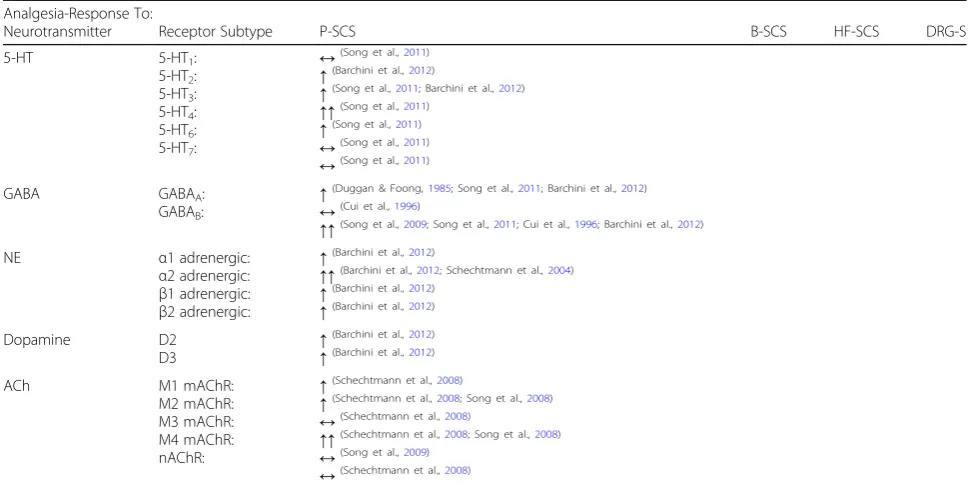
evidence for neurotransmitter receptor subtype
contribu-tion to SCS-mediated analgesia is summarized in Table2.
These preclinical models indeed demonstrate that P-SCS activates the serotonergic DAS in an orthodromic manner
(Fig.5). However, theclinical magnitude of 5-HT
contri-bution to SCS-mediated antinociception is still unknown. Nonetheless emerging reports support the construct that serotonergic mechanisms play a significant role in pain
relief. Prabhala et al. (2019), demonstrated that
Duloxe-tine, a 5-HT and NE reuptake inhibitor, combined with P-SCS therapy significantly improved pain scores at one year
compared to SCS alone (Prabhala et al., 2019). Taken
Table 1Measured Neurotransmitters in response to spinal cord stimulation (SCS)
Tissue Sampled and Measured Change To:
Neurotransmitter P-SCS B-SCS HF-SCS DRG-S
Glu SC: ↓(Cui et al.,1997)
PAG: ↓(Stiller et al.,1995)
5-HT SC: ↑(Linderoth et al.,1992; Song et al.,2009) PAG: ↔(Stiller et al.,1995)
GABA SC: ↑(Cui et al.,1997; Stiller et al.,1996) PAG: ↓(Stiller et al.,1995)
RVM: ↓(Song et al.,2013c) Plasma:↑(Crosby et al.,2015a)
Plasma: ↔(Crosby et al.,2015a)
NE SC: ↔(Song et al.,2013b)
CSF: ↑(Levin & Hubschmann,1980; Liu et al.,2008) Plasma:↑(Levin & Hubschmann,1980)
ACh SC: ↑(Schechtmann et al.,2008)
SP SC: ↑(Linderoth et al.,1992)
PAG: ↔(Stiller et al.,1995)
together, preclinical and emerging clinical investigations suggest the significance of P-SCS mediated orthodromic activation of the serotonergic DAS for analgesia. However, further work is needed to determine if 5-HT may play a role in other paradigms, such as PF-SCS or
DRG-S (Tables 1 and2).
Gamma-aminobutyric acid
The principle inhibitory neurotransmitter of the CNS, GABA binds to two known GABA receptor classes:
GABAA and GABAB (Olsen & DeLorey, 1999). The
postsynaptic GABAA receptor complex consists of a
multi-target binding domain linked with a chloride
ionophore (Olsen & DeLorey, 1999). GABAA receptor
agonism increases inward Cl− current, leading to
post-synaptic hyperpolarization and increases the firing
threshold (Olsen & DeLorey, 1999). The metabotropic
postsynaptic GABAB receptor indirectly opens
potas-sium channels though a G-protein-coupled mechanism, leading to membrane hyperpolarization and a similar
increase in firing threshold (Olsen & DeLorey, 1999).
Uniquely, presynaptic GABAB receptors inhibit
neuro-transmitter release via a Ca2+dependent mechanism and
are critical in presynaptic inhibition (Todd, 2010; Olsen
& DeLorey, 1999). Receiving projections from Aβ, Aδ
and C fibers, GABAergic interneurons populate laminae I-III and demonstrate GABA positive terminals
synap-sing on PN (Todd, 2010; Zhang et al., 2014; Sivilotti &
Woolf, 1994; Lekan & Carlton, 1995). Of all DH
neu-rons, GABAergic signaling is present in 25% of lamina I,
30% of lamina II and 40% of lamina III (Todd, 2010).
Dysfunction of the spinal GABA circuitry in addition to increased excitatory neurotransmitter release is
corre-lated with WDR neuron hyperexcitability (Fig.4) (Cui et
al.,1997; Stiller et al.,1996). Yakhnitsa et al. (1999)
dem-onstrated P-SCS decreases WDR neuron hyperexcitabil-ity, noted by depressed evoked potentials and decreased spontaneous discharge on these neurons (Yakhnitsa et
al., 1999). This P-SCS-induced suppression of WDR
neurons is thought to be due to an increase in DH GABA and concurrent decrease in excitatory glutamate
(Cui et al., 1997). SCS modulation of GABA in the DH
and other CNS locations is a well-defined phenomenon
(Table1). Contributing mechanisms of analgesia utilizing
specific GABAA and GABAB receptor pathways have
been clarified using agonist-antagonist experiments
Table 2Spinal cord stimulation exerts analgesia by activating specific receptor subtypes
Analgesia-Response To:
Neurotransmitter Receptor Subtype P-SCS B-SCS HF-SCS DRG-S
5-HT 5-HT1:
5-HT2: 5-HT3: 5-HT4: 5-HT6: 5-HT7:
↔(Song et al.,2011)
↑(Barchini et al.,2012)
↑(Song et al.,2011; Barchini et al.,2012)
↑↑(Song et al.,2011)
↑(Song et al.,2011)
↔(Song et al.,2011)
↔(Song et al.,2011)
GABA GABAA:
GABAB:
↑(Duggan & Foong,1985; Song et al.,2011; Barchini et al.,2012)
↔(Cui et al.,1996)
↑↑(Song et al.,2009; Song et al.,2011; Cui et al.,1996; Barchini et al.,2012)
NE α1 adrenergic:
α2 adrenergic: β1 adrenergic: β2 adrenergic:
↑(Barchini et al.,2012)
↑↑(Barchini et al.,2012; Schechtmann et al.,2004)
↑(Barchini et al.,2012)
↑(Barchini et al.,2012)
Dopamine D2
D3 ↑
(Barchini et al.,2012)
↑(Barchini et al.,2012)
ACh M1 mAChR:
M2 mAChR: M3 mAChR: M4 mAChR: nAChR:
↑(Schechtmann et al.,2008)
↑(Schechtmann et al.,2008; Song et al.,2008)
↔(Schechtmann et al.,2008)
↑↑(Schechtmann et al.,2008; Song et al.,2008)
↔(Song et al.,2009)
↔(Schechtmann et al.,2008)
Activation of specific receptor subtypes contributes to P-SCS mediated analgesia. Experiments used receptor antagonists and agonists to clarify the role of particular receptors to SCS-mediated analgesia. 5-HT3receptor, GABABreceptor,α2 adrenergic receptor and M4 mAChR pathways were found to contribute to this analgesia to a
greater extent than other receptor subtypes. An“up”arrow indicates that activation of that particular receptor subtype contributed to analgesia.‘Double up’arrows represent a greater increase and the predominant pathway.‘Sideways’arrows represent no contribution to analgesia. Acronyms: Glu (Glutamate), 5-HT (5-hydroxytryptamine, serotonin), GABA (Gamma-aminobutyric Acid), NE (Norepinephrine), ACh (Acetylcholine), 5-HT1(5-hydroxytryptamine receptor 1), 5-HT2(5-hydroxytryptamine
receptor 2), 5-HT3(5-hydroxytryptamine receptor 3), 5-HT4(5-hydroxytryptamine receptor 4), 5-HT6(5-hydroxytryptamine receptor 6), 5-HT7(5-hydroxytryptamine receptor 7),
(Table 2). Converging lines of evidence confirm that P-SCS employs a GABAergic mechanism in the DH, as evi-denced by inhibition of PN firing in response to P-SCS
treatment (Sivilotti & Woolf, 1994; Duggan & Foong,
1985). Specifically, administration of a GABAA receptor
antagonist reversed PN inhibition produced by P-SCS, demonstrating the importance of DH GABA to P-SCS
mediated antinociception (Sivilotti & Woolf, 1994;
Dug-gan & Foong,1985). Notably, P-SCS responders in an
allo-dynic rat model were observed to have increased levels of GABA in the dorsal horn, whereas non-responders and sham animals exhibited no change, indicating that P-SCS mediated DH GABA release may prevent allodynia (Stiller
et al., 1996). At the segmental level, suppression of
glu-tamate release is dependent on presynaptic activation of
GABABreceptors, which is likely more important to the
P-SCS interneuron-mediated inhibition of PN firing than
activation of postsynaptic GABAA receptors (Cui et al.,
1997; Stiller et al.,1996; Cui et al., 1996). Expanding on
these findings, Cui et al. (1997) found that administration
of an intrathecal GABAB receptor agonist would
trans-form P-SCS non-responding animals into responders (Cui
et al.,1997). This finding is similar to the work by Song et
al. (2011) that shows greater analgesia with 5-HT3
receptor-mediated GABABsignaling than GABAA
signal-ing (Song et al.,2011). It remains unclear which
GABA-mediated effects are due to local interneuron circuitry and which are the result of activation of the serotonergic DAS. As noted previously, P-SCS recruits the serotonergic DAS, which employs downstream GABAergic mechanisms of antinociception. Using P-SCS and implanted microdialysis
catheters, Stiller et al. (1995, 1996) and Linderoth et al.
(1993) demonstrated an increase in extracellular GABA in
the DH and decreased levels in the PAG (Stiller et al.,
1996; Linderoth et al., 1993; Stiller et al., 1995). This
decrease in PAG GABA argues that P-SCS relieves inhib-ition of the serotonergic DAS, which relays through the RVM, allowing the system to exert descending control over DH PN.
In clinical studies, Lind et al. (2004,2008) showed that
administration of an intrathecal GABAB receptor
agon-ist (Baclofen) significantly enhanced the analgesia of P-SCS and rescued non-responders, echoing results of
pre-clinical animal models (Lind et al., 2008; Lind et al.,
2004). Cortical and subcortical pain circuits may also be
modulated by P-SCS utilizing a GABAergic mechanism.
Moens et al. (2013) studied 20 FBSS patients who were
treated with P-SCS and underwent functional magnetic
resonance imaging (fMRI) 7–10 days post-implantation
(Moens et al., 2013). They discovered increased GABA
and decreased glucose signals in the ipsilateral thalamus, potentially explained by orthodromic activation of the paleospinothalamic pathway. They hypothesized that projections from the reticular formations to GABAergic
nuclei in the thalamus, hypothalamus and limbic system may indicate an interference with the affective
compo-nent of pain (Moens et al., 2013; Moens et al., 2012).
This would represent an additional mechanism of P-SCS efficacy. In summary, P-SCS mediated GABAergic mech-anisms have been described at three targets. First, P-SCS activates GABAergic inhibitory interneurons at the dor-sal horn, either directly or by recruiting the serotonergic DAS. Second, P-SCS results in decreased GABAergic signaling in the PAG, which results in disinhibition and thus activation of the serotonergic DAS. Lastly, P-SCS orthodromically activates thalamic GABAergic neurons, which may modulate cortical processing and thalamo-cortical dysrhythmia. Converging lines of evidence derived from preclinical and emerging clinical work suggest a central role for GABAergic P-SCS mediated an-algesia. These GABAergic pathways likely contribute to the observed clinical analgesic effects of P-SCS at both the segmental and supraspinal levels. Clearly, more work is needed to clarify these pathways in emerging paradigms,
including DRG-S and PF-SCS (Tables1and2).
Norepinephrine
Norepinephrine (NE), often referred to as Noradren-aline, is a catecholamine neurotransmitter produced pri-marily in the LC and released in the spinal cord DH
(Hayashida et al., 2008a). In neuropathic pain, not only
is there an increase in DH NE release and adrenergic axon sprouting, but corroborative evidence suggests an inability to recruit the DAS may contribute to chronic
pain states (Hayashida et al.,2008b; Witting et al.,2003).
NE has antinociceptive effects through presynaptic
inhi-bition of primary Aδand C fibers, postsynaptic inhibition
of WDR and NS neurons, and activation of inhibitory
interneurons (Hayashida et al.,2007; Pertovaara,2006). In
a rat model, the activation of inhibitory interneurons by NE increases GABAergic and glycinergic postsynaptic
currents as measured in the SG (Baba et al.,2000a; Baba
et al., 2000b). While this supports the segmental role for
NE-mediated analgesia in the DH, converging lines of evidence now suggest that local DH NE release is unlikely
to be augmented by P-SCS (Tazawa et al.,2015; Song et
al.,2013b). Song et al. demonstrated that while the LC is activated in a neuropathic rat model treated with P-SCS,
no change was observed in DH NE (Song et al., 2013b).
Tazawa et al. (2015) supported this conclusion and further
clarified that no increase in DH NE was observed with
P-SCS (Tazawa et al.,2015). However, scant clinical reports
confirmed concentrations of NE increased in CSF sampled after P-SCS therapy. Two separate human studies to date show an immediate increase in CSF NE concentration
pre-to-post P-SCS (Levin & Hubschmann,1980; Liu et al.,
2008). However, while Levin et al. (1980) showed an
baseline after 5 min, calling into question the durable role of the noradrenergic DAS in P-SCS. In total, the current literature suggests that rather than recruiting descending
NE fibers,orthodromic activation of the LCby P-SCS likely
relays through the PAG, in essence reinforcing descending antinociception through the serotonergic DAS previously
discussed (Fig.5). The clinical longevity and significance of
potential increases in CSF NE mediated by P-SCS, PF-SCS, and DRG-S remains to be determined.
Acetylcholine
Though the mechanism is not clearly defined, cholinergic inputs to PN play a role in modulating nociceptive signals
via spinal interneuron circuitry (Foreman, 2012).
Cloni-dine, a presynaptic alpha 2 (α2) adrenergic receptor
agon-ist, exerts its analgesic effect largely through a cholinergic
mechanism at the spinal level (Foreman, 2012). After
discovering that clonidine may potentiate the analgesic
effect of P-SCS in a rat model, Schechtmann et al. (2008)
demonstrated lower basal DH ACh in nerve-lesioned animals and an increase in DH ACh in P-SCS responding
animals (Schechtmann et al., 2008). Using
agonist-antagonist studies, their group further noted reversal of analgesia with administration of selective and non-selective muscarinic acetylcholine receptor (mAChR)
antagonists (Table 2). Specifically, they noted that
mus-carinic 1 (M1) mAChR and M4 mAChR contributed to P-SCS mediated analgesia while nicotinic and mus-carinic 3 (M3) mAChrR antagonism had no effect on PWT. Supporting this finding and further clarifying
the receptor subtypes involved, Song et al. (2008)
found that intrathecal muscarinic 2 (M2) mAChR and M4 mAChR agonists reversed P-SCS non-responders
(Song et al., 2008). However as noted above, Song et al.
(2009) showed that 5-HT signaling in the serotonergic
DAS did not involve the M4 mAChR pathway (Song et al.,
2009). Thus, cholinergic signaling in P-SCS may represent
a novel, independent mechanism exclusive of the seroto-nergic DAS or a parallel, redundant pathway. In a
subse-quent randomized clinical trial, Schechtmann et al. (2010)
delivered sub-analgesic doses of intrathecal clonidine or baclofen combined with P-SCS in non-responding pa-tients with prior P-SCS devices. Similar to the work by
Lind et al. (2004, 2008) (Lind et al., 2008; Lind et al.,
2004), Schechtmann et al. (2010) showed intrathecal
clo-nidine significantly improved pain scores (in two patients) when combined with P-SCS. Together, this provides some clinical evidence for cholinergic augmentation in P-SCS
non-responders (Schechtmann et al., 2010). In sum,
cholinergic mechanisms play a role in P-SCS mediated segmental antinociception. However, as with other neuro-transmitters, work is needed to clarify the clinical contri-bution of cholinergic mechanisms to the analgesic effects of newer paradigms, including PF-SCS and DRG-S.
Endogenous opioids
Endogenous peptides of the brain and spinal cord, β
-endorphins, enkephalins and dynorphins are ligand
classes that activate μ, κ and δ-opioid receptors. These
ligands exert analgesia segmentally through direct receptor activation in addition to recruitment of the serotonergic and noradrenergic DAS (Benzon et al.,
2014). A well-known opioidergic center, the PAG
com-municates with the DAS, medulla, dorsal raphe nucleus
and the LC (Benzon et al., 2014). Causing
hyperpolari-zation and signal inhibition through a G-protein coupled
mechanism, μ, κ and δ-opioid receptors are widely
present on pre and post-synaptic neurons of the DH
(Benzon et al.,2014). As the μ-opioid receptor is known
to play a role in the development of windup, modulation of the endogenous opioidergic system may potentially
explain analgesia with SCS treatment (Guan et al.,2006).
Providing a physiologic basis for this hypothesis, Wang
et al. (2003) has shown that activation of the
serotoner-gic DAS increases enkephalin and dynorphins in the DH
(Wang et al.,2003). In line with this finding, Ding et al.
(2008) showed that P-SCS resulted in an increase in
thoracic DH dynorphin in a rat angina model (Ding et
al., 2008). This suggests the possibility that κ opioid
re-ceptor activation may contribute to the analgesic effects
of P-SCS (Ding et al., 2008). In a subsequent SNI rat
model, Sato et al. (2013) observed improved PWT after
frequency-specific P-SCS at 4 Hz and 60 Hz. Notably, they observed that the improvement with 4 Hz and 60 Hz was reversed upon administration of naloxone and
naltrindole, respectively (Sato et al., 2013). Similar to
what has been observed with transcutaneous electrical
nerve stimulation (TENS), this finding indicates
frequency-specific activation of μ and δ-opioid
receptors with P-SCS (Sato et al., 2013; Chandran &
Sluka, 2003). Despite frequency-specific activation of
opioidergic pathways, the role of this mechanism in P-SCS is likely temporally limited to the first few days of stimula-tion. In an SNI model treated with P-SCS at 3 and 7 days, naloxone administration attenuated early but not late
re-versal of hyperalgesia (Sun et al., 2017). Postulating that
the temporal reduction in efficacy occurred through endo-genous opioid tolerance, Chandran et al. (2002), observed similar findings in patients treated with both low and high
frequency TENS (Chandran & Sluka, 2003). Limited
evidence from clinical studies appears to mirror the re-sults of these preclinical investigations. In 17 patients with chronic pain admitted to the neurosurgical service, Tonelli
et al. (1988) implanted and initiated single lead P-SCS
during hospital admission, sampling their CSF one to two days prior and again one day after implantation and
initi-ation of therapy (Tonelli et al., 1988). Responders were
noted to have a significant increase in CSF β-endorphin
peptide. Unfortunately, CSF was not sampled at any additional timepoints to clarify whether this increase
in β-endorphin was sustained (Tonelli et al., 1988). In
other clinical work, Freeman et al. (1983) seemed to
agree with preclinical models regarding the lack of sustained opioidergic mechanisms with P-SCS (Freeman
et al., 1983). Specifically, naloxone did not reverse
anal-gesia from P-SCS after 30 days of stimulation in patients having relief with TENS or P-SCS. This continues to suggest that recruited opioidergic mechanisms may contribute to the initial analgesic efficacy of P-SCS (during P-SCS trial and subsequent implant) but the longevity of this mechanism is questionable. There is likely an anal-gesic ceiling effect in regard to opioidergic pathways re-cruited by P-SCS. In a rat SNI model treated with P-SCS, administration of the opioidergic enhancer proglumide had no additive or synergistic effect on PWT or physical
activity levels (Inoue et al., 2017). Tolerance and
time-limited opioidergic analgesia that contribute to the clinical efficacy of P-SCS may also apply to PF-SCS. To date, one study of non-lesioned rat lumbar spinal cord slices dem-onstrated frequency-dependent opioid release from DH neurons, with a maximal release at 500 Hz (Song &
Mar-vizón,2003). Further work is needed to determine the role
and longevity of opioidergic mechanisms in all modes of SCS. Moreover, additional studies are needed to evaluate frequency-specific endogenous opioid release in P-SCS and PF-SCS and whether these mechanisms are predom-inantly segmental or supraspinal.
SCS modulates Neuroinflammatory pain regulation
Signatures of glial activation
Critical to the structure, metabolism and immunity of both the CNS and peripheral nervous system (PNS), glia
are non-neuronal cells intimately associated with
neurons. Glia of the CNS include microglia, astrocytes, oligodendrocytes and ependymal cells while those of the PNS include satellite glial cells (SGCs) and Schwann
cells (Ji et al., 2013). It is now understood that glia also
provide a functional microenvironment modulating sig-nal transduction, neuroplasticity and synaptic pruning
(Tremblay et al., 2011). In response to nerve injury,
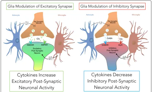
striking changes are seen in glial morphology, concentra-tion, cellular signaling, receptor regulation and mediator release. Termed gliosis, these responses together consti-tute a phenotypic transformation that alters signaling by changing the synaptic neuron-glia mediator balance
(Fig. 6) (Ji et al.,2013). Temporally correlating with the
onset of neuropathic pain, PNS and CNS insults includ-ing nerve injury and cord hypoxemia lead to reactive gliosis. Specifically, reactive gliosis leads to increased post-synaptic potentials at excitatory synapses and de-creased post-synaptic potentials at inhibitory synapses
(Fig. 6). Through complex signaling mechanisms, these
changes result in increased PN firing and pathologic
nociception (Ji et al., 2013). While microglial activation
occurs 24 h after injury and is limited to 3 months, astro-cyte activation occurs 3 days after injury and is maintained
(Coyle, 1998; Ledeboer et al., 2005; Mika, 2008; Mika et
al.,2009). Activation and intracellular signaling culminate
with the release of glial mediators, which exhibit their downstream effect on pre and post-synaptic targets
(Fig. 6) (Ji et al., 2013; Mika et al., 2013). Mediator
classes known to influence nociceptive transmission in-clude nitric oxide (NO), cytokines, chemokines, comple-ment components and other bioactive factors, some of
which have been examined in response to SCS (Table 3)
(Mika et al.,2013). Given that glial activation is important
to the development and maintenance of neuropathic pain and that glial inhibition improves mechanical allodynia, one potential mechanism of SCS is glial modulation
(Wat-kins et al.,2001a; Watkins et al.,2001b). In a seminal
pre-clinical study, Sato et al. (2014) showed decreased glial
ac-tivation after P-SCS therapy correlating with improved
PWT (Sato et al.,2014). Specifically, P-SCS decreased
im-munostaining of microglia marker OX-42 and astrocyte markers GFAP and MCP-1 in the superficial and deep DH
lamina (Sato et al., 2014). Changes in glial activation are
also observed at the DRG.
DRG SGCs are perfectly positioned within the sand-wich synapse to regulate nociceptive transmission
(Todd, 2010; Rozanski et al., 2013). Encircling a single
neuron with a thin sheath intrasynaptically, SGCs are
implicated in nociceptive modulation (Hanani, 2005).
Having multiple bioactive receptors, SGC modulate neural activity through the release of mediators that have been shown to alter neural activity via the P2X3 pathway. The upregulation of this pathway promotes
abnormal nociception in rats (Hanani,2005; Chen et al.,
2008). In a L5 SNI model, SGC activation was observed
while glial inhibitors administered to the ipsilateral DRG provided alleviation of mechanical allodynia (Liu et al.,
2012a). Moreover, SGC reorganization and the for-mation of new neural contacts and gap junctions has been observed after peripheral nerve axotomy (Hanani
et al., 2002). Given the proximity to the DRG, DRG-S
may modulate SGC activation. In a tibial nerve injury
model, Pan et al. (2016) first showed that DRG-S
nor-malized glial fibrillary acidic protein (GFAP) and acti-vating transcription factor 3 (ATF-3), markers of astrocytic activation and neuronal injury (Pan et al.,
2016). Moreover DRG-S was noted to reverse cold and
mechanical hypersensitivity, suggesting that DRG-S impacts SGC-mediated neuroinflammation (Pan et al.,
2016). While evidence is still limited, SGCs clearly play a
efficacious at the DRG. To date, no human clinical work has yet been completed that characterizes these promising observations seen in DRG-S treated preclinical models. Taken together, neuroinflammatory glial mechanisms are critical in chronic pain maintenance while glial-mediated mechanisms of P-SCS, PF-SCS and DRG-S remain to be elucidated. While a comprehensive examination of glial activation is beyond the scope of this review, the evidence for glial mediator response to modes of SCS are carefully
summarized in Table3and reviewed below.
Cytokines, neurotrophic factors and biomarkers
Critical to the induction and maintenance of chronic pain, dysregulation of local cytokine and neurotrophic factor signaling directly and indirectly influences AP generation in the PN. The release of inflammatory cytokines, neurotrophic factors and other mediators operates under a positive feed-back mechanism of autocrine and paracrine regulation. While likely evolutionarily advantageous in recruiting the inflammatory cascade, gliosis and the resultant local inflam-matory response becomes a maladaptive and injurious
process in chronic pain. While evidence for glial modulation in response to SCS is mounting, the effect of SCS on the local and systemic inflammatory responses remains unclear.
Upregulated in gliosis after nerve injury, the
pro-inflammatory cytokines interleukin-1β (IL-1β), IL-6, IL-8
and TNF-αexert potent pronociceptive action (Bjurstrom
et al., 2016). Of particular importance, IL-1βand TNF-α
increase PN excitability, reinforce glial activation and further recruit the inflammatory response (Bjurstrom et
al., 2016). Concentrations of CSF proinflammatory
cytokines are increased in multiple chronic pain states including osteoarthritis, CRPS, postherpetic neuralgia and
fibromyalgia (Bjurstrom et al., 2016). Patients with
indi-cations for SCS therapy including chronic low back pain, lumbar radiculopathy and failed back surgery syndrome (FBSS) were also noted to have elevations in
proinflamma-tory cytokines (Bjurstrom et al.,2016). Post P-SCS effects
in pre-clinical models have been evaluated with regard to
cytokine levels, summarized in Table 3. In non-nerve
injury models treated with P-SCS, Tilley et al. (2009)
de-monstrated increased expression of meningeal TNF-αand
IL-10, but no change was noted in IL-6 or IL-1β(Tilley et
al.,2009). Later, Tilley et al. carefully separated the
menin-ges into its three layers and measured cytokine expression
following P-SCS (Tilley et al.,2019). They found increases
in IL-1βand IL-10 within the dura mater, correlated in a
dose-dependent manner to the delivered current (Tilley et
al.,2019). Further, the concentration of IL-6, a pleiotropic
cytokine, was notably related to delivered current with a
Table 3Evidence for the response of cytokines and neurotrophic factors to spinal cord stimulation (SCS) paradigms
Tissue Sampled and Measured Change with SCS
Neuromediator WDR/NS Firing Nociceptive Effect P-SCS B-SCS HF-SCS DRG-S
Measured Centrally:
BDNF Increased Algesia DRG: ↔(Tilley et al.,2017) C3 Increased Algesia SC: ↑(Stephens et al.,2018) c-Fos Increased Algesia SC: ↑(Tilley et al.,2016)
IL-1β Increased Algesia SC: ↑(Stephens et al.,2018; Vallejo et al.,2016) DM: ↑(Tilley et al.,2019)
DRG: ↑(Tilley et al.,2017) IL-6 Increased Algesia DRG: ↓(Tilley et al.,2017) TNF-a Increased Algesia SC: ↑(Tilley et al.,2016) VEGF Increased Algesia CSF: ↓(McCarthy et al.,2013) CXCL16 Decreased Analgesia SC: ↑(Vallejo et al.,2016)
DRG: ↑(Vallejo et al.,2016) GDNF Decreased Analgesia CSF: ↑(McCarthy & McCrory,2014a) IL-1α Decreased Analgesia SC: ↑(Vallejo et al.,2016) IL-10 Decreased Analgesia DM: ↑(Tilley et al.,2019) NpY Decreased Analgesia DRG: ↔(Tilley et al.,2017) VIP Decreased Analgesia DRG: ↔(Tilley et al.,2017) Measured Peripherally
CXCL10 Increased Algesia IF: ↓(Kriek et al.,2018) IFN-γ Increased Algesia IF: ↓(Kriek et al.,2018)
Plasma: ↔(Kamieniak et al.,2019a) IL-1β Increased Algesia Plasma: ↔(Kamieniak et al.,2019a) IL-2 Increased Algesia IF: ↓(Kriek et al.,2018) IL-6 Increased Algesia IF: ↔(Kriek et al.,2018)
Plasma: ↔(Kamieniak et al.,2019a) IL-12 Increased Algesia IF: ↓(Kriek et al.,2018) IL-15 Increased Algesia IF: ↓(Kriek et al.,2018) TNF-a Increased Algesia IF: ↔(Kriek et al.,2018)
Plasma: ↔(Kamieniak et al.,2019a) VEGF Increased Algesia IF: ↓(Kriek et al.,2018) IL-4 Decreased Analgesia IF: ↓(Kriek et al.,2018) IL-5 Decreased Analgesia IF: ↓(Kriek et al.,2018) IL-10 Decreased Analgesia IF: ↓(Kriek et al.,2018)
Plasma: ↔(Kamieniak et al.,2019a)
Plasma:↑(Kinfe et al.,2017)
TGF-β Decreased Analgesia Plasma: ↔(Kamieniak et al.,2019a)