BioMed Central
BMC Biology
Open Access
Research article
Systematic comparison and reconstruction of sea urchin
(Echinoidea) internal anatomy: a novel approach using magnetic
resonance imaging
Alexander Ziegler*
1, Cornelius Faber
2,3, Susanne Mueller
4and
Thomas Bartolomaeus
1Address: 1Institut für Biologie, Freie Universität Berlin, Königin-Luise-Straße, 14195 Berlin, Germany, 2Experimentelle Physik 5, Universität
Würzburg, Am Hubland, 97074 Würzburg, Germany, 3Institut für Klinische Radiologie, Universitätsklinikum Münster, Waldeyerstraße, 48149
Münster, Germany and 4Berlin NeuroImaging Center, Charité-Universitätsmedizin Berlin, Charitéplatz, 10117 Berlin, Germany
Email: Alexander Ziegler* - [email protected]; Cornelius Faber - [email protected]; Susanne Mueller - [email protected]; Thomas Bartolomaeus - [email protected] * Corresponding author
Abstract
Background: Traditional comparative morphological analyses and subsequent three-dimensional reconstructions suffer from a number of drawbacks. This is particularly evident in the case of soft tissue studies that are technically demanding, time-consuming, and often prone to produce artefacts. These problems can partly be overcome by employing non-invasive, destruction-free imaging techniques, in particular micro-computed tomography or magnetic resonance imaging.
Results: Here, we employed high-field magnetic resonance imaging techniques to gather numerous data from members of a major marine invertebrate taxon, the sea urchins (Echinoidea). For this model study, 13 of the 14 currently recognized high-ranking subtaxa (orders) of this group of animals were analyzed. Based on the acquired datasets, interactive three-dimensional models were assembled. Our analyses reveal that selected soft tissue characters can even be used for phylogenetic inferences in sea urchins, as exemplified by differences in the size and shape of the gastric caecum found in the Irregularia.
Conclusion: The main focus of our investigation was to explore the possibility to systematically visualize the internal anatomy of echinoids obtained from various museum collections. We show that, in contrast to classical preparative procedures, magnetic resonance imaging can give rapid, destruction-free access to morphological data from numerous specimens, thus extending the range of techniques available for comparative studies of invertebrate morphology.
Background
For centuries, comparative zoomorphological analyses have formed the backbone of phylogenetic inferences. Data on the external and internal morphology of
speci-representative species. These traditional procedures are technically demanding as well as time-consuming and irretrievably alter or even destroy the specimen. Further complications arise from the difficult three-dimensional
Published: 23 July 2008
BMC Biology 2008, 6:33 doi:10.1186/1741-7007-6-33
Received: 12 February 2008 Accepted: 23 July 2008
This article is available from: http://www.biomedcentral.com/1741-7007/6/33
© 2008 Ziegler et al; licensee BioMed Central Ltd.
for example deformation or compression of histological slices, and the problematic alignment of sections make 3D reconstructions a laborious and often highly subjec-tive procedure. The problems encountered are further aggravated by the logistic challenges that arise when com-parative studies on selected species of a whole taxon are to be carried out. For example, in case of sea urchins (Echi-noidea, Echinodermata), some species are deep-sea dwell-ers or confined to remote regions of the world. It is therefore a considerable problem to obtain these animals in a freshly fixed state. Understandably, the desirable use of specimens from museum collections is usually not per-mitted for invasive and destructive analyses.
The application of non-invasive imaging techniques such as micro-computed tomography (μCT; see, for example, [1-8]) or magnetic resonance imaging (MRI; see below) has substantially increased in basic morphological research. Whereas μCT is mainly used to display hard tis-sue such as bone or calcite structures, MRI provides images depicting primarily soft tissue anatomy. MRI is based on the principles of nuclear magnetic resonance (NMR) and is predominantly used to measure the distri-bution of hydrogen protons within a sample. Image con-trast is achieved due to the different physical properties of tissues. Both μCT and MRI produce standardized digital data, principally permitting an interactive access through virtual collections or voxel libraries as envisaged previ-ously [9,10]. This data represents the unaltered structural composition of the specimen, in contrast to classical his-tological or dissection techniques and apart from fixation. In the light of these and other recent technological advances, Budd and Olsson [11] refer to the renaissance of morphology, calling for a more extensive use of non-invasive and non-destructive imaging techniques and 3D applications. Since many of the specimens in natural his-tory collections are conserved in alcohol, forming the so-called wet collections, non-invasive imaging techniques might be employed to reveal the internal and external anatomy of preserved, scientifically irreplaceable speci-mens. This extended use of museum specimens for com-parative anatomical studies would allow for a substantial increase in taxon sampling on one hand, and on the other would minimize the ecological impact of such analyses.
The feasibility of MRI studies to reveal soft tissue charac-teristics of invertebrates was demonstrated by studies on crabs [12,13], squid [14], crayfish [15,16], oysters [17,18], spiders [19], demosponges [20], and insects [21-29] (see also the review by Hart et al. [30]). Ziegler and Angenstein [31] used living and freshly fixed specimens of the sea urchin Psammechinus miliaris (Müller, 1771) and showed that members of this taxon are likely to be suitable for detailed MRI studies. These studies yielded imaging results comparable to manual dissection and were
corre-lated with histological data. To achieve better resolved images of the complex internal anatomy of sea urchins, the application of higher magnetic field strengths was pro-posed [32]. However, all of the studies mentioned above were restricted to the analysis of single specimens or spe-cies in a non-systematic context.
For systematic analyses, however, sea urchins appear par-ticularly suitable. Like sea stars or sea cucumbers, they are part of a major deuterostome taxon, the Echinodermata, which shares a common ancestor with diverse groups such as acorn worms (Enteropneusta), tunicates (Tuni-cata), and vertebrates (Vertebrata) [33]. Owing to the cal-cified skeleton found in sea urchins, palaeontologists have access to a rich fossil record permitting thorough ground-truthing of phylogenetic hypotheses that are based upon molecular or morphological datasets [34,35]. Recently, the importance of these animals for evolution-ary and developmental inferences has been further emphasized by results obtained through the sequencing of the genome of the purple sea urchin, Strongylocentrotus purpuratus (Stimpson, 1857) [36].
Sea urchins are spherical, oval or flattened free-moving echinoderms covered with spines. Their soft tissue anat-omy is characterized by a dominant digestive tract, a var-ying number of gonads, and the highly specialized water vascular system with its external appendages, the tube or ambulacral feet. The space between the major organ sys-tems is composed of the main body cavities (the so-called perivisceral coeloms) that are filled with coelomic fluid [37]. Therefore, echinoids possess several of the internal structures encountered in most other taxa, although a number of highly specialized organs are present as well. This taxon can thus serve as a model for analyses using non-invasive imaging techniques on a broad systematic scale.
The purpose of our study was to explore the possibility of generating high-resolution 3D MRI datasets from selected specimens of a major invertebrate taxon. For the first time, a non-invasive imaging technique is employed for system-atic comparative analyses. We show that MRI permits the rapid acquisition of digitized morphological data on a broad scale and the construction of 3D models that can be made interactively accessible on the Web. In addition, our approach can be employed to compare the internal anat-omy of sea urchins to evaluate such datasets for evolution-ary inferences.
Results
Establishment of imaging conditions
Initially, freshly fixed as well as museum specimens of
BMC Biology 2008, 6:33 http://www.biomedcentral.com/1741-7007/6/33
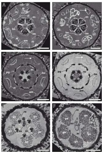
Establishment of imaging conditions using specimens of Psammechinus miliaris Figure 1
Establishment of imaging conditions using specimens of Psammechinus miliaris. (A), (B) Comparison of two freshly fixed specimens. Magnetic resonance imaging (MRI) sections at the height of Aristotle's lantern and digestive tract. Resolution: (81 μm)3, no contrast agent added. The two specimens show a high degree of similarity in their overall internal architecture.
Arrows indicate paramagnetic gut content. (C), (D) Effects of a contrast agent on image quality. MRI sections at the height of perignathic girdle and lower stomach. Resolution: (81 μm)3. This freshly fixed specimen was scanned (C) before and (D) after
the application of a contrast agent, Magnevist. Arrows indicate susceptibility artefacts. (E), (F) Comparison of a freshly fixed and a museum specimen. MRI sections at the height of gonads and upper oesophagus. Resolution: (81 μm)3. The 135-year-old
and contrasting protocols for broader systematic analyses. Living specimens of P. miliaris were made available through the Biologische Anstalt Helgoland, Germany (BAH). Several dozen sea urchins were kept in the aquar-ium facilities of the Institut für Biologie, Freie Universität Berlin, Germany, for subsequent scanning and contrasting experiments.
The first set of analyses was conducted to compare differ-ent specimens of freshly fixed sea urchins. Imaging was performed initially at a resolution of (81 μm)3 using dif-ferent imaging protocols with specimens of almost identi-cal size. This analysis showed that although slight differences in anatomical makeup did occur, localization, arrangement and overall shape of the internal organs did not differ significantly. In the specimens depicted (Figure 1A and 1B), this is exemplified by the almost identical shapes of both Aristotle's lanterns and the localization and shape of the digestive tract components. The structure of the wall of the sea urchin lower gut loop (also termed the stomach) was undulated in both specimens, whereas the horizontal sections of the upper gut loop (also termed the intestine) showed a smooth structure. The digestive tract of one specimen was partly filled (Figure 1A), while the digestive tract of the other specimen was empty, apart from tiny objects (Figure 1B). Another striking similarity was the almost exactly identical location of the inner and outer marginal sinus of the haemal system in both speci-mens. However, some differences were discernable, for example the distinct shapes of the ampullae and the thick-ness of the interpyramidal muscles, indicating that, ide-ally, several specimens of a given species should be scanned. Similar comparative experiments were per-formed on a total of 18 echinoid species (data not shown). In all cases, the overall internal architecture of two specimens of a given species resembled each other more closely than that of other species.
The next set of experiments served to assess the effects of a gadolinium-based contrast agent (Magnevist, a standard in clinical and pre-clinical MRI studies) on image quality. Using the same specimen of P. miliaris and the same imag-ing protocol, we found that the application of the contrast agent resulted in enhanced contrast, although some thin-walled structures, such as the ampullae or the oesophagus, became less visible (Figure 1C and 1D). Another advan-tage of the use of this contrast agent was that some of the artefacts caused by paramagnetic gut content were reduced due to the stronger water signal. Therefore, most specimens were scanned both before and after the appli-cation of the contrast agent (see Additional file 1 for details).
The final set of experiments for establishing a standard set of imaging conditions was designed to compare imaging
results obtained from freshly fixed and museum speci-mens of the same species. Prior to the present MRI studies, several museum specimens from the wet collection of the Systematische Zoologie am Museum für Naturkunde, Ber-lin, Germany (ZMB) were dissected in order to assess the state of preservation after decades or even more than a century of alcohol conservation (Strongylocentrotus drö-bachiensis (Müller, 1776), Sphaerechinus granularis
(Lamarck, 1816), Schizaster lacunosus (Linnaeus, 1758), and Brissopsis lyrifera (Forbes, 1841)). Their state of preser-vation was surprisingly good and seemed mainly to depend on the mode of collection, the fixation mode, and the subsequent preservation in alcohol. Following the dis-sections, MRI experiments using freshly fixed and museum specimens of P. miliaris were carried out. Struc-tures visible in the freshly fixed specimen (Figure 1E) were recognizable also in the 135-year-old museum specimen (Figure 1F). The size and location of the anatomical fea-tures varied slightly, but this had been observed among different specimens of freshly fixed animals as well. Despite these differences, the results confirmed our assumption that museum material, even if more than a hundred years old, can be used for systematic MRI studies.
Scanning of selected members of high-ranking echinoid subtaxa
After an initial assessment of the suitability (that is, size, state of preservation, and availability) of specimens in natural history collections worldwide, MRI studies were carried out using selected species of the 14 currently rec-ognized echinoid subtaxa (orders). In three cases, freshly fixed animals were employed for the analysis (see Addi-tional file 2 for details).
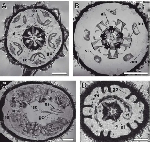
The full datasets from two 'regular' sea urchins (Eucidaris metularia (Lamarck, 1816) and P. miliaris) as well as two irregular sea urchins (Echinoneus cyclostomus (Leske, 1778) and Echinocyamus pusillus (Müller, 1776)) are presented here as interactive videos (Additional files 3, 4, 5, 6; Figure 2 shows images of selected sections). The four image data-sets range in spatial resolution from 20 × 18 × 18 μm3 to (86 μm)3 and represent the current state-of-the-art in high-field MRI at a field of view of up to (3.3 cm)3. In E.
resi-BMC Biology 2008, 6:33 http://www.biomedcentral.com/1741-7007/6/33
Selected horizontal magnetic resonance imaging sections of different sea urchin species taken from Additional files 3, 4, 5, 6
Figure 2
Selected horizontal magnetic resonance imaging sections of different sea urchin species taken from Additional files 3, 4, 5, 6. (A) Eucidaris metularia. Resolution: (81 μm)3. Aristotle's lantern, gonads, Stewart's organs, and stomach can be
seen. (B) Psammechinus miliaris. Resolution: (44 μm)3. Aristotle's lantern, lantern muscles, stomach, and ampullae are visible. (C)
Echinoneus cyclostomus. Resolution: (86 μm)3. Digestive tract with oesophagus, gastric caecum, stomach, and rectum are shown.
(D) Echinocyamus pusillus. Resolution: 20 × 18 × 18 μm3. Aristotle's lantern, gonads, stomach, and rectum are represented.
dues) that caused artefacts by altering the homogeneity of the magnetic field inside the MRI instrument.
In E. cyclostomus (Figure 2C) and E. pusillus (Figure 2D), the secondarily developed bilateral symmetry found in the Irregularia is obvious when looking at the gonads (reduced to four). The digestive tract of E. cyclostomus is characterized by a dominant gastric caecum, localized at the beginning of the stomach, and a long rectum, leading down to the anus which is situated at the oral side of the animal. E. pusillus, in common with all members of the Clypeasteroida, the so-called sand dollars, possesses a modified Aristotle's lantern. Other structures that can be identified in the irregular species include relatively small spines, one dominant (E. pusillus) or two huge (E. cyclos-tomus) gut loops, filled with debris and detritus, and an axial complex running straight down from the madreporic plate to the adoral part of the oesophagus (E. cyclos-tomus). In addition to soft tissues, the delineation of cal-careous structures caused by the signal-providing surrounding fluids and tissues adds some information on hard-part anatomy to the virtual sections as well.
Although several specimens of the echinoid taxon missing in our study, the Echinothurioida (or leather urchins), were found in museum collections, their shape prevented successful soft tissue imaging. These sea urchins possess a flexible calcite endoskeleton as well as specialized internal muscles that contract when the animal is fixed, resulting in a pancake-like shape of the organism. For a reliable comparison of internal structures it would be necessary to scan specimens prior to contraction of these powerful muscles.
3D reconstruction of selected internal organs
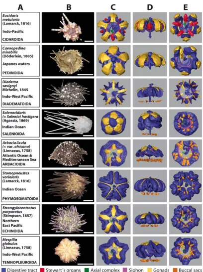
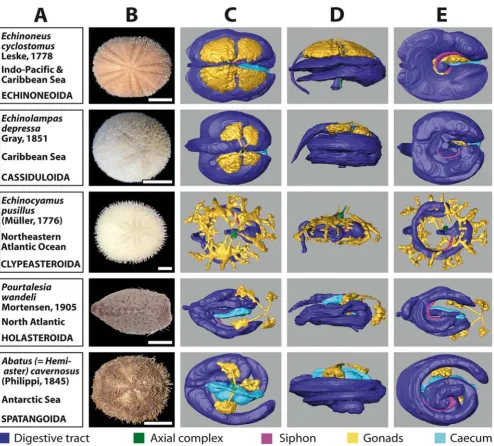
The 3D reconstructions presented here are limited to the major soft tissue structures identifiable in all datasets, if present in the respective species (Figure 3C–E and 4C–E): digestive tract, Stewart's organs, axial complex, siphon, gonads, buccal sacs (or 'gills') and gastric caecum.
Two of the immediately recognizable features are size and shape of the gonads. Both differ substantially between the analyzed species and usually constitute a character set of high intraspecific variability. This becomes evident from the analysis of animals from a given species in distinct developmental stages (data not shown). The specimen of
Stomopneustes variolaris (Lamarck, 1816) (Figure 3) shown here was fixed before gonad maturation. In the case of the highly branched gonads found in E. pusillus (Figure 4), depicted here for the first time, the advantages offered by the digitized reconstruction of morphological structures become obvious: a 3D reconstruction of such complex structures would be particularly demanding using tradi-tional techniques. In contrast to the gonads, clearly
dis-tinct shapes are revealed when the guts of 'regular' (Figure 3) and irregular (Figure 4) sea urchin specimens are com-pared. After the scanning process, a digital photograph was taken of every specimen selected for this study (Fig-ures 3B and 4B).
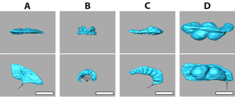
MRI also revealed a striking difference regarding the archi-tecture of the gastric caecum. Except for the two clypeas-teroid subtaxa Laganina and Scutellina, this structure can be found in all irregular taxa (Figure 4C–E), located at the beginning of the stomach. Its presence correlates with the feeding habits of this group of sea urchins: most of these animals are partially or completely covered by sediment which they ingest to filter out organic material. Although the precise function of the gastric caecum is still a matter of discussion, it appears to have a major role in digestion (see, for example, [38,39]). In specimens of comparable size, substantial differences in its size and shape were found (Figure 5). The highly reduced gastric caecum con-sisting of numerous smaller blindly ending sacs that can be observed in Echinolampas depressa Gray, 1851 (Figure 5B) appears to be a common characteristic which sets the Cassiduloida apart from other taxa (Figure 5A,C and 5D) of the Irregularia. This is emphasized by the presence of a similar, highly reduced gastric caecum in the cassiduloid species Cassidulus caribearum Lamarck, 1801 (data not shown; as described also by Gladfelter [40]) and Rhyn-cholampas pacificus Agassiz, 1863 (as shown in [41]). A comprehensive description and comparative analysis of sea urchin internal anatomy will be published elsewhere (Ziegler et al., publication in preparation).
Interactive viewing of selected species in three dimensions
To illustrate the opportunities offered by digitized recon-structions, Figure 6A–C shows different views of the 3D model of a member of the Cidaroida (or pencil urchins),
BMC Biology 2008, 6:33 http://www.biomedcentral.com/1741-7007/6/33
Overview chart showing analyzed specimens of 'regular' sea urchins and corresponding 3D reconstructions of selected internal organs
Figure 3
an unobstructed view on all organs they may occlude. Sev-eral pre-installed views can be selected as well. The crea-tion of this model, including MRI data acquisicrea-tion, took less than three full work days. All other models shown in Figures 3 and 4 are available for download and interactive viewing on The Echinoid Directory [43].
Discussion
MethodologyThe initial phase of our study served to determine the proper imaging conditions using high-field MRI instru-ments. In order to achieve resolutions below (100 μm)3, we employed 7 T and 17.6 T small animal scanners. We were able to obtain resolutions ranging from 20 × 18 × 18
μm3 to (86 μm)3 with specimens ranging from 5 mm to Overview chart showing analyzed specimens of irregular sea urchins and corresponding 3D reconstructions of selected inter-nal organs
Figure 4
BMC Biology 2008, 6:33 http://www.biomedcentral.com/1741-7007/6/33
3.4 cm in size. Therefore, in terms of resolution, our data is comparable to results derived from manual dissection techniques in combination with stereomicroscopic obser-vation. Susceptibility artefacts were primarily seen in the sediment-feeding irregular species, with the gut content being the source of these artefacts (see, for example, Addi-tional file 6, Echinocyamus pusillus (Müller, 1776)). What sediment component is causing which type of artefact needs to be determined in mineralogical studies on freshly fixed specimens. The effects of the contrast agent made it possible to reduce the negative effects of these artefacts in some cases (Figure 1C and 1D), but a thor-ough investigation of the potential of selective and non-selective contrast agents [44] is essential for their future application in invertebrate morphology. Other major sources for artefacts were the tendency of spatangoid sea urchins to accumulate ferric iron phosphate in the con-nective tissues of their digestive tracts [45], and the diges-tive tract diverticula of scutelline sand dollars known to harbour magnetite [46]. However, the scanning of several specimens of a given species and the matching of their internal structures did in some cases help to overcome these problems.
One of the big advantages of MRI techniques is the extremely simple preparation of fixed specimens, being
presence of a contrast agent) and later putting them back into alcohol for storage. No detrimental effects of this pro-cedure were noted during our studies. Apart from mor-phological studies on fixed specimens, MRI can also be used for in vivo analyses of invertebrates where, in contrast to μCT techniques that are based on X-rays, the living specimen is not harmed during the scanning process. Extensive morphological or developmental studies can thus be carried out, although anaesthetic substances may have to be applied to reduce movement artefacts during the long scanning times that are needed for high-resolu-tion datasets and 3D reconstruchigh-resolu-tions. Another very impor-tant advantage of non-invasive imaging techniques in general is the fact that the digitally acquired image stacks do not have to be manually aligned, as is the case with dig-ital pictures from histological or electronmicroscopical sections. Although these datasets can be used to generate highly resolved digitized 3D models of invertebrates as well (see, for example, [47,48]), these analyses are extremely demanding in terms of specimen preparation, slice alignment and 3D reconstruction. In comparison, 3D reconstruction of MRI data is simple and highly accu-rate, but the segmentation process carried out for the present work is still subjective and our primary hypothe-ses regarding organ designation had to be evaluated against the information available from classical histologi-3D reconstructions of the gastric caecum of selected irregular sea urchin species
Figure 5
mentation [49] as well as artefact and noise reduction [50] algorithms have recently become available, and may fur-ther simplify the task of 3D reconstruction. The highly demanding step of obtaining a completely automated seg-mentation and reconstruction of an entire animal would be very desirable and appears technically feasible, although it is not yet available for comparative morpho-logical studies.
In spite of the many positive features, a number of nega-tive aspects are currently inherent to morphological
stud-ies of small animals using MRI. The achievable resolution is comparatively low, and results derived from the recon-struction of internal structures are currently comparable only to manual dissection. Furthermore, an exact outline of how future experiments on other invertebrate taxa should be performed can only be partially derived from our study, since imaging results of fixed specimens depend on several factors, some of them still not fully understood (for example, susceptibility artefacts or changes to organ systems after fixation). Another draw-back is the fact that only limited in vitro staining possibil-Comparison of a digital 3D model with a traditional anatomical sketch
Figure 6
Comparison of a digital 3D model with a traditional anatomical sketch. (A)-(C) Eucidaris metularia. Selected views taken from the interactive 3D model: (A) external view; (B) external view with transparent test, internal organs visible; (C) external view with transparent test and all internal organs removed except for Stewart's organs and Aristotle's lantern. (D)
BMC Biology 2008, 6:33 http://www.biomedcentral.com/1741-7007/6/33
ities are available for MRI studies, although some substances (for example manganese) can be used for in vivo staining [28]. A further disadvantage of MRI is that specimens cannot currently be scanned in alcohol, since this would require spectroscopic imaging techniques resulting in much longer scanning times at the desirable resolutions below (100 μm)3. In addition, in the case of sea urchins, the 3D models generated (see, for example, Figure 6) are practically useless for taxonomic purposes since sea urchin taxonomy is currently based entirely on hard parts. Resolutions well below (10 μm)3 would be necessary to differentiate between the often miniscule characters of the sea urchin skeleton. However, using μCT, these datasets can potentially be gathered, as demon-strated by μCT scans of two sea urchin species, Strongylo-centrotus purpuratus (Stimpson, 1857) [51] and Encope michelini Agassiz, 1841 [52], presented on the Digital Morphology Library (DigiMorph) website [53].
On the other hand, the access to MRI instruments does not appear to be a limiting factor. There are currently more than 1000 experimental small animal MRI instru-ments worldwide available for pre-clinical imaging. Since all high-field MRI scanners consist of super-conducting electromagnets, overnight and weekend scans as per-formed during this study would permit an optimal use of this resource.
Internal morphology of sea urchins
Despite the existence of extensive fossil, morphological and molecular datasets for this taxon, comparatively little systematic knowledge has been gathered on the internal anatomy of sea urchins. However, some authors have greatly extended our knowledge about the internal anat-omy of selected, readily available sea urchin species such as Cidaris cidaris (Linnaeus, 1758) [54], Arbacia punctulata
(Lamarck, 1816) [55], Paracentrotus lividus (Lamarck, 1816) [56,57], Echinus esculentus Linnaeus, 1758 [58],
Sphaerechinus granularis (Lamarck, 1816) [57,59], Spatan-gus purpureus Müller, 1776 [57] or Echinocardium cordatum
(Pennant, 1777) [38], not to mention the extensive works of Alexander E Agassiz, Hubert L Clark and Theodor J Mortensen. A common drawback of these studies is that a given author rarely dissected and analysed several species of a single taxon using one consistent method. Only in very few cases have histological sections, dissected speci-mens and so on, been preserved. The conclusions drawn from the original data can therefore not always be scruti-nized.
Furthermore, the results were sometimes biased by pre-vailing morphological hypotheses. For example, recent histological studies [60] as well as our findings (Figure 3) indicate that the siphon, a canal bypassing the sea urchin
matoida (see, for example, [61]), is indeed also present in this group of animals. Distinct features of internal sea urchin anatomy may extend to the sizes and shapes of digestive tract, gonads, ampullae and muscles. A thorough revision of internal structures in the light of recent techno-logical advances will therefore almost certainly change our view of facts that, for years, were taken for granted. Scanning of sea urchins has so far been performed by us on more than 50 species (Ziegler et al., publication in preparation).
The feasibility of comparative morphological studies using MRI is further exemplified by our description of the highly variable shape and size of the gastric caecum found in the Irregularia (Figure 5). Our findings were correlated with the little information available on this structure; the combined data led us to assume that a reduced gastric cae-cum consisting of several smaller blindly ending sacs might constitute a soft tissue character common to all cas-siduloid sea urchins. We therefore predict the presence of a similarly shaped structure in all species of the Echi-nolampadidae and Cassidulidae. Furthermore, it would be of interest to extend our analyses to members of the Apatopygidae and Neolampadidae, the other two extant families currently assigned to the Cassiduloida.
Although for some organs high intraspecific variability was detected, in particular in the case of organs with a development-dependent size and shape such as the gonads (Figure 1E and 1F), shape and localization of other organs (for example the digestive tract) did not dif-fer significantly among specimens from a given echinoid species, allowing comparison on various taxic levels (Fig-ures 3 and 4). However, intraspecific variability of internal organs can also be observed in other taxa, for example gas-tropods (sinistral freak) or humans (situs inversus vis-cerum). Therefore, like larval and hard-part morphology, which are continuously updated [62,63], morphological character sets derived from internal organs of sea urchins will serve as a useful systematic reference for future stud-ies.
Non-invasive imaging techniques other than MRI
dure reveals internal structures only in rare cases and is limited to tiny objects.
In contrast to CLSM, μCT has already become a standard tool for detailed non-invasive studies of whole specimens, especially in studies on fossils [67]. It is based on the anal-ysis of a sample by means of X-rays and can primarily be used to reveal hard tissue structures such as bone or cal-cite. Moreover, recent studies using more advanced tech-niques such as X-ray synchrotron microtomography and a new setup called very-high-resolution X-ray computed tomography have been employed to display soft tissues,
in vitro as well as in vivo [6,7,68-70] (see also the review by Attwood [4]). Using μCT, isotropic resolutions of less than (5 μm)3 have been achieved on whole specimens, although, technically, resolutions in the nanometre scale are within reach. However, whether these new μCT meth-ods can compete with the ability of MRI to display soft tis-sue at high resolutions still needs to be evaluated. Our own preliminary studies to generate images of internal organs performed on several sea urchin museum speci-mens (Echinoneus cyclostomus Leske, 1778, Psammechinus miliaris (Müller, 1771), Mespilia globulus (Linnaeus, 1758), and Moira atropos (Lamarck, 1816)) using a desk-top high-resolution μCT scanner (Skyscan 1172, Skyscan, Kontich, Belgium) at the Max-Planck-Institut für evolu-tionäre Anthropologie, Leipzig, Germany were not suc-cessful, although these μCT datasets can be used for the reconstruction of hard tissue structures. 3D models based on this data will be made available on the DigiMorph website [53].
These results demonstrate that complementary imaging techniques should ideally be used for comparative mor-phological analyses, an approach commonly referred to as multimodality.
Conclusion
Our results extend the use of MRI in invertebrate mor-phology to a systematic approach using museum speci-mens of a major marine invertebrate taxon, the sea urchins (Echinoidea). MRI can be employed for the rapid, non-destructive and unbiased acquisition of digital mor-phological data, and the creation of interactive 3D models accessible both within a publication and via the Web. The revelation of the internal anatomy of echinoid museum specimens at a resolution comparable with manual dis-section extends our knowledge of soft tissue characters, permitting phylogenetic inferences.
Methods
Specimen preparation
All specimens analyzed belong to the taxon Echinoidea (sea urchins), one of the five subtaxa of the Echinoder-mata (marine spiny-skinned animals). The museum
spec-imens were mostly fixed in formalin and all were conserved in alcohol, although in some cases the exact mode of fixation could not be determined. For MR scan-ning, the museum specimens had to be lowered down to distilled water in a gradual alcohol series. Specimens that were obtained in living state were fixed in a 7% formalde-hyde solution. For MR scanning, the freshly fixed speci-mens were kept in this solution. Results derived from specimens scanned in alcohol and formalin did not appear to differ significantly.
The specimens where placed either inside a custom-built Plexiglas chamber (Berlin), inside a 50 ml Falcon tube (Berlin), or inside NMR tubes with diameters between 5 and 20 mm (Würzburg), depending on the size of the specimen. On some specimens spines had to be dressed for tight fit inside the tubes. Magnevist(Bayer-Schering, Berlin, Germany), a gadolinium-based non-selective con-trast agent, was added at a final concentration of 2 mM. This concentration had been employed successfully in preliminary studies (results not shown). The addition of the contrast agent to the museum specimens was dis-cussed beforehand with the curators who judged the risk to the specimens' integrity as negligible. Prior to scanning, samples were degassed at 50 mbar. The freshly fixed spec-imens used in this study will be deposited as voucher material at the ZMB. Museum specimens will be stored in separate jars for potential later re-scanning. Table 2 lists detailed information on every museum and freshly fixed specimen used in this study.
Specimen scanning and photography
Berlin
Experiments were conducted at the Berlin NeuroImaging Center, Charité – Universitätsmedizin Berlin, Germany on a 7 T Pharmascan 70/16 AS rodent scanner with a 1 H-resonance frequency of 300 MHz (Bruker Biospin GmbH, Ettlingen, Germany). The system consisted of a 160 mm horizontal bore magnet, a shielded gradient set with an inner diameter of 90 mm, and a maximum gradient strength of 300 mT/m. A 1H-radio frequency linear trans-mit/receive birdcage resonator with an inner diameter of 38 mm was used for excitation and signal detection. The images described here were acquired at around 18°C. Data acquisition and image processing were carried out with Paravision 4.0.
Würzburg
BMC Biology 2008, 6:33 http://www.biomedcentral.com/1741-7007/6/33
mm linear birdcage resonator and a 5 mm linear bird cage resonator were used. The images described here were acquired at around 18°C. Image processing was carried out with Paravision 3.0.2 and involved zero filling to a (512)3 matrix prior to Fourier transformation.
Scanning protocols employed a 3D gradient echo imaging sequence (FLASH) with parameters depending on the specimen size (see Additional file 2 for details). Almost all scans resulted in datasets with isotropic spatial resolution.
The photos of museum and freshly fixed specimens were made using a digital camera (Casio Exilim) with 7.2 Meg-apixels. The acquired images were processed using Adobe Photoshop CS2 9.0.
3D visualization
3D image reconstruction and visualization were per-formed by converting the generated MRI data (DICOM standard) into an 8-bit TIFF image sequence (ImageJ 1.38w) and by using 3D imaging software (amira 3.0.2, Mercury Computer Systems, Berlin, Germany). Segmenta-tion was carried out manually by using the brush tool in the amira Image Segmentation Editor. In some cases struc-tures on every other slice were labelled, with subsequent 'interpolation' of structures on intervening slices, fol-lowed by a check and correction of segmentation results if necessary. Detection of borderlines and organ designa-tions were performed based on literature, our own histo-logical data and previously acquired MRI data [31,32].
Operations were carried out on a desktop PC (CPU: Intel Core 2 Duo, 2.67 GHz; graphics card: NVIDIA GeForce 8600 GT; operating system: Windows XP; display: WACOM Cintiq 21UX Pen Display). Image processing was performed using Adobe Photoshop CS2 9.0 and Adobe Illustrator CS2 12.0.1. The movies were produced using the ImageJ 1.38w QuickTime export function. The individual 3D structures were saved as Wavefront files and imported into Adobe 3D Toolkit 8.1.0, where they were reassembled and transformed into an interactive 3D PDF. The 3D model was embedded into this publication using the Adobe 3D Toolkit according to the procedures described in [71].
List of abbreviations
3D: three-dimensional; BAH: Biologische Anstalt Helgo-land, Germany; CAS: California Academy of Sciences, San Francisco, USA; CLSM: confocal laser scanning micros-copy; μCT: micro-computed tomography; FOV: field of view; MRI: magnetic resonance imaging; NHM: Natural History Museum, London, UK; NMR: nuclear magnetic resonance; USNM: National Museum of Natural History, Washington DC, USA; ZMB: Systematische Zoologie am
Authors' contributions
AZ designed and coordinated the experiments, carried out the dissections, prepared specimens and carried out scan-ning and 3D modelling. CF and SM prepared and scanned specimens. TB supervised the experiments. All authors contributed to writing the manuscript and approved of the final version.
Additional material
Additional file 1
Table 1 – Scanning parameters for specimens used in this study.
Click here for file
[http://www.biomedcentral.com/content/supplementary/1741-7007-6-33-S1.pdf]
Additional file 2
Table 2 – List of specimens used in this study.
Click here for file
[http://www.biomedcentral.com/content/supplementary/1741-7007-6-33-S2.pdf]
Additional file 3
Video 1 – MRI dataset of Eucidaris metularia (Lamarck, 1816). Reso-lution: (81 μm)3.
Click here for file
[http://www.biomedcentral.com/content/supplementary/1741-7007-6-33-S3.mov]
Additional file 4
Video 2 – MRI dataset of Psammechinus miliaris (Müller, 1771). Res-olution: (44 μm)3.
Click here for file
[http://www.biomedcentral.com/content/supplementary/1741-7007-6-33-S4.mov]
Additional file 5
Video 3 – MRI dataset of Echinoneus cyclostomus Leske, 1778. Reso-lution: (86 μm)3.
Click here for file
[http://www.biomedcentral.com/content/supplementary/1741-7007-6-33-S5.mov]
Additional file 6
Video 4 – MRI dataset of Echinocyamus pusillus (Müller, 1776). Res-olution: 20 × 18 × 18 μm3.
Click here for file
[http://www.biomedcentral.com/content/supplementary/1741-7007-6-33-S6.mov]
Additional file 7
3D model – 3D PDF version of the entire article. By clicking anywhere onto Figure 6 the 3D model of Eucidaris metularia (Lamarck, 1816) can be interactively accessed. The 3D model is based on the virtual MRI sections seen in Video 1. In order to view the 3D model please install (or upgrade to) Adobe Reader Version 7.1 or higher.
Click here for file
Acknowledgements
The authors thank Andrew Cabrinovic (NHM), Carsten Lüter (ZMB), David Pawson (USNM), and Rich Mooi (CAS) for the generous supply with sea urchin museum specimens. We sincerely appreciate the help with pho-tography from Peter Adam (Institut für Biologie, Freie Universität Berlin, Germany). Andrew B Smith (NHM) is thanked for introducing AZ to sea urchin systematics and commenting on the manuscript. We are grateful to Heiko Temming (Max-Planck-Institut für evolutionäre Anthropologie, Leip-zig, Germany) for carrying out the μCT scans. The authors appreciate the comments by Timothy Rowe (The University of Texas at Austin, Austin, USA) and two anonymous reviewers. We also thank Andreas Ziegler (Insti-tut für Immungenetik, Freie Universität Berlin, Germany) for critical reading of the manuscript. The 17.6 T NMR spectrometer was funded by the DFG (Ha1232/13). AZ was supported through stipends from the State of Berlin (NaFöG), Germany, and the Friedrich-Naumann-Stiftung für die Freiheit, Potsdam, Germany
References
1. Hörnschemeyer T, Beutel RG, Pasop F: Head structure of Priacma serrata Leconte (Coleoptera, Archostemata) inferred from x-ray tomography. J Morphol 2002, 252:298-314.
2. Wirkner CS, Richter S: Improvement of microanatomical research by combining corrosion casts with microCT and 3D reconstruction, exemplified in the circulatory organs of the woodlouse. Microsc Res Tech 2004, 64:250-254.
3. Burrow CJ, Jones AS, Young G: X-ray microtomography of 410 million-year-old optic capsules from placoderm fishes. Micron
2005, 36:551-557.
4. Attwood D: Nanotomography comes of age. Nature 2006,
442:642-643.
5. Betz O, Wegst U, Weide D, Heethoff M, Helfen L, Lee WK, Cloetens P: Imaging applications of synchrotron X-ray phase-contrast microtomography in biological morphology and biomateri-als science. I. General aspects of the technique and its advan-tages in the analysis of millimetre-sized arthropod structure.
J Microsc 2007, 227:51-71.
6. Dierick M, Cnudde V, Masschaele B, Vlassenbroeck J, Van Hoorebeke L, Jacobs P: Micro-CT of fossils preserved in amber. Nucl Instrum Meth Phys Res A 2007, 580:641-643.
7. Socha JJ, Westneat MW, Harrison JF, Waters JS, Lee WK: Real-time phase-contrast x-ray imaging: a new technique for the study of animal form and function. BMC Biol 2007, 5:6.
8. Wirkner CS, Richter S: The circulatory system in Mysidacea – implications for the phylogenetic position of Lophogastrida and Mysida (Malacostraca, Crustacea). J Morphol 2007,
268:311-328.
9. Jones G, Christal M: The future of virtual museums: on-line, immersive, 3D environments. 2002 [http://tinyurl.com/4vt97g]. Created Realities Group
10. Rowe J: Developing a 3D digital library for spatial data: issues identified and description of prototype. RLG DigiNews 2002, 6:5. 11. Budd GE, Olsson L: A renaissance for evolutionary
morphol-ogy. Acta Zool 2007, 88:1.
12. Brouwer M, Engel DW, Bonaventura J, Johnson GA: In vivo mag-netic resonance imaging of the blue crab, Callinectes sapidus: effect of cadmium accumulation in tissues on protein relax-ation properties. J Exp Zool 1992, 263:32-40.
13. Bock C, Frederich M, Wittig RM, Pörtner HO: Simultaneous observations of haemolymph flow and ventilation in marine spider crabs at different temperatures: a flow-weighted MRI study. Magn Reson Imaging 2001, 19:1113-1124.
14. Gozansky EK, Ezell EL, Budelmann BU, Quast MJ: Magnetic reso-nance histology: in situ single cell imaging of receptor cells in an invertebrate (Lolliguncula brevis, Cephalopoda) sense organ. Magn Reson Imaging 2003, 21:1019-1022.
15. Herberholz J, Mims CJ, Zhang X, Hu X, Edwards DH: Anatomy of a live invertebrate revealed by manganese-enhanced mag-netic resonance imaging. J Exp Biol 2004, 207:4543-4550. 16. Brinkley CK, Kolodny NH, Kohler SJ, Sandeman DC, Beltz BS:
Mag-netic resonance imaging at 9.4 T as a tool for studying neural anatomy in non-vertebrates. J Neurosci Meth 2005, 146:124-132.
17. Davenel A, Quellec S, Pouvreau S: Noninvasive characterization of gonad maturation and determination of the sex of Pacific oysters by MRI. Magn Reson Imaging 2006, 24:1103-1110. 18. Pouvreau S, Rambeau M, Cochard JC, Robert R: Investigation of
marine bivalve morphology by in vivo MR imaging: first ana-tomical results of a promising technique. Aquaculture 2006,
259:415-423.
19. Pohlmann A, Möller M, Decker H, Schreiber WG: MRI of tarantu-las: morphological and perfusion imaging. Magn Reson Imaging
2007, 25:129-135.
20. Müller WEG, Kaluzhnaya OV, Belikov SI, Rothenberger M, Schröder HC, Reiber A, Kaandorp JA, Manz B, Mietchen D, Volke F: Magnetic resonance imaging of the siliceous skeleton of the demos-ponge Lubomirskia baicalensis. J Struct Biol 2006, 153:31-41. 21. Goodman BA, Gordon SC, Chudek JA, Hunter G, Woodford JAT:
Nuclear magnetic resonance microscopy as a non-invasive tool to study the development of lepidopteran pupae. J Insect Physiol 1995, 41:419-424.
22. Mapelli M, Greco F, Gussoni M, Consonni R, Zetta L: Application of NMR microscopy to the morphological study of the silk-worm, Bombyx mori, during its metamorphosis. Magn Reson Imaging 1997, 15:693-700.
23. Jasanoff A, Sun PZ: In vivo magnetic resonance microscopy of brain structure in unanesthetized flies. J Magn Reson 2002,
158(1-2):79-85.
24. Wecker S, Hörnschemeyer T, Hoehn M: Investigation of insect morphology by MRI: assessment of spatial and temporal res-olution. Magn Reson Imaging 2002, 20:105-111.
25. Haddad D, Schaupp F, Brandt R, Manz G, Menzel R, Haase A: NMR imaging of the honeybee brain. J Insect Sci 2004, 4:7.
26. Huetteroth W, Schachtner J: Standard three-dimensional glomeruli of the Manduca sexta antennal lobe: a tool to study both developmental and adult neuronal plasticity. Cell Tissue Res 2005, 319:513-524.
27. Michaelis T, Watanabe T, Natt O, Boretius S, Frahm J, Utz S, Sch-achtner J: In vivo 3D MRI of insect brain: cerebral development during metamorphosis of Manduca sexta. Neuroimage 2005,
24:596-602.
28. Watanabe T, Schachtner J, Krizan M, Boretius S, Frahm J, Michaelis T:
Manganese-enhanced 3D MRI of established and disrupted synaptic activity in the developing insect brain in vivo. J Neu-rosci Meth 2006, 158:50-55.
29. Hörnschemeyer T, Goebbels J, Weidemann G, Faber C, Haase A:
The head morphology of Ascioplaga mimeta (Coleoptera: Archostemata) and the phylogeny of Archostemata. Eur J Entomol 2006, 103:409-423.
30. Hart AG, Bowtell RW, Köckenberger W, Wenseleers T, Ratnieks FLW: Magnetic resonance imaging in entomology: a critical review. J Insect Sci 2003, 3:5.
31. Ziegler A, Angenstein F: Analyse von Seeigeln (Echinoidea) mit Hilfe der bildgebenden Magnetresonanztomographie.
Mikrokosmos 2007, 96:49-54.
32. Ziegler A, Mueller S, Bartolomaeus T: Sea urchin (Echinoidea) anatomy revealed by magnetic resonance imaging and 3D visualization. In Proceedings of the 12th International Echinoderm Con-ference Edited by: Harris LG. Rotterdam: AA Balkema in press. 33. Dunn CW, Hejnol A, Matus DQ, Pang K, Browne WE, Smith SA,
Seaver E, Rouse GW, Obst M, Edgecombe GD, Sørensen MV, Had-dock SHD, Schmidt-Rhaesa A, Okusu A, Kristensen RM, Wheeler WC, Martindale MQ, Giribet G: Broad phylogenomic sampling improves resolution of the animal tree of life. Nature 2008,
452:745-749.
34. Littlewood DTJ, Smith AB: A combined morphological and molecular phylogeny for sea urchins (Echinoidea: Echinoder-mata). Philos Trans R Soc Lond B Biol Sci 1995, 347:213-234. 35. Smith AB, Pisani D, Mackenzie-Dodds JA, Stockley B, Webster BL,
Lit-tlewood DTJ: Testing the molecular clock: molecular and paleontological estimates of divergence times in the Echinoi-dea (Echinodermata). Mol Biol Evol 2006, 23:1832-1851. 36. Sea Urchin Genome Sequencing Consortium: The genome of the
sea urchin Strongylocentrotus purpuratus. Science 2006,
314:941-952.
Publish with BioMed Central and every scientist can read your work free of charge
"BioMed Central will be the most significant development for disseminating the results of biomedical researc h in our lifetime."
Sir Paul Nurse, Cancer Research UK
Your research papers will be:
available free of charge to the entire biomedical community
peer reviewed and published immediately upon acceptance
cited in PubMed and archived on PubMed Central
yours — you keep the copyright
BMC Biology 2008, 6:33 http://www.biomedcentral.com/1741-7007/6/33
38. DeRidder C, Jangoux M: The digestive tract of the spatangoid echinoid Echinocardium cordatum (Echinodermata): morpho-functional study. Acta Zool 1993, 74:337-351.
39. Thorsen MS: Abundance and biomass of the gut-living micro-organisms (bacteria, protozoa and fungi) in the irregular sea urchin Echinocardium cordatum (Spatangoida: Echinoder-mata). Mar Biol 1999, 133:335-360.
40. Gladfelter WB: General ecology of the cassiduloid urchin Cas-sidulus caribbearum. Mar Biol 1978, 47:149-160.
41. Agassiz A: Revision of the Echini. In Illustrated Catalogue of the Museum of Comparative Zoology at Harvard College, No. VII Volume 1872–1874. Cambridge: Cambridge University Press :1-761. 42. Stewart C: On certain organs of the Cidaridae. Trans Linn Soc –
Lond 1877, 1:569-572.
43. The Echinoid Directory [http://tinyurl.com/33bn38]
44. Shapiro MG, Atanasijevic T, Faas H, Westmeyer GG, Jasanoff A:
Dynamic imaging with MRI contrast agents: quantitative considerations. Magn Reson Imaging 2006, 24:449-462.
45. Buchanan JB, Brown BE, Coombs TL, Pirie BJS, Allen JA: The accu-mulation of ferric iron in the guts of some spatangoid echin-oderms. J Mar Biol Assoc 1980, 60:631-640.
46. Seilacher A: Constructional morphology of sand dollars. Paleo-biology 1979, 5:191-221.
47. Dacosta S, Cunha CM, Simone LRL, Schrödl M: Computer-based 3-dimensional reconstruction of major organ systems of a new aeolid nudibranch subspecies, Flabellina engeli licianae from Brazil (Gastropoda: Opisthobranchia). J Mollus Stud 2007,
73:339-353.
48. Ruthensteiner B, Lodde E, Schopf S: Genital system development of Williamia radiata (Gastropoda, Siphonariidae). Zoomorphol-ogy 2007, 126:17-29.
49. Yushkevich PA, Piven J, Hazlett HC, Smith RG, Ho S, Gee JC, Gerig G: User-guided 3D active contour segmentation of anatomi-cal structures: significantly improved efficiency and reliabil-ity. Neuroimage 2006, 31:1116-1128.
50. Vlassenbroeck J, Dierick M, Masschaele B, Cnudde V, Van Hoorebeke L, Jacobs P: Software tools for quantification of x-ray microto-mography at the UGCT. Nucl Instrum Meth Phys Res A 2007,
580:442-445.
51. The Digital Morphology Library – Strongylocentrotus purpu-ratus [http://tinyurl.com/5x9apf]
52. The Digital Morphology Library – Encope michelini [http:// tinyurl.com/4tfgbl]
53. The Digital Morphology Library [http://www.digimorph.org] 54. Prouho H: Recherches sur le Dorocidaris papillata et quelques
autres échinides de la Méditeranée. Arch Zool Exp Gén 1887,
5:213-380.
55. Coe WR: Echinoderms of Connecticut. In State Geological and Natural History Survey Bulletin No. 19 State of Connecticut; 1912. 56. Valentin G: Anatomie du genre Echinus. In Monographies
d'Echin-odermes Edited by: Agassiz L. Neuchatel; 1841:1-126.
57. Koehler R: Recherches sur les échinides des côtes de Pro-vence. Ann Mus Hist Natur Marseilles Zool 1883, 1:1-139.
58. Chadwick HC: Echinus. In L.M.B.C. Memoirs Edited by: Herdman WA. Liverpool: Liverpool Marine Biology Committee; 1900. 59. Strenger A: Sphaerechinus granularis (Violetter Seeigel),
Anlei-tung zur makroskopischen und mikroskopischen Untersuc-hung. In Großes Zoologisches Praktikum, Band 18 Edited by: Siewing R. Stuttgart: Gustav Fischer; 1973:1-68.
60. Campos LS, Moura RB: Macrostructure and evolution of the echinoid digestive system. Zoomorphology 2008, 127:135-141. 61. Holland ND, Ghiselin MT: A comparative study of gut mucous
cells in thirty-seven species of the class Echinoidea (Echino-dermata). Biol Bull 1970, 138:286-305.
62. Mooi R: Paedomorphosis, Aristotle's lantern, and the origin of the sand dollars (Echinodermata: Clypeasteroida). Paleobi-ology 1990, 16:25-48.
63. Smith AB, Stockley B: Fasciole pathways in spatangoid echi-noids: a new source of phylogenetically informative charac-ters. Zool J Linn Soc 2005, 144:15-35.
64. Klaus AV, Kulasekera VL, Schawaroch V: Three-dimensional visu-alization of insect morphology using confocal laser scanning microscopy. J Microsc 2003, 212:107-121.
65. Klaus AV, Schawaroch V: Novel methodology utilizing confocal laser scanning microscopy for systematic analysis in
athro-66. Michels J: Confocal laser scanning microscopy: using cuticular autofluorescence for high resolution morphological imaging in small crustaceans. J Microsc 2007, 227:1-7.
67. Sutton MD: Tomographic techniques fort the study of excep-tionally preserved fossils. Proc R Soc Lond B Biol Sci 2008,
275:1587-1593.
68. Nickel M, Donath T, Schweikert M, Beckmann F: Functional mor-phology of Tethya species (Porifera): 1. Quantitative 3D-analysis of Tethya wilhelma by synchrotron radiation based x-ray microtomography. Zoomorphology 2006, 125:209-223. 69. Tafforeau P, Boistel R, Boller E, Bravn A, Brunet M, Chaimanee Y,
Cloetens P, Feist M, Hoszowska J, Jaeger JJ, Kay RF, Lazzari V, Marivaux L, Nel A, Nemoz C, Thibault X, Vignaud P, Zabler S: Appli-cations of x-ray synchrotron microtomography for non-destructive 3D studies of paleontological specimens. Appl Phys A: Mater 2006, 83:195-202.
70. Penney D, Dierick M, Cnudde V, Masschaele B, Vlassenbroeck J, Van Hoorebeke L, Jacobs P: First fossil Micropholcommatidae (Ara-neae), imaged in Eocene Paris amber using x-ray computed tomography. Zootaxa 2007, 1623:47-53.