Meenakshi Dadwal
School of Pharmacy and Emerging Sciences, Baddi University of Emerging Sciences and
Technology, Baddi (173205), Distt. Solan, Himachal Pradesh, India
E-mail: [email protected] Address for correspondence
Access this article online www.japer.in
Polymeric Nanoparticles as Promising Novel Carriers for Drug
Delivery: An Overview
INTRODUCTION
Over the past few decades, there has been a
considerable research interest in the area of drug
delivery using nanoparticles as carriers for small and
large molecules. Polymeric nanoparticulate systems
from biodegradable and biocompatible polymers are
interesting options for controlled drug delivery and
drug targeting. Polymeric nanoparticles are particulate dispersions or solid particles with size in
the range of 10-1000 nm. The drug is dissolved,
entrapped, encapsulated or attached to a nanoparticle
matrix. They have been used in vivo to protect the
drug entity in the systemic circulation, restrict access
of the drug to the chosen sites and to deliver the drug at a controlled and sustained rate to the site of action.
Various polymers have been used in delivery research
as they can effectively deliver the drug to a target site
and thus increase the therapeutic benefit, while
minimizing side effects. [1] Polymer-based
nanoparticles effectively carry drugs, proteins, and DNA to target cells and organs. Their nanometer-size
promotes effective permeation through cell membranes and stability in the blood stream.
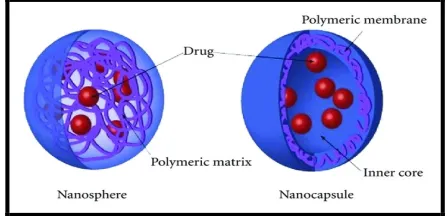
Depending upon the method of preparation
nanoparticles, nanospheres or nanocapsules can be
obtained. Nanospheres have a monolithic-type
structure (matrix) in which drugs are dispersed or
adsorbed onto their surfaces or encapsulated within
the particles. Nanocapsules are the vesicular system in which the drug is confined to a cavity consisting of an
inner liquid core surrounded by a polymeric
membrane. In this case the active substance is usually
dissolved in the inner core, but may also be adsorbed
to the capsule surface [Figure 1]. [2]
Figure 1: Types of polymeric nanoparticles
Advantages
The advantages of using nanoparticles as a drug
delivery system are as follows: [3]
1. Ease of manipulation of the particle size and surface characteristics of nanoparticles so as to Polymeric nanoparticles are particulate dispersions or solid particles with size in the range of 10-1000 nm. They represent a promising drug delivery system of controlled and targeted release. The use of polymeric nanoparticle for drug delivery is a strategy that aims to optimize therapeutic effects while minimizing adverse effects. Due to their small sizes, they exhibit unique physicochemical and biological properties like an enhanced reactive area as well as an ability to cross cell and tissue barriers, that make them a favourable material for biomedical applications. The present review focuses on methods of preparation, characterization and potential therapeutic applications of polymeric nanoparticles.
Keywords: Nanoparticles, Biodegradble polymers, Drug delivery, Characterization, Applications.
ABSTRACT ABSTRACT ABSTRACT ABSTRACT Meenakshi Dadwal
School of Pharmacy and Emerging Sciences, Baddi University of Emerging Sciences and Technology, Baddi (173205), Distt. Solan, Himachal Pradesh, India
achieve both passive and active drug targeting
after parenteral administration.
2. Controlled and sustained release of the drug during the transportation and at the site of
localization, altering organ distribution of the
drug and subsequent clearance of the drug so as
to achieve increase in drug therapeutic efficacy
and reduction in side effects.
3. Controlled release and particle degradation characteristics can be readily modulated by the
choice of matrix constituents.
4. Drug loading is relatively high and drugs can be
incorporated into the systems without any
chemical reaction; this is an important factor for preserving the drug activity.
5. Site-specific targeting can be achieved by attaching targeting ligands to surface of particles
or use of magnetic guidance.
6. Small sized nanoparticles can penetrate through
smaller capillaries, which could allow efficient
drug accumulation at the target sites.
7. The system can be used for various routes of
administration including oral, nasal, parenteral,
intra-ocular etc.
Limitations
Inspite of these advantages, nanoparticles have some
limitations [3]
1. Their small size and large surface area can lead to particle-particle aggregation, making physical
handling of nanoparticles difficult in liquid and
dry forms.
2. Small particle size results in limited drug loading
and burst release.
Polymers used in the preparation of nanoparticles The polymers used for preparation of nanoparticles
are as follows: [4]
• Natural polymers
• Synthetic polymers
Natural polymers:
Natural hydrophilic polymers such as proteins
(albumin, gelatine, legumin or vicilin) and
polysaccharides (alginate, dextran, chitosan, agarose,
pullulan) are widely used. But these polymer But
these polymers have certain disadvantages such batch-to-batch variation, conditional biodegradability,
and antigenicity.
Synthetic polymers:
These polymers are divided into two groups:
• Pre-polymerized: Poly (ε-caprolactone) (PECL), Poly (lactic acid) (PLA), Poly
(lactide-co-glycolide) (PLGA), and Polystyrene.
• Polymerized in process: Poly
(isobutylcyanoacrylates) (PICA), Poly (butylcyanoacrylates) (PBCA), Poly methyl
(methacrylates) (PMMA), and
Polyhexalcyanoacrylate (PHCA), and Copolymer
of aminoalkylmethacrylate methyl methacrylate.
Methods of Preparation of Polymeric
Nanoparticles
Nanoparticles can be prepared from a variety of
materials such as proteins, polysaccharides and
synthetic polymers. The selection of matrix materials
is dependent on many factors like:[1]
a) Size of nanoparticles required.
b) Inherent properties of the drug, e.g., aqueous solubility and stability.
c) Surface characteristics such as charge and
permeability.
d) Degree of biodegradability, biocompatibility and
toxicity.
e) Drug release profile desired.
f) Antigenicity of the final product.
Nanoparticles can be prepared by following methods:
1. Solvent Evaporation method
In this method, the polymer is dissolved in an organic
solvent such as dichloromethane, chloroform or ethyl
acetate, which is also used as the solvent for
dissolving the hydrophobic drug. The mixture of polymer and drug solution is then emulsified in an
aqueous solution containing a surfactant or
emulsifying agent to form oil in water (o/w) emulsion.
After the formation of stable emulsion, the organic
or by continuous stirring. Particle size was found to be
influenced by the type and concentrations of stabilizer, homogenizer speed and polymer
concentration. In order to produce small particle size,
often a high-speed homogenization or ultrasonication
may be employed. [5]
2. Spontaneous Emulsification or Solvent Diffusion Method
This is a modified version of solvent evaporation
method. In this method, the water miscible solvent
along with a small amount of the water immiscible
organic solvent is used as an oil phase. Due to the
spontaneous diffusion of solvents an interfacial
turbulence is created between the two phases leading to the formation of small particles. As the
concentration of water miscible solvent increases, a
decrease in the size of particle can be achieved. Both
solvent evaporation and solvent diffusion methods
can be used for hydrophobic or hydrophilic drugs. In
the case of hydrophilic drug, a multiple w/o/w
emulsion needs to be formed with the drug dissolved in the internal aqueous phase.[6]
3. Polymerization Method
In this method, monomers are polymerized to form
nanoparticles in an aqueous solution. Drug is
incorporated either by being dissolved in the
polymerization medium or by adsorption onto the nanoparticles after polymerization completed. The
nanoparticle suspension is then purified to remove
various stabilizers and surfactants employed for
polymerization by ultracentrifugation and
re-suspending the particles in an isotonic surfactant-free medium. This technique has been reported for making
polybutylcyanoacrylate or poly (alkylcyanoacrylate)
nanoparticles. [7,8]
4. Coacervation or Ionic gelation method
The method involves a mixture of two aqueous
phases, of which one is the polymer chitosan, a
di-block co-polymer ethylene oxide or propylene oxide (PEO-PPO) and the other is a polyanion sodium
tripolyphosphate. In this method, positively charged
amino group of chitosan interacts with negative
charged tripolyphosphate to form coacervates with a
size in the range of nanometer. Coacervates are formed as a result of electrostatic interaction between
two aqueous phases, whereas, ionic gelation involves
the material undergoing transition from liquid to gel
due to ionic interaction conditions at room
temperature. [9]
5. Salting out method
In this method acetone is chosen as the water miscible
organic solvent because of its pharmaceutical
acceptance with regard to toxicity. The method
consists of addition of water soluble polyvinyl alcohol
(PVA) in a highly concentrated salt solution in water
(aqueous phase) to a polymer solution in acetone (organic phase). Although acetone is miscible with
pure water in all ratios, the high salt concentration of
the aqueous phase prevents mixing of the phase. After
emulsification, addition of pure water in a sufficient
quantity causes acetone to diffuse into the aqueous
phase, resulting in the formation of nanoparticles. [10]
6. Nanoprecipitation method
Typically, this method is used for hydrophobic drug
entrapment, but it has been adapted for hydrophilic
drugs as well. Polymers and drugs are dissolved in a
polar, water-miscible solvent such as acetone,
acetonitrile, ethanol, or methanol. The solution is then
poured in a controlled manner (i.e. drop-by-drop addition) into an aqueous solution with surfactant.
Nanoparticles are formed instantaneously by rapid
solvent diffusion. Finally, the solvent is removed
under reduced pressure.[11]
7. Spray drying method
In this method, chitosan is first dissolved in acetic
acid; the drug is dissolved or dispersed in the solution
and then a suitable cross-linking agent is added; this
solution or dispersion is then atomised in a stream of
hot air. Atomisation leads to the formation of small
droplets, from which the solvent evaporates, leading
to the formation of free-flowing powders. Particle size depends upon size of the nozzle, spray flow rate,
atomisation pressure and inlet air temperature, and
8. Supercritical Fluid Technology
Conventional methods such as solvent extraction-evaporation, solvent diffusion and organic phase
separation methods require the use of organic
solvents which are hazardous to the environment as
well as to physiological systems. Therefore, the
supercritical fluid technology has been investigated as
an alternative to prepare biodegradable micro- and nanoparticles because supercritical fluids are
environmentally safe. [13]
A supercritical fluid can be generally defined as a
solvent at a temperature above its critical
temperature, at which the fluid remains a single phase
regardless of pressure. [13] Supercritical CO2 (SC CO2)
is the most widely used supercritical fluid because of
its mild critical conditions (Tc = 31.1 °C, Pc = 73.8
bars), nontoxicity, non-flammability, and low price.
The most common processing techniques involving
supercritical fluids are supercritical anti-solvent (SAS)
and rapid expansion of supercritical solution (RESS).
In the supercritical antisolvent method, both the drug and the polymer are dissolved in a suitable organic
solvent and are atomized through a nozzle into
supercritical CO2. The dispersed organic solvent phase
and the antisolvent CO2 phase diffuse into each other
and since CO2 is miscible only with the solvent, the
solvent gets extracted causing the supercritical fluid– insoluble solid to precipitate as nanoparticles. [14] In
the rapid expansion of supercritical solutions
technique, the solute is dissolved in supercritical CO2
and this solution is atomized through a nozzle into a
collection chamber at atmospheric conditions. When expanded, CO2 immediately evaporates and the solute
precipitates as a coprecipitate of the drug embedded
in the polymer matrix. [15]
Supercritical fluid technology technology technique,
although environment friendly and suitable for mass
production, require specially designed equipments
and is more expensive.
Characterization of Nanoparticles
Nanoparticles are generally characterized by their
size, morphology and surface charge, using such advanced microscopic techniques as scanning electron
microscopy (SEM), transmission electron microscopy
(TEM) and atomic force microscopy (AFM). The
average particle diameter, their size distribution and
charge affect the physical stability and the in vivo
distribution of the nanoparticles. Electron microscopy techniques are very useful in ascertaining the overall
shape of polymeric nanoparticles, which may
determine their toxicity. The surface charge of the
nanoparticles affects the physical stability and
redispersibility of the polymer dispersion as well as
their in vivo performance. Particle size
Particle size distribution and morphology are the most
important parameters of characterization of
nanoparticles. Morphology and size are measured by
electron microscopy. The major application of
nanoparticles is in drug release and drug targeting. It
has been found that particle size affects the drug release. Smaller particles offer larger surface area. As
a result, most of the drug loaded onto them will be
exposed to the particle surface leading to fast drug
release. On the contrary, drugs slowly diffuse inside
larger particles. As a drawback, smaller particles tend
to aggregate during storage and transportation of nanoparticle dispersion. Hence, there is a compromise
between a small size and maximum stability of
nanoparticles. [16]
There are a several tools for determining nanoparticle
size as discussed below:
Dynamic light scattering (DLS)
Currently, the fastest and most popular method of
determining particle size is photon-correlation
spectroscopy (PCS) or dynamic light scattering (DLS).
DLS is widely used to determine the size of Brownian
nanoparticles in colloidal suspensions in the nano and
submicron ranges .Shining monochromatic light (laser) onto a solution of spherical particles in
Brownian motion causes a Doppler shift when the
wavelength of the incoming light. This change is
related to the size of the particle. It is possible to extract the size distribution and give a description of
the particle’s motion in the medium, measuring the
diffusion coefficient of the particle and using
autocorrelation function. The photon correlation
spectroscopy (PCS) represent the most frequently
used technique for accurate estimation of the particle size and size distribution based on DLS. [17]
Scanning electron microscopy
Scanning electron microscopy (SEM) is giving
morphological examination with direct visualisation.
The techniques based on electron microscopy offer
several advantages in morphological and sizing analysis; however, they provide limited information
about the size distribution and true population
average. For SEM characterization, nanoparticles
solution should be first converted into a dry powder,
which is then mounted on a sample holder followed
by coating with a conductive metal, such as gold, using
a sputter coater. The sample is then scanned with a focused fine beam of electrons. [18] The surface
characteristics of the sample are obtained from the
secondary electrons emitted from the sample surface.
The nanoparticles must be able to withstand vacuum,
and the electron beam can damage the polymer. The
mean size obtained by SEM is comparable with the results obtained by dynamic light scattering.
Moreover, these techniques are time consuming,
costly and frequently need complementary
information about sizing distribution.
Transmission electron microscope
TEM operates on different principle than SEM, yet it
often brings same type of data. The sample
preparation for TEM is complex and time consuming
because of its requirement to be ultra thin for
the electron transmittance. The nanoparticles
dispersion is deposited onto support grids or films. To
make nanoparticles withstand the instrument vacuum and facilitate handling, they are fixed using either a
negative staining material, such as phosphotungstic
acid or derivatives, uranyl acetate, etc, or by plastic
embedding. Alternate method is to expose the sample
to liquid nitrogen temperatures after embedding in vitreous ice. The surface characteristics of the sample
are obtained when a beam of electrons is transmitted
through an ultra thin sample, interacting with the
sample as it passes through. [19]
Atomic force microscopy
Atomic force microscopy (AFM) offers ultra-high resolution in particle size measurement and is based
on a physical scanning of samples at sub-micron level
using a probe tip of atomic scale. [20] Instruments
provides a topographical map of sample based on
forces between the tip and the sample surface.
Samples are usually scanned in contact or noncontact mode depending on their properties. In contract
mode, the topographical map is generated by tapping
the probe on to the surface across the sample and
probe hovers over the conducting surface in
non-conduct mode. The prime advantage of AFM is its
ability to image non-conducting samples without any
specific treatment, thus allowing imaging of delicate biological and polymeric nano and microstructures.
[21] AFM provides the most accurate description of size
and size distribution and requires no mathematical
treatment.
Surface Charge
The nature and intensity of the surface charge of
nanoparticles is very important as it determines their
interaction with the biological environment as well as
their electrostatic interaction with the bioactive
compounds. The colloidal stability is analyzed through zeta potential of nanoparticles. This potential
is an indirect measure of the surface charge. It
corresponds to potential difference between the outer
Helmholtz plane and the surface of shear. The
measurement of the zeta potential allows for
predictions about the storage stability of colloidal
dispersion. High zeta potential values, either positive or negative, should be achieved in order to ensure
stability and avoid aggregation of the particles. The
predicted from the values of zeta potential. The zeta
potential can also provide information regarding the nature of material encapsulated within the
nanocapsules or coated onto the surface. [22]
Surface Hydrophobicity
Surface hydrophobicity can be determined by several
techniques such as hydrophobic interaction
chromatography, biphasic partitioning, adsorption of probes, contact angle measurements etc. Recently,
several sophisticated analytical techniques are
reported in literature for surface analysis of
nanoparticles. X-Ray photon correlation spectroscopy
permits the identification of specific chemical groups
on the surface of nanoparticles. [23] Drug Loading
Ideally, a successful nanoparticle system should have
a high drug-loading capacity thereby reduce the
quantity of matrix materials for administration. Drug
loading can be done by two methods:
• Incorporating at the time of nanoparticle production (incorporation method)
• Absorbing the drug after formation of nanoparticles by incubating the carrier with a
concentrated dug solution
(adsorption/absorption technique).
Drug loading and entrapment efficiency very much
depend on the solid-state drug solubility in matrix
material or polymer (solid dissolution or dispersion),
which is related to the polymer composition, the
molecular weight, the drug polymer interaction and
the presence of end functional groups (ester or carboxy). [11]
Drug Release
Drug release from the carrier-based particulate
system depends upon the cross-linking, morphology,
size, density of the particulate system and the
physiochemical properties of the drug, as well as presence of adjuvant. In vitro drug release also
depends upon pH, polarity and presence of enzymes
in the dissolution medium.
The release of drug from the particulate system
depends upon three different mechanisms: [24]
• Release from the surface of particles.
• Diffusion through the swollen rubbery matrix.
• Release due to erosion.
Various methods, which can be used to study the in
vitro release of drug, are as follows:
• Side-by-side diffusion cells with an artificial or biological membrane.
• Dialysis bag diffusion technique.
• Reverse dialysis bag technique.
• Ultracentrifugation.
• Centrifugal ultrafiltration.
Applications of Nanoparticles
Nanoparticles for drug delivery into the brain The blood-brain barrier (BBB) is the most important
factor limiting the development of new drugs for the central nervous system (CNS). [25] The BBB is
characterized by relatively impermeable endothelial
cells with tight junctions, enzymatic activity and active
efflux transport systems. It effectively prevents the
passage of water soluble molecules from the blood circulation into the CNS, and consequently only
permits selective transport of molecules that are essential for brain function. [26]
Strategies for nanoparticle targeting to the brain rely
on nanoparticle’s interaction with the specific
receptor-mediated transport systems in the BBB. For
example, polysorbate 80/LDL, transferrin receptor binding antibody (such as OX26), lactoferrin, cell
penetrating peptides and melanotransferrin have
been shown to be capable of delivery of a self non
transportable drug into
the brain via the chimeric construct that can undergo
receptor-mediated transcytosis. [28-30] It has been
reported that poly(butylcyanoacrylate) (PBCA)
nanoparticles were able to deliver hexapeptide
dalargin, doxorubicin and other agents into the brain
which is significant because of the great difficulty for
drugs to cross the BBB. [29] Despite some reported
success with polysorbate 80 coated nanoparticles, this
degradation and toxicity caused by presence of high
concentration of polysorbate 80. [31] OX26 MAbs
(anti-transferrin receptor MAbs), the most studied BBB
targeting antibody, have been used to enhance the
BBB penetration of lipsosomes. [32]
Another study by Kreuter et al. demonstrates the
delivery of several drugs successfully through the
blood brain barrier using polysorbate 80 coated PACA nanoparticles. [33] It is thought that after
administration of the polysorbate 80-coated particles,
apolipoprotein E (ApoE) adsorbs onto the surface. The
ApoE protein mimics low density lipoprotein (LDL)
causing the particles to be transported across the
blood brain barrier via the LDL receptors. The effects of polysorbate-80 on
transport through the blood brain barrier were
confirmed by Sun et al. with PLA nanoparticles. [34]
Nanoparticles were also functionalized with thiamine
surface ligands. These particles, with an average
diameter of 67 nm, were able to associate with the
blood brain barrier thiamine transporters and thereby increase the unidirectional transfer coefficient for the
particles into the brain. [35]
Nanoparticles for vaccine/gene delivery
Polynucleotide vaccines/DNA vaccines/plasmid
vaccines work by delivering genes encoding relevant
antigens to host cells where they are expressed, producing the antigenic protein within the vicinity of
professional antigen presenting cells to initiate
immune response. Such vaccines produce both
humoral and cell-mediated immunity because
intracellular production of protein, as opposed to extracellular deposition, stimulates both arms of the
immune system. [36] The key ingredient of
polynucleotide vaccines, DNA, can be produced
cheaply and has much better storage and handling
properties than the ingredients of the majority of
protein-based vaccines. Hence, polynucleotide
vaccines/DNA vaccines are set to supersede many conventional vaccines particularly for
immunotherapy. However, there are several issues
related to the delivery of polynucleotides which
limit their application. These issues include efficient
delivery of the polynucleotide to the target cell population, its localization to the nucleus of these
cells, and ensuring that the integrity of the
polynucleotides is maintained during delivery to the
target site. Nanoparticles loaded with plasmid DNA
could also serve as an efficient sustained release gene
delivery system due to their rapid escape from the degradative endo-lysosomal compartment to the
cytoplasmic compartment. [37] Hedley et al. [38]
reported that following their intracellular uptake and
endolysosomal escape, nanoparticles could release
DNA at a sustained rate resulting in continuous gene
expression. This gene delivery strategy could be applied to facilitate bone healing by using PLGA
nanoparticles containing therapeutic genes such as
bone morphogenic protein.
Tumor Targeting
The rationale of using nanoparticles for tumor
targeting is based on
1. Nanoparticles ability to deliver the requisite dose load of drug in the vicinity of the tumor due
to the enhanced permeability and retention
effect or active targeting by ligands on the
surface of nanoparticles.
2. Nanoparticles ability to reduce the drug exposure
to healthy tissues by limiting drug distribution to the target organ.
Active tumor targeting of nanoparticles may be
achieved with either direct targeting or the
pretargeting method. In direct targeting method,
nanoparticles are covalently coupled with the
ligands. The ligand coupled nanoparticles are received
by the tumor cells expressing a homologous receptor
on their surfaces. The specific ligand-receptor binding
ensures that the nanoparticles carrying drugs will get
attached specifically to the tumor cells. This will
facilitate delivery of drugs only to the cells (tumor
cells) expressing receptor and not the normal healthy cells. In the pretargeting approach, the therapeutic
administered after an appropriate delay time
following the administration of the targeting ligand. Nobs et al. explored both-approaches to target PLA
nanoparticles to tumor cells. In the direct approach,
nanoparticles with MAbs exposed on their surface
were incubated with the two tumor cells, while in the
pretargeting protocol, tumor cells were pretargeted
with biotinylated MAbs prior to the administration of avidin-labelled nanoparticles. [39]
Verdun et al. in an elegant experiment demonstrated
positive effects of using poly- isohexylcyanoacrylate-
nanospheres in the delivery of doxorubicin in mice.
The doxorubicin incorporated into poly
(isohexylcyanoacrylate) nanopsheres and delivered in mice showed higher concentrations of doxorubicin in
the liver, spleen and the lungs than in mice treated
with only free doxorubicin. [40]
Nanoparticles for oral delivery of peptides and proteins
Significant advances in biotechnology and
biochemistry have led to the discovery of a large number of bioactive molecules and vaccines based on
peptides and proteins. Development of suitable
carriers remains a challenge due to the fact that
bioavailability of these molecules is limited by the
epithelial barriers of the gastrointestinal tract and
their susceptibility to gastrointestinal degradation by digestive enzymes. Polymeric nanoparticles allow
encapsulation of bioactive molecules and protect them
against enzymatic and hydrolytic degradation. For
instance, it has been found that insulin-loaded
nanoparticles have preserved insulin activity and produced blood glucose reduction in diabetic rats for
up to 14 days following the oral administration. [41]
The surface area of human mucosa extends to 200
times that of skin. The gastrointestinal tract provides a
variety of physiological and morphological barriers
against protein or peptide delivery, for example:
proteolytic enzymes in the gut lumen like pepsin, trypsin and chymotrypsin; proteolytic enzymes at the
brush border membrane (endopeptidases); bacterial
gut flora; and mucus layer and epithelial cell lining
itself. [42] The histological architecture of the mucosa is
designed to efficiently prevent uptake of particulate matter from the environment. One important strategy
to overcome the gastrointestinal barrier is to deliver
the drug in a colloidal carrier system, such as
nanoparticles, which is capable of enhancing the
interaction mechanisms of the drug delivery system
and the epithelia cells in the GI tract. Nanoparticles for Ocular delivery
Topical ophthalmic drugs have generally poor
absorption in the eye due to the cornea’s low
permeability to drugs and noncorneal factors such as
rapid tear turnover, nasolacrimal drainage, and
systemic absorption. One of the major problems in ocular delivery is providing and maintaining an
adequate concentration of the therapeutic agent in the
precorneal area. Topical drop administration of
ophthalmic drugs in aqueous solutions results in
extensive drug loss due to tear fluid and eyelid
dynamics. Most non-invasive approaches for
enhancing ocular drug absorption involve the use of prodrugs, the use of viscosity agents designed to
prolong the drug residence time, and colloidal
systems.[43] Polymeric nanoparticles are attractive
colloidal systems because they demonstrate increased
stability and have a longer elimination half-life in tear
fluid (up to 20 min), than do conventional drugs applied topically to the eye, which have half-lives of
just one to three minutes. Nanoparticulate drug
delivery systems have been evaluated for ocular
applications to enhance absorption of therapeutic
drugs, improve bioavailability, reduce systemic side effects, and sustain intraocular drug levels. [44]
Qaddoumi et al.have studied the characteristics and
mechanism of uptake of poly(lactic-co-glycolic acid)
(PLGA) based nanoparticles for ophthalmic
application.[45] They suggested that PLGA-based
nanoparticles could be used for the enhancement of
drug absorption in the eye and the controlled release of proteins and drugs. Salgueiro et al.demonstrated
ophthalmic application of cyclophosphamide loaded
immunosuppressive agent. The morphometrical
properties such as average particle size and polydisparity index of these drug delivery systems are
adequate for ophthalmic application without induced
corneal or conjunctival irritation. [46]
Nanoparticles for Pulmonary Treatment
Nanostructured drug delivery and targeting systems
are tools to overcome the limitations of lung delivery by stabilizing and protecting the release in the bronchi
and make lung therapy through inhalation possible
and effective. Insulin-loaded PBCA nanoparticles were
studied by Zhang et al., they demonstrated that the
pulmonary administration of these nanoparticles
could significantly prolong the hypoglycemic effect of insulin. [47] An interesting study was reported by Liu et
al. incorporating estradiol and colloidal gold
nanoparticles in PLGA nanoparticles to be used as a
model for the pulmonary drug delivery systems. [48]
Vila et al. have shown that polyethylene glycol (PEG)
coating of the PLA nanoparticles increased the
absorption of drug in nasal mucosa. [49] Pandey et al.
demonstrated the application of nanoparticulate drug
delivery systems for the treatment of experimental
tuberculosis using poly(d,l-lactide-co-glycolide) as a
polymer. They used an inhalable system using the
nanoparticles and three anti-tubercular drugs
Rifampicin, Isoniazid, and Pyrazinamide. [50]
CONCLUSION
In the past few decades, researchers have studied
alternative delivery systems to improve the efficacy of
different medicines. Nanotechnology is an exciting novel field with hopes for improvements in wide
variety of uses in drug delivery in pharmaceutical
research. Polymeric nanoparticles offer a new avenue
to achieve drug delivery and drug targeting with
newly discovered disease site-specific drugs and
existing poorly soluble drugs. Overcoming the
obstacles in conventional drug delivery systems, polymeric nanoparticles are anticipated for better
application and effective drug delivery, and would
ultimately enhance treatment and patient compliance.
REFERENCES
1. Kreuter J. Nanoparticles. In: Kreuter J, editor.
Colloidal Drug Delivery Systems, New York : Marcel
Dekker; 1994, 219–342.
2. Krishna R.S., Shivakumar H.G., Gowda D.V., Banerjee
S. Nanoparticles: A novel colloidal drug delivery system. Indian J Educ Res 2006; 40:15‑21
3. Mohanraj V.J., Chen Y. Nanoparticles-A Review. Trop
J Pharm Res 2005; 5(1): 561-573.
4. Vyas S.P., Khar R.K. Targeted and controlled drug
delivery system. 1st ed. New Delhi: CBS Publication
and Distribution; 2000, 332-381.
5. Zambaux M., Bonneaux F., Gref R. Influence of
experimental parameters on the characteristics of
poly (lactic acid) nanoparticles prepared by double
emulsion method. J. Control. Release 1998; 50:31-40.
6. Niwa T., Takeuchi H., Hino T. Preparation of
biodegradable nanoparticles of water-soluble and
insoluble drugs with D,Llactide/ glycolide copolymer
by a novel spontaneous emulsification solvent
diffusion method, and the drug release behavior. J.
Contro.l Release 1993; 25: 89-98.
7. Zhang Q., Shen Z., Nagai T. Prolonged hypoglycaemic
effect of insulin-loaded polybutylcyanoacrylate
nanoparticles after pulmonary administration to
normal rats. Int. J. Pharm. 2001; 218:75-80.
8. Boudad H., Legrand P., Lebas G., Cheron M., Duchene
D., Ponchel G. Combined hydroxypropyl-[beta]-
cyclodextrin and poly (alkylcyanoacrylate)
nanoparticles intended for oral administration of
saquinavir. Int J. Pharm. 2001; 218:113-124.
9. Calvo P., Remunan-Lopez C., Vila-Jato J.L., Alonso M.J.
Novel hydrophilic chitosan-polyethylene oxide
nanoprticles as protein carriers. J. Appl. Polymer Sci.
1997; 63:125-132.
10. Mittal G., Sahana D.K., Bhardwaj V., Ravi Kumar M.N.
Estradiol loaded PLGA nanoparticles for oral
administration: Effect of polymer molecular weight
and copolymer composition on release behaviour in
vitro and in vivo. J. Control. Release 2007; 119:77-85.
11. Govender T., Stolnik S., Garnett MC., Illum L., Davis
S.S. PLGA nanoparticles prepared by
nanoprecipitation: drug loading and release studies
of a water soluble drug. J. Control. Release 1999;
12. Sinha S., Muthu M.S. Preparation and
characterisation of nanoparticles containing an
atypical anti-psychotic agent. Nanomedicine (Lond)
2007; 2:233-40.
13. Jung J., Perrut M. Particle design using supercritical
fluids: Literature and patent survey. J. Supercritical
Fluids 2001; 20:179-219.
14. Subramaniam B., Rajewski R.A., Snavely K.
Pharmaceutical processing with supercritical carbon
dioxide. J Pharm Sci 1997; 86:885-90.
15. Matson D.W., Peterson R.C., Smith R.D. Production of
fine powders by the rapid expansion of supercritical
fluid solutions. Adv Ceram 1987; 21:109.
16. Redhead H.M., Davis S.S., Illum L. Drug delivery in
poly(lactide-co-glycolide) nanoparticles surface
modified with poloxamer 407 and poloxamine 908:
in vitro characterization and in vivo evaluation. J.
Control. Release 2001; 70:353-363.
17. De Assis D.N., Mosqueria V.C., Vilela J.M., Andarde
M.S., Cardoso V.N. Release profiles and
morphological characterization by atomic force
microscopy and photon correlation spectroscopy of
99m Technetium-fluconazole nanocapsules.
International Journal of Pharmaceutics 2008;
349:152-160.
18. Jores K., Mehnert W., Drecusler M., Bunyes H., Johan
C., Mader K. Investigation on the stricter of solid lipid
nanoparticles and oil loaded solid nanoparticles by
photon correlation spectroscopy, field flow
fractionasition and transmission electron
microscopy. J. Control. Release 2004; 17:217-227.
19. Molpeceres J., Aberturas M.R., Guzman M.
Biodegradable nanoparticles as delivery system for
cyclosporine: preparation and characterization. J
Microencapsul 2000; 17: 599-614.
20. Muhlen A.Z., Muhlen E.Z., Niehus H., Mehnert W.
Atomic force microscopy studies of solid lipid
nanoparticles. Pharmaceutical Research 1996;
13:1411-1416.
21. Shi H.G., Farber L., Michaels J.N., Dickey A.,
Thompson K.C., Shelukar S.D., Hurter P.N., Reynolds
S.D., Kaufman M.J. Characterization of crystalline
drug nanoparticles using atomic force microscopy
and complementary techniques. Pharmaceutical
Research 2003; 20:479-484.
22. Pangi Z., Beletsi A., Evangelatos K. PEG-ylated
nanoparticle for biological and pharmacological
application. Adv. Drug Deliv. Rev. 2003; 24:403-419.
23. Scholes P.D., Coombes A.G., Illum L., Davis S.S., Wats
J.F., Ustariz C., Vert M., Davies M.C. Detection and
dtermination of surface levels of poloxamer and PVA
surfactant on biodegradable nanospheres using
SSIMS and XPS. J. Control. Release 1999; 59:261-278.
24. Jawahar N. and Meyyanathan S.N. Polymeric
nanoparticles for drug delivery and targeting: A
comprehensive review. Int J Health Allied Sci 2012;
1(4):217-23.
25. Chakraborty C., Sarkar B., Hsu C., Wen Z., Lin C., Shieh
P. Future prospects of nanoparticles on brain
targeted drug delivery. Journal of Neuro-Oncology
2009; 93(2):285-286.
26. Chen Y., Dalwadi G., Benson H.A.E. Drug Delivery
Across the Blood-Brain Barrier. Current Drug
Delivery 2004; 1(4):361-376.
27. Gabathuler R., Arthur G., Kennard M., Chen Q., Tsai
S., Yang J., Schoorl W., Vitalis T.Z., Jefferies W.A.
Development of a potential protein vector
(NeuroTrans) to deliver drugs across the
blood-brain barrier. International Congress Series 2005;
1277:171-184.
28. Ji B., Maeda J., Higuchi M., Inoue K., Akita H.,
Harashima H., Suhara T. Pharmacokinetics and brain
uptake of lactoferrin in rats. Life Sciences 2006;
78(8):851-855.
29. Pardridge W.M. Drug and gene targeting to the brain
with molecular trojan horses. Nat Rev Drug Discov
2002; 1(2):131-139.
30. Scherrmann J.M., Temsamani J. The use of Pep: Trans
vectors for the delivery of drugs into the central
nervous system. International Congress Series 2005;
1277:199-211.
31. Olivier J.C. Drug Transport to Brain with Targeted
Nanoparticles. NeuroRx: the journal of the American
Society for Experimental NeuroTherapeutics 2005;
2(1):108-119.
32. Pardridge W.M. Drug and gene targeting to the brain
via blood-brain barrier receptor-mediated transport
systems. International Congress Series 2005;
1277:49-62.
33. Kreuter J. Nanoparticulate systems for brain
delivery of drugs. Adv. Drug Deliv. Rev. 2001;
34. Sun W., Xie C., Wang H., Hu Y. Specific role of
polysorbate 80 coating on the targeting of
nanoparticles to the brain. Biomaterials 2004;
25(15):3065-3071.
35. Lockman P.R., Oyewumi M.O., Koziara J.M., Roder
K.E., Mumper R.J., Allen D.D. Brain uptake of
thiamine-coated nanoparticles. J. Control. Release
2003; 93(3):271-282.
36. Gurunathan S., Wu C.Y., Freidag B.L., Seder R.A. DNA
vaccines: a key for inducing long-term cellular
immunity. Current Opinion in Immunology 2000;
12(4):442-447.
37. Panyam J., Zhou W.Z., Prabha S., Sahoo S.K.,
Labhasetwar V. Rapid endolysosomal escape of
poly(DL-lactide-co-glycolide) nanoparticles:
implications for drug and gene delivery. The FASEB
Journal 2002; 16(10):1217-1226.
38. Hedley M.L., Curley J., Urban R. Microspheres
containing plasmid-encoded antigens elicit cytotoxic
T-cell responses. Nat Med 1998; 4(3):365-368.
39. Nobs L., Buchegger F., Gurny R. Biodegradable
Nanoparticles for Direct or Two-Step Tumor
Immunotargeting. Bioconjugate Chemistry 2005;
17(1):139-145.
40. Verdun C., Brasseur F., Vranckx H., Couvreur .P,
Roland M. Tissue distribution of doxorubicin
associated with polyisohexylcyanoacrylate
nanoparticles. Cancer Chemotherapy and
Pharmacology 1990; 26(1):13-18.
41. Damge C., Michel C., Aprahamian M., Couvreur P.,
Devissaguet J.P. Nanocapsules as carriers for oral
peptide delivery. J. Control. Release 1990; 13:
233-239.
42. Lee V., Yamamoto A. Penetration and enzymatic
barriers to peptide and protein absorption. Adv.
Drug Deliv. Rev. 1990; 4: 171-207.
43. Bourlais C.L., Acar L., Zia H., Sado P.A., Needham T.,
Leverge R. Ophthalmic drug delivery systems: recent
advances. Prog Retin Eye Res 1998; 17:33.
44. Zimmer A., Chetoni P., Saettone M., Zerbe H., Kreuter
J. Evaluation of pilocarpine loadedalbumin
nanoparticles as controlled drug delivery systems
for the eye. II. Coadministration with bioadhesive
and viscous polymers. J. Control. Release 1995;
33:31.
45. Qaddoumi M.G., Ueda H., Yang J., Davda J.,
Labhasetwar V., Lee H.L. The characteristics and
mechanisms of uptake of PLGA nanoparticles in
rabbit conjunctival epithelial cell layers. Pharm Res
2004; 21:641.
46. Salgueiro A., Egea M.A., Espina M., Valls O., Garcia
M.L. Stability and ocular tolerance of
cyclophosphamide loaded nanospheres. J
Microencapsul 2004; 21:213.
47. Zhang Q., Shen Z., Nagai T. Prolonged hypoglycemic
effect of insulin loaded polybutylcyanoacrylate
nanoparticles after pulmonary administration to
normal rats. Int J Pharm 2001; 218:75.
48. Liu Y., Tsapis N., Edwards D.A. Investigating
sustained-release nanoparticles for pulmonary drug
delivery. Cambridge, MA: Harvard University Press,
2003.
49. Vila A., Gill H., McCallion O., Alonso M.J. Transport of
PLA–PEG particles across the nasal mucosa: effect of
particle size and PEG coating density. J. Control.
Release 2004; 98:231.
50. Pandey R., Sharma A., Zahoor A., Sharma S., Khuller
G.K., Prasad B. Poly(d,l-lactide-coglycolide)
nanoparticles based inhalable sustained drug
delivery system for experimental tuberculosis. J
Antimicrob Chemother 2003; 10:1093.
How to cite this article: Meenakshi Dadwal; Polymeric Nanoparticles as Promising Novel Carriers for Drug Delivery: An Overview; J. Adv. Pharm. Edu. & Res. 2014: 4(1): 20-30.