This is an open access journal, and articles are distributed under the terms of the Creative Commons Attribution-Non Commercial-ShareAlike 4.0 License, which allows others to remix, tweak, and build upon the work non-commercially, as long as appropriate credit is given and the new creations are licensed under the identical terms.
© 2018 Journal of Advanced Pharmacy Education & Research | Published by SPER Publication 443
Strategic management of deep sternal wound infection using
Vacuum Assisted Closure system
Mahmoud ElDegwy
1, Alaa Omar
1, Ahmed Othman Elasheery
2, Mohamed AbdElsalam Shaaban
11Cardiothoracic Surgery Department, Faculty of Medicine, Cairo University, Egypt, 2Cardiothoracic Surgery Department, Faculty of Medicine, Fayoum University, Egypt
Correspondence: Mahmoud ElDegwy Cardiothoracic Surgery Department, Faculty of Medicine, Cairo University, Egypt, Email: [email protected]
ABSTRACT
Objectives: Vacuum-assisted closure (VAC) was primarily designed for the treatment of pressure ulcers and chronic debilitating wounds. Recently, VAC has become an encouraging treatment modality for sternal wound infection after cardiac surgery, providing an excellent result.
Methods: This was a prospective observational study that enrolled a total of 109 patients with deep sternal wound infections under usage of VAC system as a primary line of treatment. After improvement of the clinical parameters and the local wound area, the wound was closed by secondary intension or followed by conventional surgery.
Results: 107 patients (98.17 %) were treated successfully with complete healing of the wound. two patients (1.83 %) died, the first one died of septic shock and the other of sudden ventricular tachycardia and arrest. The overall length of hospitalization was 32.2 days (range 14-68), some patients discharged and complete VAC dressing at home. The median VAC treatment time until closure was 51.1 days (range 9-110). The most common organism obtained from wound culture was MRSA in 51 patients (46. 8%).VAC therapies were used as effective primary line of treatment of deep sternal wound infection (DSWI).
Conclusion: VAC therapy is a safe and reliable option in the treatment of sternal wound infection in cardiac surgery. VAC therapy should be considered as an effective primary modality of treatment of deep sternal wound infections and also effective adjunct to conventional treatment modalities for the treatment of extensive and life-threatening wound infections following cardiac surgery, particularly in the presence of risk factors.
Keywords: Vacuum assisted closure, sternal wound infection, reconstruction
Introduction
Sternal wound infection is considered a dreaded and highly lethal complication after cardiac surgery [1]
. This complication occurs in 1-5 % following cardiac surgeries with association of many problems related to cost of prolonged hospital stay,
prolonged use of antibiotics, frequent dressings and associated morbidity and mortality. There is many modalities and methods for dealing with this complication including repeated dressing, extensive wound debridement, closed irrigation with antibiotics and antiseptics or conventional surgeries using muscle oromental flaps [2]. The first use of vacuum assisted closure
system in practice was in 1993 in the treatment of chronic debilitating wound and pressure sores. Now the VAC system gaining popularity over other traditional conventional methods regarding to cost and effectiveness [3]
. the VAC system allowing marked granulation tissue growth and proliferation in acute and chronic Wounds that makes condition optimum for closure by secondary sutures or other conventional techniques specially in patients with high risk to undergo operation or failed conventional methods [4].
Access this article online
Website:
www.japer.in E-ISSN: 2249-3379
How to cite this article: Mahmoud ElDegwy, Alaa Omar, Ahmed Othman Elasheery, Mohamed AbdElsalam Shaaban. Strategic management of deep sternal wound infection using vacuum assisted closure system. J Adv Pharm Edu Res 2017;7(4):443-449.
Source of Support: Nil, Conflict of Interest: None declared.
Material and Methods
This study was a prospective observational study carried out between July 2011 and June 2017 in Cairo university hospitals. From 3123 median sternotomy operation carried on 109 patients of deep sternal wound infection occurred. Deep sternal wound infection (mediastinitis) was defined according to guidelines of the US Centers for disease Control and Prevention (CDC), which requires at least one of the following criteria: (a) an organism was isolated from culture of mediastinal tissue or fluid; (b) evidence of mediastinits during surgery; or (c) one of the following conditions, chest pain, sternal instability, or fever(>38 OC) is present and either purulent discharge from the
mediastinum or an organism isolated from the blood culture or culture of drainage of the mediastinal area.
Patients with fat necrosis, superficial wound infection involving only skin and subcutaneous tissue were excluded. Debridement carried mostly with local anesthesia with testing of sternal viability clinically. Some patients needing debridement under general anesthesia. The VAC system is the primary line of treatment which applied after debridement and irrigation of the wound. Tissue and fluid culture obtained at the time of debridement was sent for culture and sensitivity. Antibiotics given empirically consisted of vancomycin 500mg vial on 100 cc saline with imepenim 500mg IV both every 8 hours till the result of culture and sensitivity then change to the new antibiotic according to the result.
Every patient studied according to demographic data and risk factors including age, sex, diabetes, hypertension, smoking, COPD, CRF, obesity, immunosuppression, bilateral internal mammary harvesting in CABG operation, prolonged ICU and Ventilation, re-opening and blood transfusion. Computerized tomography to detect any retrosternal collection. Follow up the patients by CBC, CRP, swab for culture and sensitivity. Culture is obtained weekly in a regular manner. When the culture is negative for bacteria with gradual improvement of the wound condition and amount of discharge the antibiotic stopped.
Length of stay was recorded from the diagnosis of mediastinitis (if it occurred during the same hospitalization as the initial cardiac surgery) or the hospitalization from a separate admission for mediastinitis. Duration of vacuum system application was also recorded. In some of our patients if there is improvement of clinical parameters with improvement of local wound area patient continued on VAC system out-patient followed by health care professionalist or caregivers who is well trained to use the device and deal with its potential complications. Sternal stability assessed clinically if stable or unstable also CT scan gave information about the sternal gapping. Percentage of new granulation tissue growth and the
reduction of the wound size was weekly measured. According to the time of presentation it's classified into early presentation within 20 days postoperative and late presentation after 20 days’ post-operative. Patients were classified according to the Oakley classification system [5]
. El Oakley classification of deep sternal wound infection:
Type I: DSWI presenting within 2 weeks after operation in the absence of risk factors.
Type II: DSWI presenting at 2–6 weeks after operation in the absence of risk factors.
Type IIIA: DSWI type I in the presence of one or more risk factors.
Type IIIB: DSWI type II in the presence of one or more risk factors.
Type IVA: DSWI type I, II or III after one failed therapeutic trial.
Type IVB: DSWI type I, II or III after more than one failed therapeutic trial.
Type V: DSWI presenting for the first time > 6 weeks after operation.
VAC system composition and application:
Debridement of necrotic tissue, loose suture material, and bone is performed. Sterile polyurethane foam dressing (PFD) with an openpore in various sizes 400 to 600 µm is used then trimmed to the geometry of each wound. one or two layers of foam can be used covering whole the wound area. If sternal dehiscence present and the heart is exposed, we use Vaseline gauze on the heart and polyurethane foam layer over it and between the heart and sternum to protect the right ventricle then other layer between sternal halves and on the wound. A perforated tube then embedded in or between foam layers at which the foam, together with the first few inches of the drainage tube and the surrounding area of healthy skin, is then covered with the adhesive transparent membrane overlaps the wound margin by 5cm.
Foam used with thickness of 1-2 cm in our wounds. Foam was sterilized by autoclave at temp 121º for 30 minutes. The distal end of the drain is connected to the VAC unit, which is programmed to produce the required level of pressure between 50-125 mmHg in intermittent or continuous manner. The machine had been adjusted to be switched on for 5 minutes & switched off for 3 minutes. Continuous suction machine was used initially in case of large wounds with heavy exudates or a site of leakage.
Dressing changing:
wound, introducing sterile water or normal saline into the dressing, waiting 15-30 minutes facilitate its removal. Any necrotic, nonviable tissue, including bone, eschar, or hardened slough are debrided the surrounding skin must be clean & dry. Caution was considered to prevent foam to overlap onto intact skin.
Difficulties Encountering During VAC
Therapy
i Recurrent tubal obstruction by thick exudates in patients with heavy infection.
ii Tubal slippage during the some dressings.
Once initiated, VAC therapy was continued until granulation to skin level was complete or until the wound ceased to further contract. Sternal wounds were closed dependent upon their size, either allowed to close by secondary intention, or with direct suturing or regional flaps.
Guidelines for VAC removal and employment of definitive surgery include:
Decline of serological inflammation parameters (TLC, CRP <50mg/l).
Negative bacteriological cultures.
Resolution of local infection signs in the wound. Choice of flap, either omental or pectoralis muscle, was decided by the plastic surgeons.
Mechanism of action of VAC Therapy
The visible stretch due to negative pressure that contracts the foam called macrostrain which:
Draws wound edges together.
Removes infectious materials and exudates.
Equal distribution of negative pressure.
Gives direct and complete wound bed contact. The microdeformation at the cellular level, which leads to cell stretch, is called microstrain which [6]:
Reduces interstitial oedema.
Enhance new granulation tissue formation by
facilitating cell migration and proliferation.
Promotes local blood flow. When measured
underlying blood flow with a Doppler, the maximal flow occurred with - 125 mmHg pressure. When the pressure was above - 200 mmHg, blood flow began to decrease.
Statistical Analysis
Data were statistically described in terms of mean ± standard deviation (± SD), and range, or frequencies (number of cases)
and percentages when appropriate. All statistical calculations were done using computer program SPSS (Statistical Package for the Social Science; SPSS Inc., Chicago, IL, USA) version 15 for Microsoft Windows.
Results
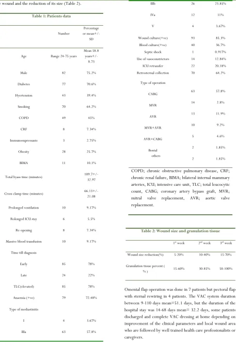
Our study including 109 patients with deep sternal wound infection in the period between July 2011 and June 2017. Most common prevalent operation was CABG (63, 57.8%), MVR (14, 12.8%), AVR (13, 11.9%), MVR+AVR (10, 9.2%), AVR+CABG (5, 4.6%), Bental (2, 1.85%), others (2, 1.85%). The age of the patients ranged from 24-75 years old with a mean of 58.8 years.27 of our 109 patients were females (24.8%) and remaining 82 patients were males (75.2%), Table 1.
Diabetes is one of the major risk factors for incidence of sternal wound infection and mediastinitis (n=77, 70.6%), hyertensives were 43 patients (39.4%). Smoking mostly associated with COPD which occurred in 70, 49 patients respectively (64.2, 45%). Obese patients were 28 (25.7%). Eight of our patients were chronic renal failure (CRF) on regular dialysis three times weekly. Three of our study groups were immunocompromized on oral corticosteroids.
There was 63 patients post CABG operation, 11 of them were subjected to bilateral internal mammary artery harvesting (17.5%). Prolonged ventilation and ICU stay represent risk factors for sternal wound infection. Prolonged mechanical ventilation was defined if more than 48 hours post-operative on ventilator. Re-exploration for bleeding occurred in 8 cases (7.3%). Massive blood transfusion was defined as transfusion of more than 3-4 units of packed RBCs which occurred in 10 (9.1%) patients of our study group. Early diagnosis within 20 days of operation occurred in 85patients (78%), but late diagnosis after 20 days of operation occurred in 29 patients (22%).
Among our study group 107 patients survived (98.2%) and complete healing achieved but two of our study group died. The first case was male patient 51 years old post MVR+AVR, diabetic, hypertensive, smoker, COPD with chronic renal failure on regular dialysis class IIIa Oakley classification died due to septic shock. the other mortality case was 62 years old male patient post CABG operation, diabetic, hypertensive, smoker, COPD, class IIIb Oakley classification, culture obtained MRSA organism. Patient died of sudden arrhythmia and arrest. All patients who had stable sternum were managed by secondary sutures (n=73, 67%).but who had unstable sternum were managed either by secondary intension sutures or by conventional flaps.
the wound and the reduction of its size (Table 2).
Table 1: Patients data
Number
Percentage or mean+/-
SD
Age Range 24-75 years
Mean 58.8 years+/-
8.73
Male 82 75.2%
Diabetes 77 70.6%
Hyertension 43 39.4%
Smoking 70 64.2%
COPD 49 45%
CRF 8 7.34%
Immunosupressants 3 2.75%
Obesity 28 25.7%
BIMA 11 10.1%
Total byass time (minutes) 109.7+/- 32.97
Cross clamp time (minutes) 66.13+/- 21.08
Prolonged ventilation 10 9.17%
Rolonged ICU stay 6 5.5%
Re-opening 8 7.34%
Massive blood transfusion 10 9.17%
Time till diagnosis
Early
Late
85
24
78%
22%
TLC(elevated) 85 78%
Anaemia (+ve) 79 72.48%
Type of mediastinitis
I
IIIa
4
63
3.67%
57.8%
IIIb
IVa
V
26
12
4
23.85%
11%
3.67%
Wound culture(+ve) 93 85.3% Blood culture(+ve) 40 36.7% Septic shock 1 0.917% Use of vasoconstrictors 14 12.84% ICU retransfer 22 20.18% Retrosternal collection 70 64.2%
Type of operation
CABG
MVR
AVR
MVR+AVR
AVR+CABG
Bental others
63
14
13
10
5
2
2
57.8%
2.8%
11.9%
9.2%
4.6%
1.85%
1.85%
COPD; chronic obstructive pulmonary disease, CRF; chronic renal failure, BIMA; bilateral internal mammary arteries, ICU; intensive care unit, TLC; total leucocytic count, CABG; coronary artery bypass graft, MVR; mitral valve replacement, AVR; aortic valve replacement.
Table 2: Wound size and granulation tissue
1st week 2nd week 3rd week
Wound size reduction(%) 5-20% 10-40% 15-70%
Granulation tissue percent (
% ) 15-60% 30-85% 50-100%
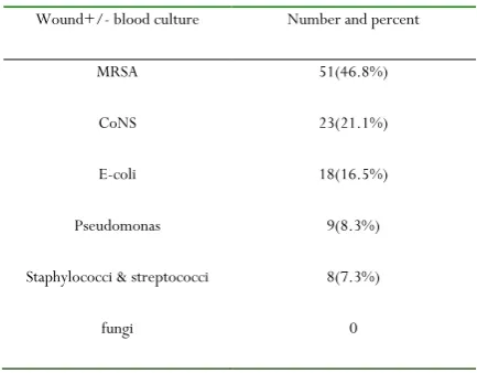
Negative pressure between 50-125mmHg did not cause any discomfort to the patients. Most of patients were culture negative between 7-10 days of VAC system application. The most prevalent organism obtained from the wound was MRSA. Other bacterial infection found including CoNS, E-coli, Pseudomonas aerigenosa, staphylococci and streptococci as (Table 3).
Table 3: Wound and blood culture
Wound+/- blood culture Number and percent
MRSA 51(46.8%)
CoNS 23(21.1%)
E-coli 18(16.5%)
Pseudomonas 9(8.3%)
Staphylococci & streptococci 8(7.3%)
fungi 0
Discussion
Following its description by Julian and colleagues in 1957, median sternotomy became the preferred incision for open cardiac procedures [7]
. Sternal wound infections are frequently seen by the plastic and cardiothoracic surgeons despite the relative low occurrence rates because of the large number of median sternotomies performed annually [8]. All procedures
designed to control sternal infections are undertaken with great risk because of possible involvement of vital underlying structures by the infectious process [9]. DSWI is still a
devastating complication after CABG [10]. The incidence of
DSWI (3.49%) is comparable to the incidence in other studies, which ranged between 0.4% and 5%. Frequent open dressing with povidoneiodine packing requires a lengthy hospital stay, and it is very demanding on patients and those treating them
[11]
. VAC is a very useful bridge to closure; it promotes formation of healthy granulation tissue, superior sternal support for improved respiratory function, and better psychological relief due to less intensive dressing every 48–96 h
[12]. Recent studies demonstrated that VAC therapy lowers the
rates of recurrent infection and therapeutic failure, and tends to decrease the morbidity and mortality associated with DSWI, compared to primary closure [10].
Retrospective study by Deniz et al., on 90 patients with DSWI, have shown significantly lower 90-day mortalityand treatment failure in patients with DSWI who were treated with VAC
system, comparedwith conventional methods of treatment. The cause of all deaths was multi-organ failureresulting from severe sepsis. Both studies also showed increased overall survival in the VAC group [13]
.
Retrospective study by De Feo et al. [14]
on 157 patients supports the finding of reduced mortality following VAC therapy compared to conventional treatment, and also showed lower reinfection rates using VAC therapy [14]
. No difference in long term survival was seen between patients treated with VAC system and closed drainage with irrigation in a recent retrospective study by Risnes and colleagues on 130 patients
[15]. They also reported failure of sternal wound healing and
reinfection to be more common in the closed drainage group. A meta-analysis by Damiani et al., of 321 patients showed no significant differences in mortality between NPWT and conventional therapy [16]
.
Another retrospective study by Sjogren and colleagues 115 showed that DSWI patients treated with NPWT may have similar long-term survival to patients without DSWI after coronary artery bypass grafting [17]
. Conflicting results have also been published regarding the length of hospital stay and length of treatment. Doss et al. described a retrospective study demonstrating a shorter length of stay and treatment after VAC compared with conventional wound management. These results were supported by a retrospective study by Simek et al.
[18].
The uniform negative pressure applied to the wound leads to arteriolar dilatation and thus increases microcirculation, thereby optimizing wound environment. By continuous suction, fluid excess and edema are decreased, thereby reducing bacterial colonization. These positive effects on the wound promote granulation, tissue proliferation, and accelerated wound healing. Therefore, definitive surgical repair, such as primary closure or plastic reconstructive surgery with omental or muscle flaps, can be accomplished safely. This reduces shear forces between the beating heart and the sternal edges, and acts as a preventive strategy against the rare, but fatal complication of right ventricular rupture. A patient on VAC feels no pain. If the patient does not tolerate this highpressure therapy, we begin with 50 mm Hg and titrate up to 125 mm Hg the following 24 to 36 hours in conjunction with adequate analgesic therapy consisting of nonsteroidal antirheumatics and opiates.
surgery. Therefore, the risk of sepsis due to swept bacteria into the circulation is markedly diminished.
With VAC therapy freedom from sternal wound microbiological cultures was achieved early, C-reactive protein level declined rapidly, in-hospital stay was short, sternal closure was achieved early and long-term survival tended to be high. VAC therapy markedly reduced required surgical intervention and reoperations for persistent infections, even in the group of high-risk patients. Traditionally, timing of the following surgical closure is based on the appearance of the wound, negativity of wound cultures and general patient condition.
Use of the VAC therapy as a definitive closure strategy has been recently reported. This approach seems to be mostly beneficial in high-risk patients, where it reduces dependence on regional flap and incidence of flap-related morbidity. The positive effect of subatmospheric pressure on the local wound healing process, active wound drainage and approximation of the wound edges was advantageous. However, the VAC therapy, guided by Creactive protein level, facilitates timing of wound closure, decreased morbidity and increased cost effectiveness of the procedure.
Conclusion
VAC system seems to significantly reduce the risk of early reinfections, increase the rate of granulation tissue formation, decrease the tissue edema, and stabilize the sternal haves, with excellent survival and low failure rate with reduction of the length and cost of hospital stay. The VAC system is a safe, reliable and effective option in the treatment of sternal wound infection in cardiac surgery, mainly promising as the first-line treatment for DSWI specially in presence of risk factors as diabetes, hypertension, smoking, bilateral IMA harvesting and others., with excellent survival and low failure rate.
References
1. Cowan KN, MD, PhD, Laura Teague, RN, MN, Sammy
C. Sue, BS, and James L. Mahoney, MDVacuum-Assisted Wound Closure of Deep Sternal Infections in High-Risk Patients After Cardiac Surgery Ann Thorac Surg
Vacuum-Assisted wound closure2005; 80:220512 doi:
10.1016/j.athoracsur.2005.04.005.
2. Ascherman JA, Patel SM, Malhotra SM, Smith CR. Management of sternal wounds with bilateral pectoralis major myocutaneous advancement flaps in 114 consecutively treated patients: Refi nements in technique and outcomes analysis. PlastReconstrSurg 2004; 114:676–83.
3. Argenta LC, Morykwas MJ. Vacuum-assisted closure. A new method for wound control and treatment: Clinical experience. Ann PlastSurg 1997; 38:563–77.
4. Raghupathy, Sabrena, Vaithiswaran A, et al. A
prospective randomized trial of vacuum-assisted closure versus standard therapy of chronic non-healing wounds. J. Evolution Med. Dent. Sci. 2016;5(49):3162-3167, DOI: 10.14260/jemds/2016/733.
5. El Oakley R, Wright J. Postoperative mediastinitis: classification and management. Ann ThoracSurg 1996; 61:1030–6.
6. Fleischmann W, Strecker W, Bombelli M, et al. Vacuum sealing as treatment of soft tissue damage in open fractures. Unfallchirurg 1993; 96(9):488-92.
7. Baskett RJ, MacDoiigall CE, and Ross DB: Is
mediastinitis a preventable complication? A 10-year Review, Ann ThoracSurg; 67: 462 - 465, 1999. 8. Blanchard A, Hurni M and Ruchat RK: Incidence of deep
and superficial sternal infection after open heart surgery. Eur J CardiothoracSurg; 9: 153- 157, 1995.
9. Wouters R, Wellens F and Geest RD: Sternitis and mediastinitis after coronary artery bypass grafting Texas Heart Inst J; 21: 183-188, 1994.
10. Mohamed AbdelrahmanBadawy, Fahad Al Shammari,
TareqAleinati, Moataz Salah Eldin, RiyadTarazi and Jamal Alfadli Deep sternal wound infection after coronary artery bypass: How to manage? Asian Cardiovascular & Thoracic Annals 2014, Vol. 22(6) 649–654.
11. Fuchs U, Zittermann A, Stuettgen B, Groening A, Minami K and Koerfer R. Clinical outcome of patients with deep sternal wound infection managed by vacuum-assisted closure compared to conventional therapy with open packing: a retrospective analysis. Ann ThoracSurg 2005; 79: 526–531.
12. Fl.eck TM, Fleck M, Moidl R, et al. The vacuum-assisted closure system for the treatment of deep sternal wound infections after cardiac surgery. Ann ThoracSurg 2002; 74: 1596–1600.
13. Deniz H, Gokaslan G, Arslanoglu Y, Ozcaliskan O, Guzel G, Yasim A, Ustunsoy H. Treatment outcomes of postoperative mediastinitis in cardiac surgery; negative pressure wound therapy versus conventional treatment. J Cardiothorac Surg. 2012; 7:67.
14. De Feo M, Della Corte A, Vicchio M, Pirozzi F, Nappi G, Cotrufo M. Is poststernotomymediastinitis still devastating after the advent of negative-pressure wound therapy? Tex Heart Inst J. 2011; 38:375-380.
traditional closed drainage on survival and reinfection rate. Int Wound J. 2012.
16. Damiani G, Pinnarelli L, Sommella L, Tocco MP, Marvulli M, Magrini P, Ricciardi W. Vacuum-assisted closure therapy for patients with infected sternal wounds:
A metaanalysis of current evidence. J
PlastReconstrAesthet Surg. 2011; 64:1119-1123. 17. Sjogren J, Malmsjo M, Gustafsson R, Ingemansson R.
Poststernotomymediastinitis: A review of conventional surgical treatments, vacuum-assisted closure therapy and presentation of the Lund University Hospital mediastinitis algorithm. Eur J Cardiothorac Surg. 2006; 30:898-905. 18. Simek M, Hajek R, Fluger I, Molitor M, Grulichova J,