Ireneusz Łątkowski
1, Mariusz Wysocki
2, Ireneusz Siewiera
234 Years of Experience in the Treatment of Patients
with Syndactyly at the Department and Clinic of Plastic
Surgery in Polanica-Zdrój
34-letnie doświadczenie w leczeniu pacjentów z palcozrostami
w Oddziale i Klinice Chirurgii Plastycznej w Polanicy-Zdroju
Clinic of Plastic Surgery, Wroclaw Medical University, Poland
Department of Plastic Surgery, Specialist Medical Center, Polanica-Zdrój, Poland
Abstract
Background. Syndactyly is the most common congenital hand malformation. Over the past two centuries 46 dif-ferent methods of syndactyly release have been described. However, the overall goal of these methods remains the same: reconstruction of the deep and wide web space with proper commissure location.
Objectives. The objective of this paper is to present the protocol for treatment used in the authors’ department in both isolated syndactyly and in syndactyly associated with other congenital hand anomalies.
Material and Methods. During the past 34 years 400 patients with syndactyly were treated in Hospital and Clinic of Plastic Surgery in Polanica Zdrój. In the analyzed population, patients with acrosyndactyly prevailed (39.5%), while 24.6% of the patients were operated on for isolated syndactyly. The basic surgical techniques of syndactyly release including operation on one side of a digit at a time, zigzag incision, commissure reconstruction using local flaps and full-thickness skin grafts for covering raw areas were used to produce a hand with as many independent and functional fingers as possible, with the fewest surgical procedures and complications.
Results. Aiming at standardizing procedures that offer the best treatment results with fewer complications, the department has used a treatment algorithm for simple syndactyly and syndactyly associated with other complex hand anomalies, in which two operation periods are distinguished: before and after two years of age. Up to age two procedures are performed that cannot be postponed without leading to hand deformities that are progressively more difficult (or even impossible) to correct, with poor treatment results. After age two, simple syndactyly release is performed between fingers of similar length, as well as corrective procedures of coexisting hand anomalies. In 90% of the patients with isolated syndactyly very good treatment results have been achieved; in 7% the result has been good; and in 3% the result was poor. We did not observe more serious complications such as necrosis of the finger secondary to vascular compromise or skin graft necrosis. Web creep and scar contracture occurred in 17% and 12% of patients respectively.
Conclusions. Surgical treatment of syndactyly associated with other hand anomalies is complex and multistaged. The surgical procedures should be performed earlier or later in the first years of the patient’s life, depending on the morphology of the anomaly. The proposed algorithm for syndactyly treatment offers good outcomes with no severe complications. Web creep and scar contractions requiring secondary correction procedures were the most frequently noted complications (Adv Clin Exp Med 2011, 20, 1, 71–78).
Key words: syndactyly, congenital hand malformation, surgical treatment.
Streszczenie
Wprowadzenie. Palcozrost jest najczęstszą wadą wrodzoną ręki. W ciągu dwóch wieków opracowano około 46 różnych technik operacyjnych rozdzielenia syndaktylii, zasadniczy cel pozostaje jednak taki sam – wytworzenie odpowiednio głębokiej i szerokiej przestrzeni międzypalcowej z prawidłowym umiejscowieniem spoidła.
Cel pracy. Przedstawienie stosowanego w Klinice Chirurgii Plastycznej w Polanicy-Zdroju protokołu leczenia pal-cozrostów izolowanych oraz towarzyszących złożonym wadom wrodzonym ręki.
Materiał i metody. W ciągu ostatnich 34 lat w Szpitalu i Klinice Chirurgii Plastycznej w Polanicy-Zdroju leczono 400 pacjentów z palcozrostami. W analizowanej populacji dominowali pacjenci z zespołem pierścieni przewężających (39,5%); 24,6% badanych operowano natomiast z powodu izolowanego palcozrostu.
Adv Clin Exp Med 2011, 20, 1, 71–78 ISSN 1230-025X
OrIgINAl PAPErS
Stosowano podstawowe techniki chirurgiczne obejmujące operowanie tylko jednej strony palca podczas jednego zabiegu, cięcia „zygzak” po stronie dłoniowej i grzbietowej, wytworzenie spoidła z użyciem tkanek miejscowych oraz stosowanie przeszczepów pełnej grubości. Celem leczenia było rozdzielenie palcozrostów z wytworzeniem ręki z możliwie jak największą liczbą wolnych i funkcjonalnych palców przy możliwie jak najmniejszej liczbie zabiegów operacyjnych i powikłań.
Wyniki. W celu standaryzacji postępowania prowadzącego do uzyskania jak najlepszych wyników leczenia przy ograniczeniu częstości powikłań opracowano protokół leczenia palcozrostów izolowanych oraz towarzyszących innym złożonym wadom ręki. Wyodrębniono dwa okresy przeprowadzania operacji chirurgicznej (przed i po 2. r.ż.). Do 2. r.ż. wykonywano zabiegi, których odroczenie skutkowałoby trudnymi, a nawet niemożliwymi do kore-kcji zniekształceniami i pogorszeniem wyników leczenia. Po 2. r.ż. rozdzielano palcozrosty proste między palcami o zbliżonej długości, a także wykonywano zabiegi korygujące towarzyszące zniekształcenia ręki. U 90% pacjentów z izolowanym palcozrostem uzyskano bardzo dobry wynik leczenia, u 7% dobry, a u 3% zły. Nie odnotowano poważniejszych powikłań w postaci martwicy palców lub przeszczepów skóry. Zaobserwowano przykurcze blizno-wate i spłycenie przestrzeni międzypalcowych odpowiednio u 12% i 17% pacjentów.
Wnioski. leczenie palcozrostów, szczególnie współwystępujących z innymi wadami rozwojowymi ręki, jest złożone i wieloetapowe. Operacje chirurgiczne powinny być przeprowadzane w pierwszych latach życia dziecka w zależności od morfologii wady. Wypracowany algorytm leczenia umożliwia uzyskanie dobrego wyniku leczenia bez poważnych powikłań pooperacyjnych. Najczęściej obserwowanymi powikłaniami były przykurcze bliznowate i spłycenia przestrzeni międzypalcowych (Adv Clin Exp Med 2011, 20, 1, 71–78).
Słowa kluczowe: palcozrost, wady wrodzone ręki, leczenie chirurgiczne.
The word syndactyly is derived from the greek
syn – meaning together – and dactylos – fingers. This deformity is the most common congenital hand anomaly, and is caused by a failure in the process of differentiation and separation of adjacent fingers. It may be an isolated form or associated with other conditions, occurring in 28 different syndromes, including Poland’s syndrome (symbrachydactyly), amniotic disruption syndrome (acrosyndactyly), Apert syndrome, Pffeifer’s syndrome, etc. Isolated hand syndactyly is estimated to occur in 1 per 2000 to 2500 children, more frequently in males than in females (56% to 44%) and both unilaterally and bi-laterally with equal frequency [1–5].
The treatment of syndactyly is dependent on the type of syndrome. Although its isolated form does not pose a great therapeutic problem, surgical treatment of syndactyly associated with other hand anomalies is a complex and multistaged process. A wide spectrum of morphologic abnormalities requires an individual approach to every patient. Moreover, the many techniques described and used over the past two centuries give surgeons the opportunity to choose the appropriate treatment method for every patient. The aim of the treatment is to achieve the best possible hand functioning with the fewest number of surgical procedures, and to avoid the risk of postoperative complications. The wide variety of operating techniques, the va-riety of anomalies and controversy concerning the age at which treatment should commence have all contributed to the establishment of numerous op-erating protocols.
On the basis of 34 years of experience in the treatment of congenital hand anomalies at the Hospital and Clinic of Plastic Surgery in Polanica Zdrój, Poland, and aiming at standardizing
pro-cedures resulting in the achievement of the best treatment results along with a reduction in the frequency of complications, the authors propose a treatment algorithm for simple syndactyly and syndactyly associated with other complex hand anomalies. The objective of this paper is to present treatment protocol.
Material and Methods
The Patients
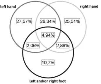
During the 34 years from January 1976 to De-cember 2009, 400 patients with syndactyly (51.3% males and 48.7% females) were treated in Hospi-tal and Clinic of Plastic Surgery in Polanica Zdrój. Their ages ranged from three months to 35 years (mean 2.9 years). In the evaluated population there was a prevalence of unilateral and bilateral syndac-tyly of the upper extremity (about 25% each), and in 5% of the population syndactyly was observed in all four extremities (Figure 1).
The patients in the study were grouped accord-ing to the type of anomaly they presented, usaccord-ing the IFSSH classification established in 1983 [6, 7]. Patients with acrosyndactyly (39.5%) were the larg-est subgroup. Syndactyly associated with various kinds of upper extremity hypoplasty was observed in 25.8%, while 24.6% of the patients were oper-ated on for isoloper-ated syndactyly (Figure 2).
Methods
The overall goal of syndactyly release is to pro-duce a hand with as many independent and func-tional fingers as possible, with the fewest number of surgical procedures and complications.
Indications for surgery were identified as func-tional and esthetic ones. generally, various forms of syndactyly were qualified for operating proce-dures if the release would offer hand function im-provement. Indications of an exclusively esthetic nature were mainly related to completely or par-tially isolated syndactyly in the feet.
Four main contraindications to surgery were highlighted, as follows:
– a polydactyl hand with syndactyly, but with functionally good syndactylized fingers, the release of which could worsen its function;
– a hand with no muscular control;
– adults with good functioning of the hand with syndactyly;
– syndactyly associated with so-called ‘super digits’ of type I or II: in type I, two metacarpals support a single oversized digit, while in type II, a single metacarpal supports two or more syndac-tylized digits distally.
The basic surgical techniques for syndactyly release were as follows:
– meticulous surgical soft tissue division to pro-tect neurovascular structures;
– operation on one side of a digit at a time; – commissure reconstruction using local flaps; – zigzag incisions on the dorsal and palmar sur-faces;
– the use of full-thickness skin grafts from the groin area to cover raw areas;
Adequate postoperative immobilization, using well-padded plaster casts.
The results of isolated syndactyly release were evaluated on a three-degree scale on the basis of commissure morphology (depth of web space), scar contracture and finger mobility. Statistical analysis was performed using Statistica 8.0 (Stat-Soft Inc.) The relationship between the results of treatment and the age of the operated patients was checked using the ANOVA test. Statistical signifi-cance was set at p < 0.05.
Results
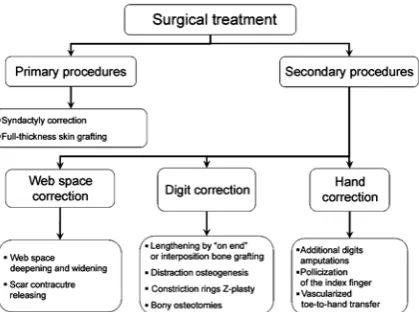
Two groups of surgical procedures were iden-tified (Figure 4): primary procedures to release syndactyly and secondary procedures to treat co-existing malformations, and to correct formed web spaces. Depending on the morphology of the anomaly, a one-stage procedure was frequently performed involving syndactyly release along with correction of a coexisting deformity.
The Timing of Surgery
Depending on the morphology of the anomaly and coexisting deformities, two periods for
sur-Fig. 1. Distribution of the analyzed population according to disease location
Ryc. 1. rozkład badanej populacji względem umiejscowienia choroby
Fig. 2. Incidence of hand anomalies in the study popu-lation according to the IFSSH classification
Ryc. 2. Procentowy rozkład badanej populacji według klasyfikacji IFSSH
Fig. 3. Incidence of web space involvement in simple syndactyly
gery have been distinguished (Table 1). The age of 24 months is the approximate ending point of the time period whenpatterns of prehensile func-tion are established in the cortex. Up to the age of two years, procedures are performed that cannot be postponed without leading to hand deformi-ties that are progressively more difficult (or even impossible) to correct, with poor treatment re-sults. After age two, simple syndactyly release is performed between fingers of similar length, as well as corrective procedures of coexisting hand anomalies.
Surgical Treatment
Algorithms
Isolated Syndactyly
In general, syndactyly between the thumb and index finger are corrected between the ages of 12 and 18 months. Wide and deep web space is obtained with a combination of Z-plasty and a dorsal hand flap. Syndactyly between the fourth and fifth fingers is released prior to the age of two years due to the difference in the fingers’ length and the risk of progression of a skeletal deformity. Other syndactylies are corrected later on (Figure 5). For commissure reconstruction, proximally pedicled triangle flaps were elevated dorsally and palmary on the level of the metacarpophalangeal joints, with a length of approximately half of the proximal phalanx (Figure 5 – chart). Syndactyly release was performed using ‘zigzag’ incisions on the dorsal and palmar surfaces. Tissue dissection was performed meticulously, preserving neurovas-cular structures.
In cases of complex syndactyly involving bones, phalanxes were cut off; and in cases of joint instability, the joint capsule was reconstructed us-ing the dorsal aponeurosis.
In multiple-digit syndactyly, due to the high risk of necrosis in case the vascular structure is altered, adjacent digits are not operated simulta-neously. reconstruction of many web spaces is performed in stages, at intervals of a minimum of six months. In cases of bilateral syndactylies, the
Fig. 4. Algorithm of the surgical treatment of hand anomalies
Ryc. 4. Algorytm leczenia chirurgicznego nieprawidło-wości budowy ręki
Table 1. Anomalies treated in two different periods
Tabela 1. Przedziały wiekowe pacjentów, u których rozpoczynano leczenie chirurgiczne poszczególnych rodzajów chorób Start of treatment
(Czas rozpoczęcia leczenia)
Malformation (rodzaj choroby)
< 24 months
(< 24 m-ce) syndactyly between fingers of unequal length – 1st and fourth web space) (palcozrost między różnej długości palcami – 1. i 4. przestrzeń międzypalcowa) syndactyly with the bridges between terminal phalanges
(palcozrost ze zrośniętymi paliczkami dystalnymi) acrosyndactyly with tethering of adjacent digits (akrosyndaktylia ze splecieniem przyległych palców) symbrachydactyly
(symbrachydaktylia)
deep constriction rings with pronounced distal edema (obrzęk limfatyczny w zespole pierścieni amniotycznych) > 24 months
(> 24 m-ce) syndactyly between fingers of equal length (prosty palcozrost między palcami podobnej długości) clinodactyly – osteotomy
(klinodaktylia – osteotomia korekcyjna)
authors recommend simultaneous operations on both hands. Incomplete isolated syndactylies are released the same way as complete syndactylies. Isolated complete and incomplete foot syndactyly that does not impair the walking process is treated only for esthetic reasons, applying the same prin-ciples as in hand syndactyly.
Postoperative dressings use medicated gauze covering the skin grafts, with compressible cotton moistened with Nitrofurazon, vaseline, etc., placed as a stent within the reconstructed web space. The dressing and hand are immobilized with a plaster cast. Particular attention is paid to keeping the finger pulps uncovered in order to monitor the blood supply. Dressings are removed on the third day following the procedure. The wounds and skin grafts are cleaned, then, after ointment application, loose protective dressing is put on and the hand is secured in loose cotton sack. The sutures are re-moved on day 9 after surgery.
Acrocephalosyndactyly
– Apert Syndrome
The complex treatment includes modeling and augmenting the cranial vault, which is performed during the first 12 months due to urgent indica-tions. The treatment of hand deformities is there-fore postponed until the age of two years.
The treatment sequence begins with the release of the border digits, converting a type III and/or type II hand to type I hand.
A spoon-shaped or hoof-shaped hand and symphalangism allow for the use of a straight inci-sion instead of typical zigzag type, without the risk of scar contractures [8]. For wide first web-space
release a dorsal transpositional flap (eg. a Buck-gramcko flap) is advocated.
In the next stages, depending on the morphol-ogy of the anomaly, syndactyly release, separation of synosthosis of the metacarpal bones and, in cas-es of decreased prehensile function, thumb clinod-actyly correction are performed (Figure 6).
The severe skeletal deformities in a type III hand in Apert syndrome prevent the reconstruc-tion of more than three digits. Hence, in planning hand reconstruction in Apert syndrome, a three-finger hand with a deep and wide first web space are advocated. Despite the accompanying sympha-langy, in many cases this appproach proves to be functionally better than a four-finger hand.
Symbrachydactyly
– “Atypical Cleft Hand”
– Poland’s Syndrome
These anomalies commonly affect fingers II-IV. In the most severe cases of Poland’s syndrome, there is a hypolpastic thumb and finger V with buds of the other fingers.
Treatment of mild cases of Poland’s syndrome with well-developed fingers consists of syndactyly release following the general principles of web-space reconstruction (Figure 7).
In patients with a severe form of the anomaly, with remnant fingers, especially II and III, transfer of the border fingers is performed, aiming at im-proving prehensile function.
Due to the undisturbed anatomy of vessels and nerves, patients with no fingers are good can-didates for toe-to-hand transfer.
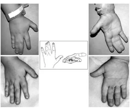
Fig. 5. Chart and clinical example of isolated simple syndactyly release
Ryc. 5. Schemat i kliniczny przykład rozdzielania pal-cozrostu izolowanego prostego palców III/IV
Fig. 6. Apert syndrome – type I hand – before and after syndactyly release
Acrosyndactyly – Constricted
Rings Syndrome
This syndrome has a varied morphology of bone and soft tissue deformities, the severity of which is correlated to the difficulty of treatment.
Mild forms comprise simple partial syndactyl-ies with superficial amniotic constriction rings and no bone deformities. The most severe cases have transverse finger amputations with no proper pha-langes and deficiencies in the metacarpal bones.
In planning the reconstructive surgery in con-stricted rings syndrome, the most essential goal is to pursue the best hand functioning, which in the most severe cases entailed the creation of a satis-factory grasping function. Surgery is commenced
in the first 12 to 18 months of life. regardless of the severity of the deformity, the first stage of surgical treatment begins with syndactyly release, excision of sinuses and meticulous reconstruction of wide and deep web spaces (Figure 8).
When correcting short and underdeveloped fingers or fingers amputated because of amniotic bands, there is a tendency to save the longest pos-sible fingers with properly modeled fingertips. In cases of finger stumps with deficient skeletal sys-tems, which aggravate hand function, stump am-putation was performed.
In patients with stumps that prevent the de-velopment of any grasping function, toe-to-hand transfers or distraction osteogenesis are performed, depending on the stump length (Figure 9).
Fig. 7. Poland’s syndrome in a male patient: the third and fourth web spaces were released. The anomaly con-sisted of short and hypoplastic rays with symphalangism
Ryc. 7. Zespół Polanda u męż-czyzny; rozdzielenie prostego palcozrostu III/IV i IV/V. Widoczne skrócone niedoro-zwinięte palce z symfalangi-zmem
Fig. 8. A patient with acrosyndactyly before and after syndactyly treatment in a one-stage operation. (Kobus K.: Atlas of Plastic Surgery)
Ryc. 8. Akrosyndaktylia przed i po rozdzie-leniu palcozrostów podczas jednego zabiegu chirurgicznego (Kobus K.: Atlas Chirurgii Plastycznej)
Fig. 9. Constricted ring syndrome: the final results after syndactyly treatment, vascular-ized toe-to-hand transfer and distraction osteogenesis of the fourth finger (X-ray, Kobus K.: Atlas of Plastic Surgery)
Results of Treatment
Due to the variety in the morphology and se-verity of the malformations treated, it is difficult to conduct a comparative analysis of the final treat-ment results within the whole group, or a compar-ative analysis according to the type of malforma-tion.
A complete functional and esthetic evaluation of the syndactyly procedure can be obtained in the case of isolated syndactyly. The criteria for treat-ment results included a properly created and locat-ed commissure (depth of web space), the presence of scar contracture and finger mobility. The results were evaluated on a three-degree scale based on these criteria (Table 2).
In 90% of the patients with isolated syndactyly, a very good treatment result was achieved; in 7% the result was good and in 3% the result was poor. There was no statistically significant correlation between the age of the operated patients and the treatment result.
Complications
In the population of patients studied, the most serious complications, such as necrosis of the fin-ger secondary to vascular compromise or skin graft necrosis, were not observed. Web creep and scar contracture occurred in 17% and 12% of patients respectively, and required secondary correction procedures.
Discussion
The treatment of simple syndactyly and syn-dactyly associated with other hand anomalies is
a multistaged and complex process. Over the past two centuries 46 different methods of syndactyly release have described [9]. However, the overall goal of these methods remains the same – recon-struction of the deep and wide web space with proper commissure location.
Although in simple syndactyly the outcomes are good, in complex syndactyly with coexisting hand deformities, the results are far from ideal.
In the treatment of severe hand deformities, a great deal of attention is paid to timing of the surgery. Despite a lack of consensus, most authors emphasize the necessity to perform the surgery during the first year of life [10]. Adequately ear-ly treatment enables proper prehensile function, which develops during the first 24 months of life (11). There have also been some reports published about surgery done in the first two weeks of life in cases of some minor deformities [12].
In the algorithm of surgical treatment presented in the current paper, procedures treating deformities requiring early intervention are performed during the first 24 months of the patient’s life. This mainly involves cases of syndactyly between fingers of dif-ferent length and with coalition, when non-equal growth may aggravate the existing deformity. More-over, early release of the first web space enables the hand to develop proper grasping function.
Complex and lengthy procedures such as toe-to-hand transfer, distraction osteogenesis and os-teotomy are performed at preschool age.
The principle goal of syndactyly release is to create the most functional hand possible. How-ever, a good functional outcome is proportionally dependent on the severity of the deformation, and the most important factor is proper innervation with good motor function of the hand.
All congenital upper extremity deformities have proper innervation, but the reinnervation
Table 2. Criteria for assessing treatment results Tabela 2. Kryteria kwalifikujące wyniki leczenia
Very good
(Bardzo dobry) good (Dobry) Poor (Zły) Finger mobility active and
passive (ruchomość pal-ców czynna i bierna)
complete range of active and passive mobility (pełny zakres ruchomości czynnej i biernej)
complete range of active and passive mobility (pełny zakres ruchomości czynnej i biernej)
impaired active and passive mobility (upośledzona ru-chomość czynna i bierna)
Web space (Przestrzeń
międzypalcowa) web space of proper depth (przestrzeń międzypalcowa prawidłowej głębokości)
web space of proper depth (przestrzeń międzypalcowa prawidłowej głębokości)
shallow web space (spłycona przestrzeń międzypalcowa)
Scar contracture
(Przy-kurcz bliznowaty) lack (brak) scar contracture with no function impairment (przykurcz bliznowaty bez upośledzenia funkcji)
after complex reconstructions with toe-to-hand transfers goes much better in younger patients [9], which constitutes a vital argument supporting early treatment.
At the same time, a meticulous surgical tech-nique is important because the main factor im-pairing proper hand development is an improp-erly performed operation. This is due to injury to the neurovascular structures and scar contractures leading to functional impairment of the hand. The frequency of complications varies from 2% to 24 % and is due to scar contractures and web creeping, requiring secondary corrective procedures [13, 14].
Flatt emphasized the fact that about 90% of ev-eryday activities might be done using one hand and that the treatment of bilateral malformation treat-ments is therefore extremely important [15]. How-ever, efforts to achieve the best possible hand func-tion in children pertain to all cases, because better function of the hand influences the child’s overall development.
Functional and esthetic outcomes are of equal importance, which is strongly highlighted by
pa-tients and their parents. Considering esthetic is-sues, the authors acknowledged that perception of the hand as normal is based more on mobility, gesticulation and the presence of the thumb than on the number of fingers and finger morphology. Nevertheless, in treating severe and complex hand malformations, often the only possible solution is restoration of the grasping function. The far-from-perfect final outcomes of complex reconstructions and multistaged treatment can be described by quoting the Flatt’s words: “functional triumphs, but esthetic disasters”[12].
The authors concluded that surgical treatment of syndactyly associated with other hand anoma-lies is complex and multistaged. Surgical treat-ment should be performed earlier or later during the first years of the patient’s life, depending on the morphology of the anomaly. The proposed al-gorithm of syndactyly treatment offers good out-comes with no severe complications. Web creep and scar contractures requiring secondary correc-tion procedures were the most frequently noted complications.
References
[1] Dobyns J: Syndactyly. [In:] Operative Hand Surgery. Ed.: green DP, Churchill livingstone, New York 1988, 346.
[2] Kelikian H: Congenital Deformities of the Hand and Forearm. WB Saunders, Philadelphia 1974, 330–407, 902–938.
[3] Woolf C, Woolf R: A genetic study of syndactyly in Utah. Soc Biol 1973, 20, 335–346.
[4] Flatt A: The Care of Congenital Hand Anomalies. CV Mosby, St. louis 1977, 99–117, 228–248.
[5] Upton J: Management of Disorders of Separation – Syndactyly. In: Plastic Surgery. Ed.: Mathes SJ, Saunders Elsevier, Philadelphia 2006; vol. 8: 139–184.
[6] Lamb DW, Wynne-Davies R, Solo L: An estimate of the population frequency of congenital malformations of the upper limb. J Hand Surg 1982, 7, 557–562.
[7] Buck-Gramcko D: Congenital malformations of the hand and forearm. Churchill livingstone, london 1998.
[8] Fearon JA: Treatment of the hands and feet in Apert syndrome: an evolution in management. Plast reconstr Surg 2003, 112, 1–12.
[9] Upton J: Congenital anomalies of the hand and forearm. [In:] Plastic Surgery, Eds.: McCarthy Jg, May JW Jr, littler JW. WB Saunders, Philadelphia, PA 1990, vol. 8, 5213–5398.
[10] Waston S: Current topic: The principles of management of congenital anomalies of the upper limb. Arch Dis Child 2000, 83, 10–17.
[11] Gesell A: The First Five Years of life: A guide to the Study of the Preschool Child. Harper & row, New York 1940.
[12] Jones N, Upton J: Early release of syndactyly within six weeks of birth. Orthop Trans 1992, 17, 360–361.
[13] Shewell PC, Nancarrow JD, Fatah F: Quantifying interdigital web morphology. J Hand Surg 1992, 17, 198–200.
[14] Brown PM: Syndactyly: A review and long-term results. Hand 1977, 9, 16.
[15] Flatt AE: growth, size, and function: Care of congenital hand anomalies. Quality Medical Publishing, St. louis, 2nd
ed. 1994.
Address for correspondence:
Mariusz Wysocki Kościelna 5/18 57-320 Polanica-Zdrój Poland
Tel./Fax: +48 74 86 21 158
E-mail: [email protected]
Conflict of interest: None declared received: 28.09.2010