R E S E A R C H
Open Access
Muscle fatigue assessment during
robot-mediated movements
Maddalena Mugnosso
1,2*, Francesca Marini
1, Michael Holmes
3, Pietro Morasso
1and Jacopo Zenzeri
1Abstract
Background: Several neuromuscular disorders present muscle fatigue as a typical symptom. Therefore, a reliable method of fatigue assessment may be crucial for understanding how specific disease features evolve over time and for developing effective rehabilitation strategies. Unfortunately, despite its importance, a standardized, reliable and objective method for fatigue measurement is lacking in clinical practice and this work investigates a practical solution.
Methods: 40 healthy young adults performed a haptic reaching task, while holding a robotic manipulandum.
Subjects were required to perform wrist flexion and extension movements in a resistive visco-elastic force field, as many times as possible, until the measured muscles (mainlyflexorandextensor carpi radialis) exhibited signs of fatigue. In order to analyze the behavior and the characteristics of the two muscles, subjects were divided into two groups: in the first group, the resistive force was applied by the robot only during flexion movements, whereas, in the second group, the force was applied only during extension movements. Surface electromyographic signals (sEMG) of both flexorandextensor carpi radialiswere acquired. A novel indicator to define theOnset of Fatigue (OF)was proposed and evaluated from theMean Frequencyof the sEMG signal. Furthermore, as measure of the subjects’ effort throughout the task, the energy consumption was estimated.
Results: From the beginning to the end of the task, as expected, all the subjects showed a decrement inMean Frequencyof the muscle involved in movements resisting the force. For theOFindicator, subjects were consistent in terms of timing of fatigue; moreover,extensorandflexormuscles presented similarOFtimes. The metabolic analysis showed a very low level of energy consumption and, from the behavioral point of view, the test was well tolerated by the subjects.
Conclusion: The robot-aided assessment test proposed in this study, proved to be an easy to administer, fast and reliable method for objectively measuring muscular fatigue in a healthy population. This work developed a framework for an evaluation that can be deployed in a clinical practice with patients presenting neuromuscular disorders. Considering the low metabolic demand, the requested effort would likely be well tolerated by clinical populations.
Keywords: Fatigue, sEMG, Robot-aided assessment, Neuromuscular disorders, Robotic rehabilitation, Upper limb
Background
Muscle fatigue has been defined as “the failure to maintain a required or expected force” [1] and it is a complex phenomenon experienced in everyday life that has reached great interest in the areas of sports, medicine and ergonomics [2]. Muscle fatigue can
*Correspondence:[email protected]
1Motor Learning, Assistive and Rehabilitation Robotics Lab, Robotics, Brain and
Cognitive Sciences unit, Istituto Italiano di Tecnologia, Genoa, Italy
2Department of Informatics, Bioengineering, Robotics and Systems
Engineering (DIBRIS),University of Genoa, Genoa, Italy Full list of author information is available at the end of the article
affect task performance, posture-movement coordina-tion [3], position sense [4] and it can be a highly debilitating symptom in several pathologies [5]. For many patients with neuromuscular impairments, tak-ing into account muscle fatigue is of crucial impor-tance in the design of correct rehabilitation protocols [6] and fatigue assessment can provide crucial infor-mation about skeletal muscle function. Specifically, sev-eral neuromuscular diseases (e.g. Duchenne, Becker Muscular Dystrophies, and spinal muscular atrophy) present muscle fatigue as a typical symptom [7], and fatigue itself accounts for a significant portion of the
disease burden. A systematic approach to assess muscle fatigue might provide important cues on the disability itself, on its progression and on the efficacy of adopted therapies. In particular, therapeutic strategies are now under deep investigation and a lot of effort has been devoted to accelerate the development of drugs targeting these disorders [8]. Therefore, the need for an objective tool to measure muscle fatigue is impelling and of great relevance.
Currently, in clinical practice muscle fatigue is evaluated by means of qualitative rating scales like the 6-min walk test (6MWT) [9] or through subjective questionnaires administered to the patient (e.g. the Multidimensional Fatigue Inventory (MFI), the Fatigue Severity Scale (FSS), and the Visual Analog Scale (VAS)) [10]. During the 6MWT patients have to walk, as fast as possible, along a 25 meters linear course and repeat it as often as they can for 6 min: ‘fatigue’ is then defined as the difference between the distance covered in the sixth minute com-pared to the first. Obviously, such a measure is only applicable to ambulant patients and this is a strong lim-itation to clinical investigation because a patient may lose ambulatory ability during a clinical trial, resulting in lost ability to perform the primary clinical endpoint [11]. It should also be considered that neuromuscular patients, e.g. subjects with Duchenne Muscular Dystrophy, generally lose ambulation before 15 years of age [12], excluding a large part of the population from the measure-ment of fatigue through the 6MWT. Since neuromuscular patients often experience a progressive weakness also in the upper limb, reporting of muscle fatigue in this region is common. A fatigue assessment for upper limb muscles could be used to monitor patients across different stages of the disease. As for the questionnaires, the MFI is a 20 items scale designed to evaluate five dimensions of fatigue (general fatigue, physical fatigue, reduced motivation, reduced activity, and mental fatigue) [13]. Similarly, the FSS questionnaire contains nine statements that rate the severity of fatigue symptoms and the patient has to agree or disagree with them [14]. The VAS is even more general: the patient has to indicate on a 10 cm line ranging from “no fatigue” to “severe fatigue” the point that best describes his/her level of fatigue [15]. Despite the ease to administer, such subjective assessments of fatigue may not correlate with the actual severity or characteristics of fatigue, and may provide just qualitative information with low resolution, reliability and objectivity. Considering various levels of efficacy among the methods currently used in clinical practice, research should focus on the development of an assessment tool for muscle fatigue, that is easy and fast to administer, even to patients with a high level of impairment. Such a tool, should provide clear results, be easy to read and understand by a clinician, be reliable
and objectively correlated with the physiology of the phenomenon.
In recent years, the assessment of sensorimotor func-tion has been deepened thanks to the introducfunc-tion of innovative protocols administered through robotic devices [28–31]. These methods have the ambition to add meaningful information to the existing clinical scales and can be exploited as a basis for the implementa-tion of a muscle fatigue assessment protocol. In order to fill the gap between the need of a quantitative clini-cal measurement protocol of muscle fatigue and the lack of an objective method which does not demand a high level of muscle activity, we propose a new method based on a robotic test, which is fast and easy to administer. Further, we decided to address the analysis of muscle fatigue on the upper limb as to provide a test suitable to assess patients from the beginning to the late stages of the disease, regardless of walking ability. Moreover, we focused on an isolated wrist flexion/extension tasks to assess wrist muscle fatigue. This ensured repeatability of the tests and prevented the adoption of compensatory movements or poor postures that may occur in multi-segmental tasks, involving the shoulder-elbow complex. In the present work, we tested the method on healthy subjects with the specific goal to evaluate when dur-ing the test the first meandur-ingful symptoms of fatigue appaered and not how much subjects are fatigued at the end of the test. The most relevant and novel fea-tures of the proposed test include the ability to per-form the test regardless of the subjects’ capability and strength, the objectivity and repeatability of the data it provides, and the simplicity and minimal time required to administer.
Methods Partecipants
Forty healthy subjects with no history of motor disor-ders were enrolled in the study. All participants were right-handed according to the Edinburgh Handedness Inventory [32]. The study was approved by the Ethics Committee of the regional health authority, Azienda Sanitaria Locale Genovese (ASL) N.3 (Protocol
num-ber 311REG2014 approved on 09/12/2015), and
all participants signed a written informed consent. Experiments were carried out at the Motor Learning, Assistive and Rehabilitation Robotics Lab of the Istituto Italiano di Tecnologia (Genoa, Italy). Participants were randomly divided into two equal groups: Flexion Group (FG) (5 male and 15 females, mean age 31.4 ± 6.3 years); and Extension Group (EG) (8 male and 12 females, mean age 25.5 ± 3.9 years). Moreover, the maximum grip force of each subject was evaluated using a hand held hydraulic dynamometer (Baseline 7-Piece Hand Evaluation Kit, Fabrication Enter-prises Inc). Subject demographics are summarized in Table1.
Table 1Subject details for theFlexion Group(FG) and the
Extension Group(EG): Sex, Age and hand grip force
FG EG
Sex Age Force [Kg] Sex Age Force [Kg]
S1 F 26 21 F 21 32
S2 F 22 24 M 30 38
S3 F 26 26 M 25 40
S4 F 34 30 M 23 40
S5 F 22 25 F 33 22
S6 F 26 32 M 22 40
S7 M 35 38 F 31 36
S8 F 27 31 F 25 40
S9 M 35 40 F 26 20
S10 F 40 24 F 30 35
S11 F 37 15 F 25 35
S12 M 40 39 F 24 36
S13 F 34 25 F 30 26
S14 F 30 35 M 19 36
S15 M 43 42 M 19 34
S16 F 33 34 M 22 43
S17 F 24 36 F 26 34
S18 M 32 39 M 28 43
S19 F 37 22 F 26 27
S20 F 25 35 F 26 30
Task and procedure
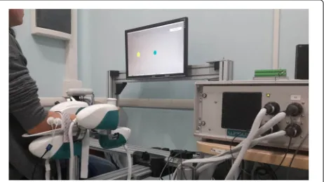
The experimental design involved a motor task where subjects were seated in front of a three degrees of free-dom wrist robotic manipulandum, called WRISTBOT and developed at Istituto Italiano di Tecnologia [33,34], holding the handle with their rigth hand (Fig. 1). Sub-jects’ forearm was strapped to the robot support in order
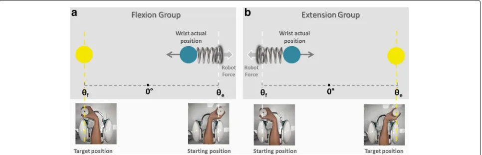
to avoid forearm movements and to have a correct align-ment between the axes of the mechanical structure and wrist. The device is a fully back-drivable manipulandum that has been specifically designed for human motor con-trol studies and for sensorimotor rehabilitation. The robot allows movements along the three wrist articulations with a range of motion (RoM) similar to a typical human sub-ject: ±62◦ flexion/extension (FE), 45◦/-40◦ radial/ulnar deviation (RUD), and±60◦pronation/supination (PS). It is powered by four brushless motors chosen in such a way to provide an accurate haptic rendering and compensate for the weight and inertia of the device. These motors can provide a maximum torque of 1.57 Nm on FE, 3.81 Nm on RUD, and 2.87 Nm on PS. Angular rotations on the three axes were acquired by means of high-resolution incremental encoders with a maximum error of ±0.17◦. A visual virtual reality environment was integrated in the system in order to provide visual feedback to the users while they complete the tasks. The experimental setup was complemented by a 6-axis force/torque sensor (Optoforce HEX-58-RE-400N) mounted on the handle in order to evaluate the efficiency of the robot in terms of provided torque. The task consisted of a series of con-tinuous target reaching movements interacting with a visco-elastic force generated by the WRISTBOT (Fig.2). Grasping the handle of the robot, subjects were requested to perform flexion and extension movements with their wrist in order to reach Flexion/Extension targets, pre-sented alternately at an angular distance ofθe = 48◦ or θf = −48◦with respect to the neutral wrist position. The
visco-elastic force experienced by FGsubjects opposed flexion movements and facilitated extension movements and it was implemented as a virtual spring whose equilib-rium angle wasθ = θe = 48◦. In the case of theEG
sub-jects the visco-elastic force opposed extension movements and facilitated flexion movements, by means of a virtual
spring whose equilibrium angle was θ = θf = −48◦.
In both cases, a small viscous force was added, in order to provide a minimal degree of damping of the inertia of the hand:
FFG= −k(θ−θe)−bθ˙
FEG= +k(θ−θf)+bθ˙ (1)
where kis the stiffness coefficient of the force-field and
b the corresponding viscous coefficient. The stiffness parameter k was set, prior to the experiment, taking into account the significant grip force difference between male and female subjects (see Table1). Mean female grip force was 30% less than that of males, which is in agree-ment with what is reported in the literature [35, 36]. More specifically, the following values of the visco-elastic parameters were chosen:k= 22.2 N/rad for female sub-jects,k= 27.7 N/rad for males, andb= 1.77 Ns/rad for all subjects. In order to avoid rest or recovery between each movements and to limit the variability between move-ment duration, targets had to be reached within a fixed time. In particular, visual and auditory feedback was pro-vided: the target color changed and a sound was produced if the subject did not reach the target within a duration of 1.5 s. Besides, the minimum time was not imposed, we allowed subjects to modulate their pace as to investigate if any change in kinematic parameters occurred with mus-cle fatigue. Subjects were instructed to perform the target reaching task until they could no longer do the task. At this time, we considered the level of fatigue experienced by the subject to be maximal (at least by our definition) and this corresponds to the maximum score on the VAS scale [15]. During the execution of the task, the experimenter encouraged participants to perform as many repetitions as possible to assure the maximum level of acceptable fatigue had been reached. Therefore, for each subject the
number of repeated ‘task-movements’ N could be dif-ferent and the sequence duration for each subject was normalized, 0–100% (rather than 1-N movements) to compare subjects. During task execution, the RUD and PS degrees of freedom were haptically blocked in order to constrain movements only to the flexion/extension. Throughout the task, we recorded electromyographic sig-nals fromextensor carpi radialisandflexor carpi radialis
muscles of the right arm, using a multichannel surface electromyography system (OTBiolab EMG-USB2+). For each muscle, two Ag/AgCl electrodes with an interelec-trode distance of 26 mm were placed in parallel with the muscle fibers on the belly of the muscle. In an attempt to reduce crosstalk, standard electrode palcements were followed, as recommended [37]. The sampling frequency was 2048 Hz, with a gain of 1000, and an internal band pass filter with cut-off frequencies of [10–900] Hz. sEMG signals and kinematic data were synchronized through a trigger signal sent from the robot to the sEMG base unit to assure association between each muscle activation and the corresponding movement. The preparation for the test (namely, electrodes placement, grip force recording, adjustment of WRISTBOT height and oral instructions) required about 180 s; the duration of the test itself varied among subjects, mainly as a function of the total num-ber of movements performed, with an average value of 80s (maximum duration in the overall population of sub-jects was 180 s). Globally, the assessment protocol could be performed with an average duration of 260 s and never exceeded 360 s.
Data analysis
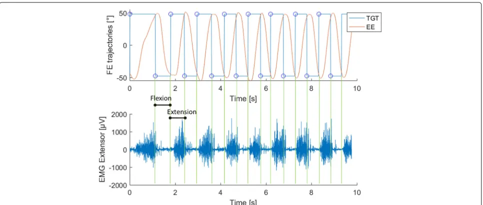
Wrist joint rotations, recorded from the robot encoders (data collection frequency set at 100 Hz), were converted to angular displacements and used to compute angu-lar velocities. All kinematic data were processed with a sixth-order Savitzky-Golay low-pass filter (10 Hz cut-off frequency) and re-sampled at the sEMG sample rate (2048 Hz) by linear interpolation while sEMG data were band-pass filtered (5–350 Hz). sEMG and kinematic data were segmented to focus the analysis on the concentric phase for each group. The analysis of the trajectory data recorded by the robot allows for the extraction of each single flexion or extension movement as shown in Fig.3 (Panel a and b). Accordingly, we analyzed the sEMG sig-nal offlexor carpi radialisduring flexion movements and the signal of theextensor carpi radialisduring extension. Next, we computed a single value of theMean Frequency
for each of the obtained intervals of the sEMG signal. Therefore, N values of Mean Frequency (FMean(k),k =
1...N) were obtained for each subject with N being the total number of movements performed by the subject. In particular theMean Frequency(FMean) of a sEMG signal
was calculated as follows:
FMean=
fs 2 0 fP(f)df fs
2 0 P(f)df
(2)
wherefsis the sampling frequency, andP(f)is the power
spectral density (PSD) of the signal. In order to trans-form the sEMG signal from the time-domain to the frequency-domain, a Fourier transform of the autocorre-lation function of the signal was employed and the PSD computed using the periodogram. The obtained N values ofMean Frequencyof each subject, were fitted with a third order polynomial function based on mean least square approximations in order to calculate theOnset of Fatigue (OF). This indicator was defined as follows: OF is the k-movement at which the initial FMean value of the
sequence decreases by a given percentage (P%) (i.e.
FMean(k=1)). More precisely we used the following
equation:
OFP%=k:FMean(k)≤FMean(1)−P%·(FMean(1)−min(FMean))
(3)
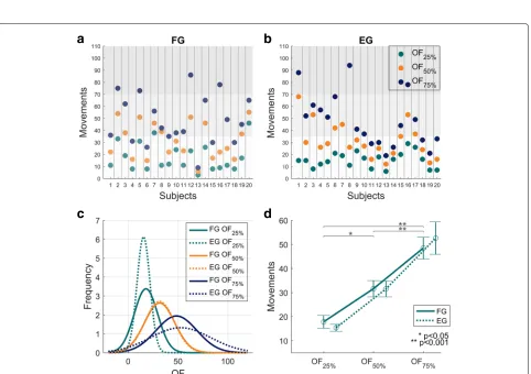
In order to choose the most appropriate value ofP%, the acquired data were analyzed with three reference values, namely 25%, 50% and 75%, thus yieldingOF25%,OF50%and
OF75% respectively. Please note also that the decrement
(P%), in Eq.3, is calculated with respect to the minimum value in theFMeansequence which may not correspond, as
will become evident in the “Results” section, with the final element of the sequence (min(FMean)=FMean(k=N)).
To ascertain that muscular behavior was not due or related to changes in motor strategy we calculated for each movement two additional indicators based on movement kinematics: theTime to velocity peak ratio(TPR) and the
Mean speed(m/s). TheTPRis defined as the ratio between the time to velocity peakTP, i.e. the time from the begin-ning of the movement and the main peak of the speed profile, and the total duration of the main movement (T):
TPR= TP
T (4)
Correlation analysis was performed to investigate the relationship between Mean Frequency and Mean Speed
by evaluating the correlation coefficient (CI) between the two metrics. To constantly monitor the required effort throughout the task we evaluated the mechanical energy consumption in Joules for each subject for each move-ment, consisting ofN samples, from the torque exerted
(τk)and the angular position(θk)recorded by the robot,
according to the following equation:
E=
N
k=1
τk·(θk+1−θk) (5)
Fig. 3Example of data segmentation.a. Red lines represent an example of the end-effector trajectory in the flexion-extension plane to reach the target at±48◦(blue lines).bExample of sEMG signal of theextensor carpi radialisduring the task. The signal was segmented according to the trajectory shown in Panela
analysis was performed to investigate the possible signifi-cance of differences on theOF.OFdata for the 40 subjects were z-transformed before the analysis. A post-hoc two-way ANOVA was chosen to investigate any difference in the OF between the three percentage levels in the two groups. The group factor (FG/EG) was set as “between” while, theOFpercentage level (25/50/75) as “within”. Sig-nificant main and interaction effects were evaluated using a two tailed t-test with Bonferroni correction for multiple comparisons. Significance was set at p<0.05.
Results
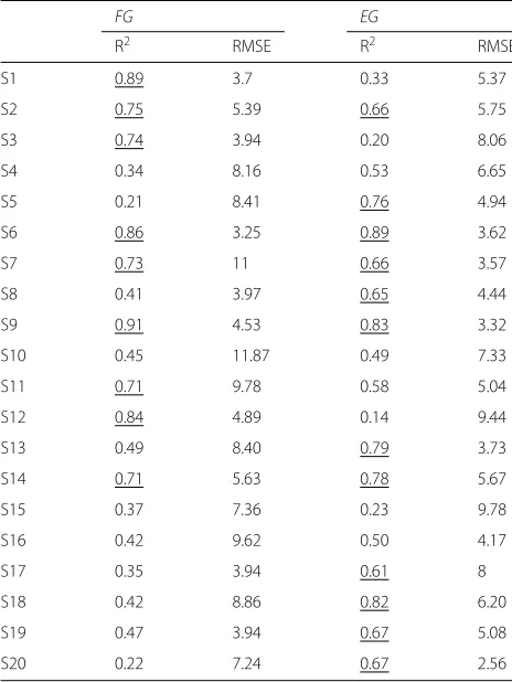
As expected, the Mean Frequency of both the Flexion Group (FG)and theExtension Group (EG)decreased dur-ing the first half of the task execution, and eventually reached a plateau (Fig.4, Panel a and Panel b respectively), although in a few cases the decrease was not monotonic: consider, in particular, subjects S2-S4-S5-S6-S13 of theFG
and subjects S13-S18-S19 of theEG. The fitting analysis indicated that the third-order polynomial model is a good predictor of theMean Frequencytrend, especially for the
Extension Group. Indeed, 12 subjects out of 20 from the
EG presented a goodness of fit higher than 0.6; such a goodness of fit was reached for 9 out of 20 from theFG
(See Table2).
Additionally, comparing the two groups, we found that the reduction inMean Frequencywas greater for theflexor carpi radialis, fatigued in the FG, than for the extensor carpi radialis, fatigued in theEG. Specifically, Fig.5 rep-resents the evolution of the averaged Mean Frequency
acrossFG andEGsubjects (panel a and b respectively),
normalized with respect to the initial value. From such a curve, it is possible to evaluate the percentual decrease of the averagedMean Frequencyof the fatigued muscles, with respect to the beginning of the task execution: 31% for theflexorand 24% for theextensor.
The next step of the analysis was to compare the three criteria for the identification of the Onset of Fatigue
Fig. 4Mean Frequencyresults. NormalizedFMean(Hz), of theFG(Panela) andEG(Panelb), fitted with a third order polynomial function. Each line
and color indicates a different subject
from the group. There was no interaction between OF
and the group (F(2, 114)=0.34286, p=0.71046). There were no significant differences between the two groups for the number of movements to OF (shown in Fig. 6 Panel d) (F(1, 114)=0.02614, p=0.87183). However, post-hoc anal-ysis demonstrated a significant difference between
Table 2Goodness of fit (R2) and RMSE ofMean Frequencycurves for each subject
FG EG
R2 RMSE R2 RMSE
S1 0.89 3.7 0.33 5.37
S2 0.75 5.39 0.66 5.75
S3 0.74 3.94 0.20 8.06
S4 0.34 8.16 0.53 6.65
S5 0.21 8.41 0.76 4.94
S6 0.86 3.25 0.89 3.62
S7 0.73 11 0.66 3.57
S8 0.41 3.97 0.65 4.44
S9 0.91 4.53 0.83 3.32
S10 0.45 11.87 0.49 7.33
S11 0.71 9.78 0.58 5.04
S12 0.84 4.89 0.14 9.44
S13 0.49 8.40 0.79 3.73
S14 0.71 5.63 0.78 5.67
S15 0.37 7.36 0.23 9.78
S16 0.42 9.62 0.50 4.17
S17 0.35 3.94 0.61 8
S18 0.42 8.86 0.82 6.20
S19 0.47 3.94 0.67 5.08
S20 0.22 7.24 0.67 2.56
Goodness higher than 0.6 is underlined
OF25% andOF50% (p=0.001), betweenOF25% andOF75% (p<0.001), and betweenOF50%andOF75%(p<0.001).
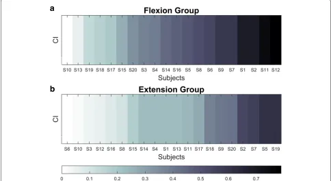
The kinematic analysis of the performed experiments should demonstrate any potential relationship between a subject’s motor strategy and patterns of muscle activity. In particular, we evaluated the correlation coefficient between the Mean speedand theMean Frequency (CI), and this is reported in Table4and Fig.7(absolute values). Overall, the correlation is negative, namely higherMean speed implies lower Mean Frequency, but, in general, it was a weak relationship: on average, the absolute correla-tion value was 0.42 for theFGpopulation and 0.27 for the
EGpopulation, and it never exceeded 0.8.
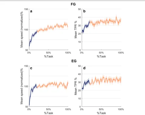
In spite of the low correlation between speed and fre-quency, the kinematic analysis was valuable for demon-strating the consistency of the designed protocol. This is shown in Fig.8which plots the evolution during task exe-cution of the normalizedMean speed(panels a and c) and theTime to velocity peak ratio(panels b and d). TheMean speedgraph is characterized, for both groups of subjects, by a steady increase in the first 20% of the task execu-tion (blue porexecu-tion of the curve) followed by a plateau in the remaining 80% of the task. The graph of the Time to velocity peak ratio (TPR) is characterized by a simi-lar trend, with an initial transient related to the first 20% of the task execution, followed by a plateau for the rest of the task. This suggests that there were no changes in wrist kinematic strategies. It is worth noting, there was a difference between the two groups. The FG subjects exhibited a speed profile with higher symmetry as indi-cated by the fact thatTPRis closer to 50% than for theEG
subjects.
a
b
Fig. 5AveragedMean Frequencyresults. NormalizedFMean(Hz), of theFG(Panela) andEG(Panelb), fitted with a third order polynomial function.
Frequency was normalized to the initial frequency of each sequence and averaged across subjects. Shaded area represents standard error
a
b
c
d
Fig. 6OFresults. Results related toOFindicators (Onset of Fatigue). Panelsaandbshow, for eachFGsubject (panela) andEGsubject (panelb), the values of the threeOFindicators:OF25%,OF50%,OF75%, expressed as number of “movements” that satisfy Eq.3. Grey areas divide the task in three
Table 3Mean speed - Mean Frequencycorrelation coefficients
FG EG
S1 -0.68 -0.23
S2 -0.68 -0.54
S3 -0.37 0.04
S4 -0.39 -0.22
S5 -0.49 -0.61
S6 -0.54 0.01
S7 -0.58 -0.56
S8 -0.51 0.09
S9 -0.58 -0.56
S10 -0.01 -0.02
S11 -0.74 0.24
S12 -0.79 -0.06
S13 -0.06 -0.23
S14 -0.44 -0.22
S15 -0.26 -0.17
S16 -0.46 0.07
S17 -0.19 -0.28
S18 -0.17 -0.37
S19 0.15 -0.61
S20 0.34 -0.42
Discussion
The importance of muscle fatigue assessment in patients with neuromuscular disorders has long been recognized. Specifically, a precise measure of muscle fatigability could provide crucial information in diagnosis, treatment plan-ning and evaluation of therapy efficacy. Yet, there is a lack of quantitative standardized and reliable methods for fatigue assessment used in clinical practice and these may not be feasible for all pathological populations at vari-ous stages of a disease. Muscle fatigue assessment using surface electromyography signals has been widely studied under isometric conditions [38] and in particular during maximum voluntary contractions [18, 20, 21]. However, the capacity to voluntarily generate and maintain a max-imal force in isometric conditions might be limited by a lack of motivation and it could represent a highly demand-ing task for people affected by severe weakness or injury. When dealing with adolescent populations, it is common to see a lack of full cooperation, which makes it chal-lenging to measure (in a reliable manner) maximum con-traction force and consequently muscle fatigue [25]. Fur-thermore, it is worth highlighting that subjects with low residual motor functions are hardly ever able to perform MVCs. Hence, few tests based on dynamic exercises have been proposed and validated [39,40], thus representing a consistent alternative to isometric contractions. Based
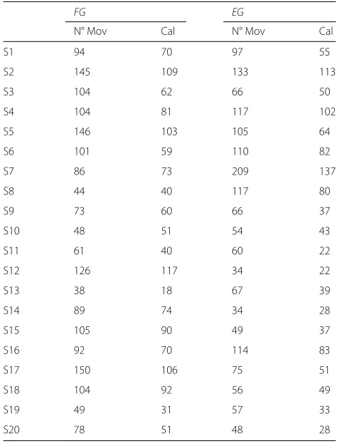
Table 4Number of movements performed by each subject and total calorie consumption
FG EG
N° Mov Cal N° Mov Cal
S1 94 70 97 55
S2 145 109 133 113
S3 104 62 66 50
S4 104 81 117 102
S5 146 103 105 64
S6 101 59 110 82
S7 86 73 209 137
S8 44 40 117 80
S9 73 60 66 37
S10 48 51 54 43
S11 61 40 60 22
S12 126 117 34 22
S13 38 18 67 39
S14 89 74 34 28
S15 105 90 49 37
S16 92 70 114 83
S17 150 106 75 51
S18 104 92 56 49
S19 49 31 57 33
S20 78 51 48 28
reli-a
b
Fig. 7CI results. Absolute values of theMean speedand theMean Frequencycorrelation for theFG(Panela) and theEG(Panelb. Subjects are sorted by correlation, as indicated in the bottom panel legend
able measure of muscle fatigue and can be used in a clini-cal setting. It has also to be mentioned that the use of the robotic device adds the ability to measure subjects’ perfor-mances in terms of kinematic parameters, thus resulting in a more detailed assessment of the patient. In the present study, the kinematic analysis demonstrated the stability of theOF indicators and it appears robust, given differ-ent motor control strategies. Regarding the applicability of the method, the experimental setup is minimal, requiring sEMG from two target muscles and the correct alignment between the human wrist and the robotic device. From a clinical perspective, test duration is also important and our test never lasted more than 3 min. It is even reason-able to expect a shorter test time in clinical populations compared to our healthy participants. We chose to base our indicator ofOnset of Fatigueon theMean frequency
since its variance is tipically lower than that of Median frequency [41]. The shift inMean frequency towards the lower frequency spectrum was noticeable in both the flex-ion andextension groups, however the shift was greater in the former. This may be due to different physiological properties of the two muscles: I) from a biomechanical perspective of the human wrist joint, the amplitude of the range of motion in flexion is higher than that achiev-able in extension (peak flexion moment is approximately 70% higher than peak extension moment [42]); II) the percent decrement of Mean Frequency is proportional
to the amount of catabolites produced by muscle fibers during activity [43]. In particular, the quantity of catabo-lites depends on the average number of muscle fibers per square unit of the muscle crossection [43] and conse-quently the higher the crossection, the higher the amount of catabolites and the greatest the rate of decrease of the Mean Frequency ; III) muscle fiber type will influ-ence sEMG parameters, in particular, a greater percent-age of type II fibers leads to a greater rate of decrease of the Mean Frequency [44]. As reported from other studies, muscles belonging to the extensor group fatigue more and faster than flexors [45], therefore, EG popu-lation was expected to exhibit more fatigue. However, directly comparing the single muscles extensor and
flexor carpi radialis, the physiological cross-sectional area (PCSA) offlexor carpi radialisis about half the size of the
extensor carpi radialis[46]. Therefore, we can assume that
flexor carpi radialishas a lower force generating capac-ity. We can speculate that, on a whole, when using the same force field intensity for both flexion and extension, the extensor muscles would fatigue more than the flex-ors. It should be noted that we measured only one wrist extensor muscle. The most important contribution of this method is the development of and evaluation of theOF
a
b
c
d
Fig. 8Kinematics results. Panelaandc:Mean speednormalized by the mean of each subject and averaged overFGandEGsubjects respectively. Panelbandd:Time to peak ratioaveraged overFGandEGsubjects respectively. %Task identifies the relative ordinal number of the task-movements of all the sequence. In all panels shaded areas indicate the standard error, the blue portions refer to the first 20% of the task while the red
corresponds to the remaining 80%
forOnset of Fatigue, the comparison amongOF25%,OF50% and OF75% revealed that OF25% is the more consistent and less variable. We can postulate that after an initial decrease, which is very similar in both groups, theMean Frequencycurves decreased with different slopes due to different subject training levels and muscle physiological properties. Our OF25% is also more inline with previ-ous studies that have suggested that a mean frequency decrease of 8% is representative of muscle fatigue onset [47,48]. Moreover, our subjects’ ability to perform the task correctly from the very beginning and the consequent sta-bility of the kinematic parameters, support the adoption of
OF25%as an indicator of fatigue.Mean speedstabilized in the first 20% of the task, suggesting thatMean Frequency
andOF25%are not related to kinematic changes. It is worth noting that, in some cases, during the last part of the task, the trend was inverted. From a physiological perspective,
an increase in temperature and the inversion of theMean Frequencytrend observed in our study. Regarding kine-matic measurements, we found that the Mean Speed
stabilized and remained constant after an initial phase, corresponding to the first 20% of the task, in which it increased up to a plateau level. This suggests thatMean Speedwas not affected by muscle fatigue (and vice-versa), which is in line with previous finding [52–54]. Conversely from what was expected, we did not find changes in kine-matic strategies that correlated with increasing fatigue level. Specifically, theTPRdid not show a shift in the peak of the bell-shaped speed profile [55] from early trials to the late trials in which fatigue appeared. A final aspect to consider was task duration. In the proposed proto-col, the number of repetitions performed, was decided by the subjects and not superimposed by the experimenter. Thus, subjects were instructed to stop when they felt tired, which is crucial in a clinical scenario. The num-ber of repetitions, therefore, could also be considered as an additional measure of performance [56], especially for populations with neuromuscular impairments, where kinematics and sEMG might have to be cautiously inter-preted. Our results in healthy participants demonstrate thatOFwas independent from the amount of repetitions of the reaching movements performed. This may be a con-sequence of the population studied, who could tolerate a high level of resistance and may not stop the test when they feel fatigue. To conclude, the developed algorithm could be improved in the future by measuring individ-ual wrist strength and grip force throughout the task [45]. Our approach used two levels of force, according to sex, but customizing the force and normalizing to individual maximal force production, could improveOFresults. This aspect is particularly relevant and needs to be considered in the application of this method to pathological sub-jects. Lastly, this study suggested that a fatigue assessment coupling a robotic task and sEMG recordings is highly feasible and practical. The present study provided a good starting point for the application of the test in clinical practice, however pilot experiments with neuromuscular and age-matched healthy subjects are necessary to con-firm the results. Finally, wrist robotic device guarantees the repeatability of the task, providing the same force and trajectory. Moreover without the addition of further mea-surement tools we attempted to exploit the torque and angular position data recorded by the robot as a simple measure of the mechanical work performed by the sub-ject during the test. Although such an approach does not provide specific information about actual internal muscle work or physiological work, it allowed for an estimate of the total energy required by the task. Our test has been demonstrated to require little effort, so the impact on daily energy expenditure (avg 2500–3000 kcal) would be minimal [57].
Conclusion
This test will provide clinicians with an objective and easily readable indicator of muscle fatigue. The method is simple, easy to administer and suitable for patients/participants who are not able to generate high levels of muscle contractions. This overcomes the prob-lem of administering maximal contraction tests in neuro-logical or injured populations. If the same robust results could be obtained from pathological population, then this method could be used as a standard muscle test procedure that is independent of the subjects ability or willingness to perform voluntary efforts. In this context, the final per-spective of the use a robotic device is to assess muscle fatigue in very controlled conditions and with the pos-sibility to change or adapt the task to the population needs.
Abbreviations
CI: Correlation index; DoF: Degree of freedom; EG: Extension group; FE: Flexion/extension; FG: Flexion group; OF: Onset of fatigue; PS:
Pronation/supination; RoM: Range of motion; RUD: radial/ulnar deviation; TPR: Time to velocity peak ratio
Acknowledgements
The authors wish to thank all the subjects who volunteered with enthusiasm to take part to the experiment.
Funding
The study was founded by Istituto Italiano di Tecnologia, Geova, Italy and partially by the European CODEFROR project (FP7-PIRSES-2013-612555).
Availability of Data and Materials
Full data set and software analysis will be provided in case of acceptance.
Authors’ contributions
MM helped in designing and implementing the experiment, acquiring the data, analyzing and interpreting the data, and drafting the manuscript. FM helped in conceiving the idea and concept, designing the experiment, interpreting the data, drafting the manuscript and helped in the statistical analysis of the data. JZ helped in conceiving the idea and concept, drafting the manuscript and supervising the study. PM and MH helped in critically revising the manuscript content. All authors read and approved the final manuscript.
Ethics approval and consent to participate
Written informed consent was obtained from the subjects for publication of this study and any accompanying images. The study was approved by the Ethics Committee of the regional health authority, Azienda Sanitaria Locale Genovese (ASL) N.3 (Protocol number 311REG2014 approved on 09/12/2015), and all participants signed a written informed consent.
Consent for publication
Consent to publish was obtained from that adult participants.
Competing interests
The authors declare that they have no competing interests.
Publisher’s Note
Springer Nature remains neutral with regard to jurisdictional claims in published maps and institutional affiliations.
Author details
2Department of Informatics, Bioengineering, Robotics and Systems Engineering (DIBRIS),University of Genoa, Genoa, Italy.3Department of Kinesiology, Brock University, St. Catharines, ON, Canada.
Received: 5 March 2018 Accepted: 19 November 2018
References
1. Silverthorn DU. Human Physiology : an Integrated Approach. 1st Ed. Upper Saddle River: Pearson Prentice Hall; 1998.
2. Ma L, Chablat D, Bennis F, Zhang W, Hu B, Guillaume F. A novel approach for determining fatigue resistances of different muscle groups in static cases. Int J Ind Ergon. 2011;41(1):10–8.https://doi.org/10.1016/j. ergon.2010.11.005.
3. Monjo F, Terrier R, Forestier N. Muscle fatigue as an investigative tool in motor control: A review with new insights on internal models and posture–movement coordination. Hum Mov Sci. 2015;44:225–33.https:// doi.org/10.1016/j.humov.2015.09.006.
4. Vafadar AK, Côté JN, Archambault PS. The Effect of Muscle Fatigue on Position Sense in an Upper Limb Multi-joint Task. Mot Control. 2012;16(2): 265–83.https://doi.org/10.1123/mcj.16.2.265.
5. Zwarts MJ, Bleijenberg G, van Engelen BGM. Clinical neurophysiology of fatigue. Clin Neurophysiol. 2008;119(1):2–10.https://doi.org/10.1016/j. clinph.2007.09.126.
6. Thacham-Poyil A, Amirabdollahian F, Steuber V. Study of Gross Muscle Fatigue During Human-Robot Interactions. IARIA. 2017. http://vuh-la-uhra.herts.ac.uk/handle/2299/18352http://uhra.herts.ac.uk/handle/2299/ 18352.
7. Angelini C, Tasca E. Fatigue in muscular dystrophies. Neuromuscul Disord. 2012;22:214–20.https://doi.org/10.1016/j.nmd.2012.10.010. 8. Shimizu-Motohashi Y, Miyatake S, Komaki H, Takeda S, Aoki Y. Recent
advances in innovative therapeutic approaches for Duchenne muscular dystrophy: from discovery to clinical trials,. Am J Transl Res. 2016;8(6): 2471–89.
9. McDonald CM, Henricson EK, Han JJ, Abresch RT, Nicorici A, Elfring GL, Atkinson L, Reha A, Hirawat S, Miller LL. The 6-min walk test as a new outcome measure in Duchenne muscular dystrophy. Muscle Nerve. 2010;41(4):500–10.https://doi.org/10.1002/mus.21544.
10. Lou J-S, Weiss MD, Carter GT. Assessment and Management of Fatigue in Neuromuscular Disease. Am J Hosp Palliat Med. 2010;27(2):145–57. https://doi.org/10.1177/1049909109358420.
11. Mayhew A, Mazzone ES, Eagle M, Duong T, Ash M, Decostre V, Vandenhauwe M, Klingels K, Florence J, Main M, Bianco F, Henrikson E, Servais L, Campion G, Vroom E, Ricotti V, Goemans N, Mcdonald C, Mercuri E. Development of the Performance of the Upper Limb module for Duchenne muscular dystrophy. Dev Med Child Neurol. 2013;55(11): 1038–45.https://doi.org/10.1111/dmcn.12213.
12. Bushby K, Finkel R, Birnkrant DJ, Case LE, Clemens PR, Cripe L, Kaul A, Kinnett K, McDonald C, Pandya S, Poysky J, Shapiro F, Tomezsko J, Constantin C. Diagnosis and management of Duchenne muscular dystrophy, part 2: implementation of multidisciplinary care. Lancet Neurol. 2010;9(2):177–89.https://doi.org/doi:10.1016/S1474-4422(09)70272-8. 13. Smets EM, Garssen B, Bonke B, De Haes JC. The Multidimensional
Fatigue Inventory (MFI) psychometric qualities of an instrument to assess fatigue. J Psychosom Res. 1995;39(3):315–25.
14. Krupp LB, LaRocca NG, Muir-Nash J, Steinberg AD. The fatigue severity scale. Application to patients with multiple sclerosis and systemic lupus erythematosus. Arch Neurol. 1989;46(10):1121–3.
15. Wolfe F, Hawley DJ, Wilson K. The prevalence and meaning of fatigue in rheumatic disease,. J Rheumatol. 1996;23(8):1407–17.
16. Cifrek M, Medved V, Tonkovi´c S, Ostoji´c S. Surface EMG based muscle fatigue evaluation in biomechanics. Clin Biomech. 2009;24(4):327–40. https://doi.org/10.1016/j.clinbiomech.2009.01.010.
17. Carr JC, Beck TW, Ye X, Wages NP. Intensity-dependent EMG response for the biceps brachii during sustained maximal and submaximal isometric contractions. Eur J Appl Physiol. 2016;116(9):1747–55.https:// doi.org/10.1007/s00421-016-3435-6.
18. Kahl L, Hofmann UG. Comparison of algorithms to quantify muscle fatigue in upper limb muscles based on sEMG signals. Med Eng Phys. 2016;38(11):1260–9.https://doi.org/10.1016/j.medengphy.2016.09.009.
19. González-Izal M, Malanda A, Navarro-Amézqueta I, Gorostiaga EM, Mallor F, Ibañez J, Izquierdo M. EMG spectral indices and muscle power fatigue during dynamic contractions. J Electromyogr Kinesiol. 2010;20(2): 233–40.https://doi.org/10.1016/j.jelekin.2009.03.011.
20. Hug F, Nordez A, Guével A. Can the electromyographic fatigue threshold be determined from superficial elbow flexor muscles during an isometric single-joint task?. Eur J Appl Physiol. 2009;107(2):193–201.https://doi.org/ 10.1007/s00421-009-1114-6.
21. Oda S, Kida N. Neuromuscular fatigue during maximal concurrent hand grip and elbow flexion or extension. J Electromyogr Kinesiol. 2001;11(4): 281–9.https://doi.org/doi:10.1016/S1050-6411(01)00004-9.
22. Oliveira ASC, Gonçalves M, Cardozo AC, Barbosa FSS. Electromyographic fatigue threshold of the biceps brachii muscle during dynamic contraction. Electromyogr Clin Neurophysiol. 2005;45(3):167–75. 23. Ratel S, Duché P, Williams CA. Muscle Fatigue during High-Intensity
Exercise in Children. Sports Med. 2006;36(12):1031–65.https://doi.org/10. 2165/00007256-200636120-00004.
24. Vøllestad NK. Measurement of human muscle fatigue. J Neurosci Methods. 1997;74(2):219–27. https://doi.org/doi:10.1016/S0165-0270(97)02251-6.
25. Halin R, Germain P, Bercier S, Kapitaniak B, Buttelli O. Neuromuscular response of young boys versus men during sustained maximal contraction. Med Sci Sports Exerc. 2003;35(6):1042–8.https://doi.org/10. 1249/01.MSS.0000069407.02648.47.
26. Gregory CM, Bickel CS. Recruitment Patterns in Human Skeletal Muscle During Electrical Stimulation. Phys Ther. 2005;85(4):358–64.https://doi. org/doi:10.1093/ptj/85.4.358.
27. Naughton G, Carlson J, Fairweather I. Determining the Variability of Performance on Wingate Anaerobic Tests in Children Aged 6-12 Years. Int J Sports Med. 1992;13(07):512–7. https://doi.org/10.1055/s-2007-1021308.
28. Marini F, Squeri V, Morasso P, Konczak J, Masia L. Robot-Aided Mapping of Wrist Proprioceptive Acuity across a 3D Workspace. PLOS ONE. 2016;11(8):0161155.https://doi.org/10.1371/journal.pone.0161155. 29. Marini F, Squeri V, Morasso P, Campus C, Konczak J, Masia L.
Robot-aided developmental assessment of wrist proprioception in children. J NeuroEngineering Rehabil. 2017;14(1):3.https://doi.org/10. 1186/s12984-016-0215-9.
30. De Santis D, Zenzeri J, Casadio M, Masia L, Morasso P, Squeri V. A new method for evaluating kinesthetic acuity during haptic interaction. Robotica. 2014;32(08):1399–414.https://doi.org/10.1017/ S0263574714002252.
31. Mugnosso M, Marini F, Gillardo M, Morasso P, Zenzeri J. A novel method for muscle fatigue assessment during robot-based tracking tasks. In: Proc. IEEE Int. Conf: Rehab. Robot (ICORR); 2017. p. 84–9.https://doi.org/10. 1109/ICORR.2017.8009226.
32. Oldfield RC. The assessment and analysis of handedness: The Edinburgh inventory. Neuropsychologia. 1971;9(1):97–113.https://doi.org/doi:10. 1016/0028-3932(71)90067-4.
33. Masia L, Casadio M, Giannoni P, Sandini G, Morasso P. No Title. J NeuroEngineering Rehabil. 2009;6(1):44. https://doi.org/10.1186/1743-0003-6-44.
34. Marini F, Contu S, Morasso P, Masia L, Zenzeri J. Codification mechanisms of wrist position sense. In: Proc. IEEE Int. Conf. Rehab. Robot (ICORR); 2017. p. 44–9.https://doi.org/10.1109/ICORR.2017.8009219. 35. Phillips BA, Lo SK, Mastaglia FL. Muscle force measured using “break”
testing with a hand-held myometer in normal subjects aged 20 to 69 years. Arch Phys Med Rehabil. 2000;81(5):653–61.https://doi.org/doi:10. 1016/S0003-9993(00)90050-9.
36. Bäckman E, Johansson V, Häger B, Sjöblom P, Henriksson KG. Isometric muscle strength and muscular endurance in normal persons aged between 17 and 70 years. Scand J Rehabil Med. 1995;27(2): 109–17.
37. Holmes MWR, Tat J, Keir PJ. Neuromechanical control of the forearm muscles during gripping with sudden flexion and extension wrist perturbations. Comput Methods Biomech Biomed Eng. 2015;18(16): 1826–34.https://doi.org/10.1080/10255842.2014.976811.
39. Dimitrov G, Arabadzhiev T, Mileva K. Muscle fatigue during dynamic contractions assessed by new spectral indices. In: Medicine and science; 2006.
40. Potvin JR, Bent LR. A validation of techniques using surface EMG signals from dynamic contractions to quantify muscle fatigue during repetitive tasks. J Electromyogr Kinesiol Off J Soc Electrophysiological Kinesiol. 1997;7(2):131–9.
41. Knaflitz M, Merletti R, De Luca CJ. Inference of motor unit recruitment order in voluntary and electrically elicited contractions,. J Appl Physiol (Bethesda, Md. : 1985). 1990;68(4):1657–67.https://doi.org/10.1152/jappl. 1990.68.4.1657.
42. Gonzalez R, Buchananf T, Delp S. How muscle architecture and moment arms affect wrist flexion-extension moment. J Biomech. 1997;30(7): 705–12.
43. Merletti R, Knaflitz M, De Luca CJ. Myoelectric manifestations of fatigue in voluntary and electrically elicited contractions. J Appl Physiol. 1990;69(5):1810–20.
44. Gerdle B, Henriksson-Larsén K, Lorenzton R, Wretling M-L. Dependence of the mean power frequency of the electromyogram on muscle force and fibre type. Acta Physiologica Scandinavica. 1991;142(4):457–65. https://doi.org/10.1111/j.1748-1716.1991.tb09180.x.
45. Hggg ’ GM, Milerad2 E. Forearm extensor and flexor muscle exertion during simulated gripping work -an electromyographic study. Clin Biomech Clin Biomech. 1997;1212(1):39–43.
46. Mirakhorlo M, Visser JMA, Goislard de Monsabert BAAX, van der Helm FCT, Maas H, Veeger HEJ. Anatomical parameters for musculoskeletal modeling of the hand and wrist. Int Biomech. 2016;3(1):40–9.https://doi. org/10.1080/23335432.2016.1191373.
47. Oberg T, Sandsjo L, Kadefors R, Öberg T, Sandsjö L, Kadefors R. Electromyogram mean power frequency in non-fatigued trapezius muscle. Eur J Appl Physiol Occup Physiol. 1990;61(5-6):362–9.https://doi. org/10.1007/BF00236054.
48. Szucs K, Navalgund A, Borstad JD. Scapular muscle activation and co-activation following a fatigue task. Med Biol Eng Comput. 2009;47(5): 487–95.https://doi.org/10.1007/s11517-009-0485-5.
49. Croce RV, Miller JP. The effect of movement velocity and movement pattern on the reciprocal co-activation of the hamstrings. Electromyogr Clin Neurophysiol. 2003;43(8):451–8.
50. Marbini A, Ferrari A, Cioni G, Bellanova MF, Fusco C, Gemignani F. Immunohistochemical study of muscle biopsy in children with cerebral palsy. Brain Dev. 2002;24(2):63–6.
51. Madigan M, science in sports And PP-M. A muscle temperature compensation technique for EMG fatigue measures. 2002. undefined.europepmc.org.
52. Gates DH, Dingwell JB. The effects of neuromuscular fatigue on task performance during repetitive goal-directed movements. Exp Brain Res. 2008;187(4):573–85.https://doi.org/10.1007/s00221-008-1326-8. 53. Lucidi CA, Lehman SL. Adaptation to fatigue of long duration in human
wrist movements. J Appl Physiol (Bethesda, Md. : 1985). 1992;73(6): 2596–603.
54. Selen LPJ, Beek PJ, van Dieën JH. Fatigue-induced changes of impedance and performance in target tracking. Exp Brain Res. 2007;181(1):99–108.https://doi.org/10.1007/s00221-007-0909-0. 55. Abend W, Bizzi E, Morasso P. Human arm trajectory formation,. Brain.
1982;105(Pt 2):331–48.
56. Octavia JR, Feys P, Coninx K. Development of Activity-Related Muscle Fatigue during Robot-Mediated Upper Limb Rehabilitation Training in Persons with Multiple Sclerosis: A Pilot Trial. Mult Scler Int. 2015;2015: 650431.https://doi.org/doi:10.1155/2015/650431.