R E S E A R C H
Open Access
Nr2f1b control venous specification and
angiogenic patterning during zebrafish
vascular development
Ru-Fang Li
1†, Ting-Yun Wu
1†, Yu-Zheng Mou
1, Yi-Shan Wang
1, Chun-Lin Chen
1and Chang-Yi Wu
1,2,3*Abstract
Background:The specification of vein and the patterning of intersegmental vessels (ISV) controlled by transcription factor is not fully characterized. The orphan nuclear receptor Chicken ovalbumin upstream promoter transcription factor II (CoupTFII, a.k.a NR2F2) positively regulates vein identity in mice. In this study, we show thatnr2f1bis important for vein and tip cell identity during zebrafish development.
Results:Nr2f1b mRNA is expressed in ventral lateral mesoderm at 15S stage and in vessels at 24 hpf
consistent with a role in early vascular specification. Morpholino knockdown ofnr2f1b results in a decrease in both vein cell number and expression of the vein specific marker flt4 and mrc1, suggested its role in venous specification. We also show loss of nr2f1breduced ISV cell number and impairs ISV growth, which is likely due to the impairment of angiogenic cells migration and/or proliferation by time-lapse imaging.
Consequently, nr2f1bmorphants showed pericardial edema and circulation defects. Overexpression of nr2f1b under the fli promoter increases the number of venous cells and ISV endothelial cells indicated the function of nr2f1b is required and necessary for vascular development. We further showed thatnr2f1b likely interact with Notch signalling. nr2f1b expression is increased in rbpsuh morphants and DAPT-treatment embryos suggested nr2f1b is negatively regulated by Notch activity.
Conclusions: We show nr2f1b control venous specification and angiogenic patterning during zebrafish vascular development, which is mediated by Notch signalings.
Keywords: Nr2f1b, Angiogenesis, vein and tip cell identity, ISV (intersegmental vessel)
Background
Vasculogenesis and angiogenesis are two processes to establish the pattern of blood vessels network in ver-tebrates [1, 2]. In the developing zebrafish trunk, a stereotypic pattern of vascular development begins with dorsal aorta (DA) and posterior cardinal vein (PCV) formation at the midline by fusion of angio-blast progenitors migrating from the lateral posterior mesoderm by the 17 somite (17S) stage [3]. Develop-ment of the intersegDevelop-mental vessels (ISVs) of the trunk
begins with an angioblast sprouting from the DA, proliferating and migrating dorsally until it reaches the dorsal aspect of the embryo and connects with adjacent ISV cells to form the dorsal longitudinal anastomotic vessels (DLAVs). The leading cells to mi-grate from the vessel are called tip cells, are prolifera-tive and show multiple filopodia, while the less proliferative, stationary cells which lumenize behind the tip cell are called stalk cells [4, 5].
Many genes and signal pathways have been identified that regulate the specification and maintenance of arter-ial identity during vasculogenesis, such as vascular endo-thelial growth factor (VEGF), nrp1, delta-like 4 (dll4), gridlock, foxc1 and foxc2 (reviewed in [6, 7]), Signalling through the Notch receptor is a major contributor to ar-terial identity as loss of Notch leads to a decrease in the number of cells expressing arterial markers and an * Correspondence:[email protected]
†Equal contributors
1
Department of Biological Sciences, National Sun Yat-sen University, Kaohsiung, Taiwan
2
Doctoral Degree Program in Marine Biotechnology, National Sun Yat-sen University and Academia Sinica, Kaohsiung, Taiwan
Full list of author information is available at the end of the article
increase in cells expressing venous markers [5]. While a number of signaling molecules have been identified to promote arterial identify, there is less description about transcription factors that promote a venous identity. In mice study, the orphan nuclear receptor NR2F2 is expressed in venous endothelial cells [8–10]. Loss of NR2F2 in mouse results in ectopic expression of arterial markers in the vein with loss of the venous endothelial cell identity and acquisition of arterial phenotypes. Thus, NR2F2 functions as the key regulator of venous identity [9]. Recent studies in zebrafish and xenopus showed that nr2f2and SoxF regulated venous differentiation [11, 12].
During the development of intersegmental vessel (ISV) sprouts from the dorsal aorta, angioblasts will specify two cell identities, the migratory tip cell that senses attractive and repulsive through the extension of filopodia, or the stalk cell that lumenizes to form an intersegmental vessels (ISVs) [13–15]. ISV angiogenesis has been shown that reg-ulated by VEGF and Notch-flt4 (Fms-related tyrosine kin-ase 4) signalings. Knockdown of VEGFR2 disrupt ISV formation and loss of Notch signaling results in a signifi-cant increase ISV cells and increase in Flt4 expression [5]. Conversely, loss of Vegfc or Flt4 impaired ISV growth and a decreased number of angioblasts in each ISV [16]. Moreover, activation of Notch signaling also results in stalled ISV growth mid-somite, suggesting that Notch re-presses the Vegfc-Flt4 signaling cascade [17]. In recent years, many molecules, such as cxcr4, UNC5B, angio-motin, pdgfb and trpc1 have been shown involved in angiogenesis [16, 18–21]. Thus, genetic interaction and coordination contributes to the control of endothelial tip-stalk cell behaviors during angiogenesis. Of those factors, NR2F2 in mice has been shown function in angiogen-esis mediated by the upregulation of Angiopoietin-1 in addition to its role in venous differentiation [8]. However, we still have limited knowledge of the role of transcription factors in tip cell specification. Surprisingly, nr2f2, which plays a major role in venous specification in mice, acts only a minor role in zebrafish vascular development based on our study and recent publications ([11, 12] and our un-published results).
During the vascular development in mouse, NR2F2 acts as a major regulator in venous identity and in an-giogenic growth [8, 9]. In addition, Nr2f2 interacts with Prox1 physically to specific lymphatic endothelial fate and promote the formation of lymphatic vessels [22, 23]. Our previous study in zebrafish showed that nr2f2 in zebrafish plays a minor role in venous identity and ISV growth but functions critically in lymphogenesis, similar to recent reports. On the other hand, the related tran-scription factor NR2F1 is a critical regulator of CNS and peripheral nervous system development and controls cell differentiation in the inner ear [24–26]. However, a vas-cular function of NR2F1 has not yet been documented.
In this study, we hypothesized thatnr2f1bhas a critical role in blood vessel formation in zebrafish. We showed that loss ofnr2f1b reduced venous cells and the expres-sion of vein specific markers. We also showed thatnr2f1b morphant reduces ISV cells and impairs ISV growth. While overexpression ofnr2f1b, we observed the increase of vein and ISV cells, suggesting thatnr2f1bplay a role in promoting vein and tip cell identity. We further showed that nr2f1b functions in vascular development mediated by Notch signalling.
Methods
Zebrafish strains and husbandry
Zebrafish (Danio rerio) wild-type Tupfel Long Fin (TL) or transgenic lines: Tg(kdrl:eGFP) la116, Tg(kdrl:mCherry)ci5, Tg(gata:dsRed), Tg(fli1a:egfp)y1 and Tg(fli1a:negfp)y7 have been described [27–30]. Zebrafish were raised and main-tained at the 28.5 °C fish room in a 20 L circulating system with filtered fresh water and aeration under the 14 hr: 10 hr (light: dark) lighting conditions. Zebrafish embryos were raised in E3 embryo media (5 mM NaCl, 0.17 mM KCl, 0.33 mM CaCl2, 0.33 mM MgSO4and supplemented
with 0.25 mg/L methylene blue) at 28.5 °C according to the Zebrafish Book [31]. Embryo development and stages were measured in hour post-fertilization (hpf). Chorions were removed by incubation in 20 mg/ml pronase (Sigma) and endogenous pigmentation was blocked by adding 0.003 % N-phenylthiourea (PTU; Sigma) to E3 media at 6 hpf. All animal experiments are approved from the National Sun Yat-sen University Animal Care Commit-tee (approval reference #10109)
Whole-mount in situ hybridization
Whole-mount in situ hybridization was performed as described in [32]. nr2f1b probe template was amplified by PCR using primers described in Additional file 1: Table S1 and in vitro transcription using T7 Polymerase (Roche) with DIG-labeled UTP. The flt4, mrc1, notch3 and ephrinb2 probes have been described [33–35]. Whole-mount in situ hybridization (WISH) was per-formed as previously described [35, 36]. Briefly, Embryos were fixed in 4 % paraformaldehyde in phosphate buff-ered saline (PBS), permeabilized in 10μg/mL Proteinase K, hybridized with DIG-labeled probes, washed, reacted with AP-conjugated anti-Dig antibody (Roche) and then proceeded to react with NBT/BCIP substrate (Roche). The reaction was stopped and embryos were fixed with PFA. Embryos were embedded in 3 % methylcellulose (Sigma) and photographed.
Imaging
(Carl Zeiss) on a Zeiss Lumar V12 stereomicroscope. Confocal images were collected on a Zeiss LSM510 or LSM700 microscope, and stacked images generated by ImageJ or ZEN 2012 software (Carl Zeiss). The number of cells in the vein and ISVs was determined by counting from individual slices of confocal stacks. The counting area is between 5thand 15thISVs of the embryo.
For histology, embryos were sectioned at 5-7 μM in JB-4 plastic medium (Polysciences, Warrington, PA) and photographed with a Magnafire camera (Optronics, Galeta, CA). Alternatively, embryos were fixed with Tek OCT freezing medium and cryo-sectioned at 10 μM using a Leica CM3050S cryostat and photographed with an SPOT RT3 camera (DIAGNOSTIC Inc.).
Morpholino and Tol2 DNA Injections
Morpholinos fornr2f1bandrbpsuhgenes were designed and ordered from Gene-Tools, LLC (Philomath, OR), dissolved in H2O to a 2 mM stock and further diluted to
the working concentration with 0.5 % phenol red (Sigma). Sequences are listed in Additional file 1: Table S1. Microinjections were performed to manipulate gene expression. MOs or expression vectors were injected into 1-2-cell-stage embryos on a 3 % agar plate. After in-jection, embryos were cultured in E3 buffer. The Tol2kit was used to generate nr2f1b overexpression driven by 0.8kb fli1a promoter construct [37]. Approximately 100 pg of plasmid DNA was co-injected with 50 pg of Tol2 mRNA into 1-cell embryos. Success of transient Tg(fli1:nr2f1b)overexpressing embryos can be verified by GFP signal expression driven by cmlc (cardiomyocyte light chain) promoter from the vector backbone.
RNA extraction, cDNA synthesis and Quantitative RT-PCR (qPCR)
Total RNA was extracted from embryos at desired develop-mental stages and purified using the Qiagen RNeasy Mini Kit (Qiagen) according to the manufacturer’s instructions. Complementary DNA (cDNA) was synthesized with Superscript III reverse transcriptase and oligo-dT primer (Invitrogen) according to the manufacturer’s instructions. Quantitative RT-PCR was performed using the DNA En-gine Opticon System (MJ Research Inc.) with iQ SYBR Green Supermix (BioRad) or using the LightCycle 96 in-strument (Roche Inc.) with SYBR Green I Master (Roche). qPCR primers are listed in Additional file 1: Table S1. Rela-tive gene expression levels were analyzed by the ΔΔ Ct
method, with elongation factor 1α (EF1α) as a reference gene All reactions were performed as biological triplicates.
DAPT (N-[N-(3,5-Difluorophenacetyl)-L-alanyl]-S-phenylglycine t-butyl ester) treatment
Embryos were treated with DAPT (Sigma), γ-secretase specific inhibitor to block Notch signalling, 75 μM for
the working concentration in E3 medium at 6 hpf. Control embryos were treated with an equivalent con-centration 0.3 % of DMSO (Dimethyl sulfoxide, Sigma).
Results
nr2f1bmRNA is expressed in vessels during zebrafish development
We sought to understand the role of the orphan nuclear re-ceptors in venous angioblast development. Loss of NR2F2 in mouse leads to an almost complete loss of the cardinal vein, however, morpholino knockdown of nr2f2 in zebrafish leads to only a reduction in venous marker expression with-out obvious defects in vein and ISVs ([11, 12] and our unpublished result). Since nuclear receptors subfamily 2 group f members (NR2F2) in mammalian and vertebrates are highly conserved, we hypothesized thatnr2f1bin zebra-fish might play an important role in vasculature.
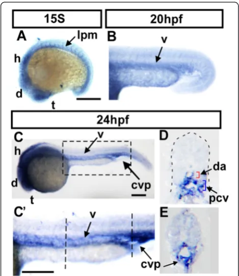
To examine the role ofnr2f1bin vascular development, we first analyze the expression ofnr2f1bby whole-mount in situ hybridization during zebrafish development. At the 15 somite stage (S),nr2f1bis expressed in the telenceph-alon (t), ventral medial diencephtelenceph-alon (d), hindbrain rhom-bomeres (h) and lateral plate mesoderm (Fig. 1a). The lateral plate mesoderm is the location of vascular precur-sors. At 20 hpf (~24S), we observed that nr2f1b is expressed in the vessels (Fig. 1b). At 24 hpf, nr2f1b is expressed in the telencephalon, diencephalon, hindbrain, as well as vessels of the trunk and caudal vein plexus (CVP) (Fig. 1c, c’). Transverse sections of embryos con-firm this localization (Fig. 1d, e). The expression ofnr2f1b in vasculature during embryonic development and sug-gests that it may play an important role.
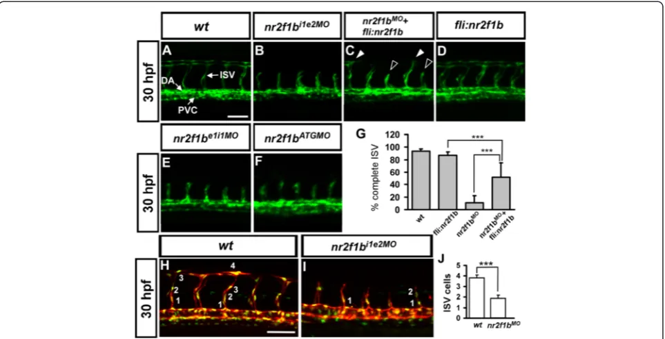
specificity of the morpholino knockdown. To further con-firm the specificity of our morpholino experiments, we performed rescue experiments by overexpression of nr2f1b in wild-type and nr2f1bi1e2 morphant embryos. Transient transgenic overexpression ofnr2f1bin endothe-lial cells under thefli1promoter rescues ISV stalling by 40 % innr2f1b morphants (n = 220 from 22 embryos) com-pared to injection ofnr2f1bmorpholino alone (Fig. 2c, g), while overexpression ofnr2f1b in wild-type embryos has no obvious defect on vascular development (Fig. 2d, n = 250 from 25 embryos).
We tested the efficiency and specificity of nr2f1bi1e2 morpholino knockdown. Injection of 1.7 ng or 3 ng of nr2f1bi1e2 morpholino showed dose-dependent disrup-tion of normal splicing of nr2f1b as determined by RT-PCR (Additional file 1: Figure S1A, B), suggesting the efficiency of nr2f1b knockdown. Sequence comparison of nr2f1bi1e2 morpholino targeting to nr2f1a and nr2f2 showed only 52 % and 28 % identity, respectively. More-over, injection ofnr2f1bi1e2morpholino greatly decrease
the amount of thenr2f1b product, butnr2f1aand nr2f2 levels were not decreased compared to uninjected con-trols as determined by RT-PCR, indicating the specificity of the morpholino knockdown of nr2f1b (Additional file 1: Figure S1C, D).
Further, nr2f1b morphant phenotypes do not result from morpholino-induced non-specific cell death as there is no significant increase in apoptosis in the trunk of morphants compared to wild type embryos by TUNEL staining (Additional file 1: Figure S2). These re-sults suggested the phenotypes of stalled ISV growth and decreased venous kdrl transgene expression are specific to the down-regulation of nr2f1b and indicate that nr2f1b plays a key role in zebrafish vascular develop-ment. To test if loss ofnr2f1bwould decrease cell prolif-eration, we counted the numbers of endothelial cells per ISV in the Tg (kdrl:mCherryci5; fli1a:negfp y7) embryos, where GFP was expressed in the nucleus of endothelial cells and the mCherry tag in the cytoplasm. Loss of nr2f1b showed significantly reduced ISV cell numbers compared to uninjected wild type embryos (1.8 ± 0.8 cells per ISV, n = 107 ISV from 18 embryos of nr2f1b morphants and 3.8 ± 0.8 cells, n = 108 ISV from 16 wt embryo,p< 0.0001) (Fig. 2 H-J). These data suggest that nr2f1b is required for ISV cell growth to contribute to the vascular development, likely by regulation of the proliferation or migration of the cells. Finally, a third phenotype was observed that loss of nr2f1b results in pericardial edema, absent parachordal vessels, mispat-terned subintestinal vessel plexus and circulation defects at later stages from 48 hpf to 72 hpf (Additional file 1: Figure S3). Since edema and lack of circulation are com-mon secondary consequences of defective blood vessel formation. The circulation defects consistent with the role ofnr2f1bin vascular development.
Nr2f1b promotes vein identity
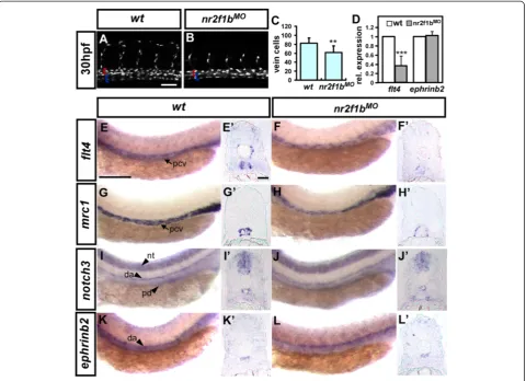
Reducedkdrl-transgene expression in the PCV ofnr2f1b morphant embryos could result from decreased number of cells contributing to the PCV or decreased venous endothelial marker expression of resident endothelial cells. Thus, we analyzed whether the number of endo-thelial cells is reduced in the PCV in nr2f1bmorphants by using Tg(fli1a:neGFP)y7 embryos which expressing GFP in endothelial cell nuclei and counting venous cells in the PCV in the region of the yolk extension (i. e. be-tween 5th to 15th ISVs) at 30 hpf. Uninjected control embryos have an average of 82.5 ± 11.5 cells (n = 14 em-bryos) in this region while nr2f1bi1e2 morphants have a significantly decreased number of cells with an average of 61.8 ± 14.7 cells (n = 12 embryos; p < 0.0001; Fig. 3a-c). Decreased PCV cell number suggests that loss of nr2f1b leads to a defect in the specification of venous cells con-tributing to the PCV but without a fate switch to an
Fig. 1Expression ofnr2f1bin during zebrafish development.aAt 15S,
nr2f1bis expressed in the telencephalon (t), ventral medial diencephalon (d), hindbrain rhombomeres (h) and lateral plate mesoderm (lpm,arrow) corresponding in location to the developing vasculature. At 20hpf (~24S),nr2f1bis expressed in the vessels (b).c,c'At 24 hpf,nr2f1bis expressed in the telencephalon (t), diencephalon (d), hindbrain (h), as well as in vessels (v), and caudal vein plexus (CVP) of the trunk.c'is an enlargement of C.d,eCross sections of embryos fromc’show that
arterial fate. Instead, we observed slightly decreased num-ber of cells contributing to the DA in the same region (67.5 ± 7.3 cells in wt and 57.9 ± 8.3 cells, p < 0.01), however, overexpression of nr2f1b did not increase the arterial cells (69.1 ± 10.9 cells, n = 10, p = 0.68), suggesting the minor necessary role of nr2f1b in aorta differentiation (data not shown).
To determine if loss of nr2f1b results in altered ex-pression of arterial and venous markers, we examined the expression flt4, mrc1, notch3, and ephrinb2 by ISH in nr2f1b morphants. No obvious differences in the ex-pression of the arterial markers notch3 and ephrinb2 were observed in nr2f1b morphants compared to con-trols at 24 hpf by both lateral view (Fig. 3i-l) and cross-section images (Fig. 3i’-l’). Conversely, expression of the venous markersflt4 andmrc1was diminished innr2f1b morphants compared to wild type controls at 24 hpf by both lateral view (Fig. 3e-h) and cross-section images (Fig. 3e’-h’). To determine the extent of decreased marker expression, we quantifiedflt4andephrinb2 tran-script levels by qPCR and identified a 60 % decrease in flt4 expression but no change inephrinb2 expression in nr2f1b morphants (Fig. 3d). These results suggest that the decrease in venous markers expression most likely due to a decreased number of vein cells.
To test whether excessnr2f1bmight increase the number of cells contributing to the PCV, we over-expressednr2f1b under control of the fli1a promoter. Overexpression of nr2f1b in transient transgenic embryos has a slight but significant increase on PCV cell number (100.3 ± 17.7 cells, n = 10) compared to uninjected controls (82.5 ± 11.5 cell; p < 0.01 by studentt-test) (Fig. 4a-c). Taken all together, the our data suggests thatnr2f1bhas a role in venous endothe-lial cell specification as loss and over-expression of nr2f1b results in decreased and increased numbers of endothelial cells contributing to the PCV, respectively.
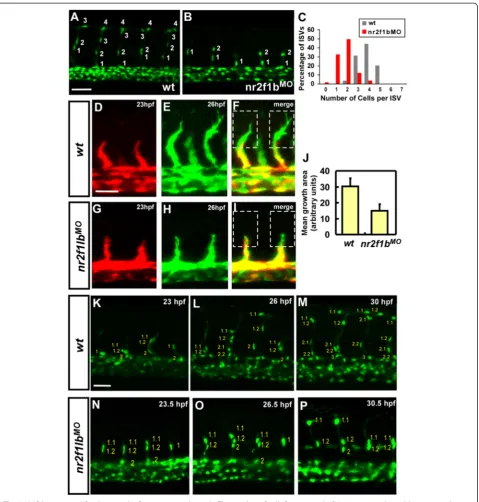
nr2f1bmodulates the number and migration of angioblasts in intersegmental vessels
The number of cells comprising the trunk intersegmen-tal vessels is regulated by a number of pathways where reduced numbers of cells per ISV can result in ISV growth defect. To determine if a reduced number of cells was present in stalled ISVs, the number of cells per ISV was assessed after knockdown ofnr2f1bin Tg(fli1a:-negfp)y7 or Tg (kdrl:mCherryci5; fli1a:negfp y7) embryos (Fig. 5a-b and Fig. 2h-i). We observed a reduced ISV cells innr2f1bmorphants compared to wild-type control (Fig. 2j) and the distribution of ISV cells showed at less cells area in nr2f1b MO as compared to uninjected
Fig. 2Morpholino knockdown ofnr2f1bcauses defects in vascular development.aIn uninjected control embryos, the arota (da) and posterior cardinal vein (pcv) have formed by 30 hpf and intersegmental vessels (isv) have reached the DLAV at the dorsal aspect of the embryo. At the same stage ISVs are stalled mid-somite in nr2f1bi1e2(b), nr2f1be1i1(e) and nr2f1bATG(f) morphants. Overexpression of nr2f1b has no obvious
embryos (Fig. 3c). Moreover, endothelial-specific over-expression of nr2f1b using the fli1 promoter results in an increased average number of cells per ISV (4.3 ± 0.9; n = 88 ISVs) in transient transgenic embryos compared to 3.8 ± 0.7 cells (n = 86 ISVs) in wild type (p< 0.0005) (Fig. 4d-f ). These data suggest thatnr2f1bis ne-cessary and sufficient to promote a tip cell identity for ISV growth.
To examine whether migration of ISV angioblasts is deficient, time-lapse imaging was performed in wild type Tg(kdrl:eGFP) la116 and nr2f1b knockdown embryos. Superimposition of images of 23hpf embryo (red) on the same embryo at 26hpf (green) shows an average of an over 50 % decreased area of extension/migration of ISVs innr2f1bmorphants as compared to wild type embryos (Fig. 5d-j). The decrease in extension could represent decreased protrusive activity of angioblasts, or could
result from fewer angioblasts migrating. We therefore examined ISV cell number in Tg(fli1a:neGFP)y7 trans-genic embryos on these time-lapse images. We found cells showed slower migration and ISVs that eventually migrate to the DLAV in nr2f1b morphants have fewer cells per ISV (Fig. 5k-p) than wild type, suggesting that nr2f1b regulates the number of cells in an ISV and the migration of ISV. Together, these data suggest that nr2f1b modulates the number and migration of angio-blasts during ISV growth.
Interaction betweennr2f1band Notch signaling
We have demonstrated that knockdown of nr2f1b ex-pression by morphlino injection results in ISV stalling at the midline with a decrease in the number of cells per ISV, while over-expression of nr2f1b leads to an in-creased number of cells per ISV. These observations are
Fig. 3Nr2f1b modulates vein cell number and marker expression.a-cAt 30 hpf, loss ofnr2f1bfunction inTg(fli1a:negfp)y7embryos results in reduced vein cell number (b, blue bracket) as compared to uninjected wild type controls (a). The artery is marked by a red bracket.cQuantitative analysis shows a significant reduction in vein cell number innr2f1bmorphants. Compared to wild type controls.e,g,i,k, expression of the venous markersflt4(f) andmrc1(g) is reduced in the trunk ofnr2f1bmorphants at 24 hpf while there is no obvious change in the expression of arterial markersnotch3(i) andephrinb2(k).e’-l’are cross sections of embryos in (e-l).dQuantification by qPCR shows a 60 % reduction inflt4
qualitatively similar to those seen with modulation of Notch signaling where activation of Notch signaling gives ISV stalling at the midline, while loss ofnotch sig-naling leads to an increased number of cells per ISV (Siekmann 2007). Thus, we sought to determine if Notch regulates nr2f1b expression. Notch signaling involves two sequential proteolytic processing events (ADAM protease andγ-secretase) that release the Notch intracel-lular domain (NICD) into cytoplasm followed by trans-location to the nucleus where it interacts with the transcription factor rbpsuh (recombination binding protein/ suppressor of hairless) to activation of target genes. DAPT specifically inhibitγ -secretase involved in the cleavage of NICD and prevents Noch activation. Therefore, to test if nr2f1binteracts with Notch signaling, we suppressed Notch signaling byrbpsuhMOinjection (Fig. 6a-c) or DAPT treat-ment (Fig. 6d-f). We found nr2f1b expression is upregu-lated when Notch signals were inhibited, with a 2-2.5 fold increase by using in-situ hybridization and qPCR (Fig. 6). These results suggest that nr2f1b might act downstream of Notch signals. We next asked whether nr2f1b and Notch genetically interact to control ISV growth. To test this, we performed a rescue experiment by injectingnr2f1b morpho-lino in an rbpsuh morphants (rbpsuhMO). Wild type em-bryos at 30 hpf have an average of 3.6 ± 0.6 cells per ISV (Fig. 6g, k).nr2f1bmorphants have an average of 1.7 ± 0.7 cells per ISV, while rbpsuh morphants have an average of 6.2 ± 1.6 cells per ISV (Fig. 6h, i, k). Knockdown ofrbpsuh in combined with nr2f1b morpholino injection reduces the number of cells per ISV to wild type levels 3.8 ± 1.2 (Fig. 6j, K; n = 25 ISV from 4 fish; p = 0.56, unpaired
student t-test). Together, these data suggest nr2f1blikely acts downstream of Notch signaling and antagonizes Notch signals to control ISV growth.
Discussion
nr2f1bexpressed in developing vessels functioning in vasculature
In this study, we observed the expression ofnr2f1bin lateral plate mesoderm at early stage and developing vessels (Fig. 1) corresponding to the location in which primitive angioblasts are developing and acquiring arterial and venous identity. At 24hpf, the expression pattern ofnr2f1bcontinues in the vasculature, suggested the role ofnr2f1bin vascular devel-opment. We further have shown that knockdown ofnr2f1b results in vascular defects, including ISV growth impair-ment, pericardial edema, less venous cells and results in loss of circulation (Fig. 2 and Additional file 1: Figure S3). In fact, there is no parachorda vessel (PCV, pre-structure of lymphatic duct) formation and defects in caudal vessel plexus (CVP) (data not shown) suggested that the impair-ment of venous angiogenesis at late developimpair-mental stage, which is likely the later effects of loss ofnr2f1b.
nr2f1bfunctions in vein identity and the growth of intersegmental vessels
Here, we explored the function ofnr2f1b modulates vein identity. We showed the decrease of endothelial venous cells and the expression of venous markersflt4andmrc1; however, there is no obvious change in the expression of arterial markers notch3 and ephrinb2 (Fig. 3) although there is a slight decrease of arterial cells. This suggested
Fig. 4Overexpression ofNr2f1bunder the fli promoter increases the number of venous cells and endothelial cells per ISV.a-bThe number of vein cells was counted in the region beneath ISV 5-15 from 14 wild type or 10Tg(fli1a:nr2f1b)transient transgenic, overexpressing embryos. The artery is marked by a red bracket, and the vein by a blue bracket.cUninjected control embryos have 82.5 (± 11.5) cells per vein segment while
that nr2f1b is not involved in artery-vein fate switching. Thus, the reduction in cell number and the loss of vein marker expression together suggest that nr2f1bis neces-sary for normal vein development. We also showed that overexpression of nr2f1b has a slight but significant in-crease on PCV cell number (Fig. 4). Together, those data
suggest the role of nr2f1b is necessary and sufficient for vein development.
Intersegmental vessels form from angioblasts sprouting from the dorsal aorta and vein. Stalling of intersegmental vessel growth at the mid somite might therefore either occur through defective proliferation or defective
Fig. 5Nr2f1b is required for the growth of intersegmental vessels. The number of cells forming each ISV were counted in wild type control
migration of cells. Here, we showed innr2f1bmorphants contains less cells compared to in wild-type (Fig. 5). We also showed a significant increase in ISV cell numbers suggesting thatnr2f1b is necessary and sufficient to pro-mote a proliferation of ISV cells. Further examining the migration of ISV angioblasts in nr2f1b knockdown Tg(fli1a:eGFP)y1 and Tg(fli1a:neGFP)y7embryos, we showed that the decrease in extension represent the de-creased protrusive activity of angioblasts and fewer angio-blasts in migrating (Fig. 5), suggesting that nr2f1b regulates the number of cells in an ISV and the migration of ISV. Loss of nr2f1b in zebrafish leads to decreased numbers of cells in the posterior cardinal vein and in ISVs, but the ultimate fate of these cells remains unknown. TUNEL analysis suggests that cell death is not increased in the trunk region and we did not observe additional cells contributing to the artery or ISVs. This suggests there may be a lack of proliferation of venous precursors or these cells may adopt a closely related fate in the mesoderm lineage, such as blood.
Interaction betweennr2f1b,nr2f2andisl2in regulating vein identity and ISV growth?
We previous identified isl2 promotes vein and tip cell identity (paper under revision) and nr2f2 also plays minor role on that although NR2F2 is a major determin-ant of venous identity in mouse. In this study, we showed that zebrafish Nr2f1b ortholog to mouse NR2F2 plays a major role in vein and tip cell identity. It has been shown that LIM-homeodimer transcription factor isl1 (an ortholog of Isl2) and CoupTFI physically bind together in vitro and in vivo to activate transcription [38], suggesting the possible interaction between isl2 and nr2f1b to control vascular development. Thus, whether Nr2f1b/Nr2f2 and Isl2 also physically interact at the pro-tein level to activate target genes and regulate endothelial cell identity remains an interesting avenue to explore in the future. Meanwhile, it would be also intriguing to address if any other signaling molecules in addition to notch, such as vegf, wnt or BMP etc. that interact with nr2f1b and/or isl2. The molecular mechanisms that how nr2f1b regulate its
Fig. 6The expression ofNr2f1bis upregulated by notch signaling.a,bnr2f1bexpression is increased at 24 hpf embryos after treatment with DAPT as compared to DMSO control embryos by in situ hybridization.d,enr2f1bexpression is upregulated inrbpsuhmorphants at 24 hpf embryos.c,fQuantification by qPCR showed the increased expression ofnr2f1binrbpsuhmorphants or DAPT treated embryos at 24 hpf significantly.g-jRepresentative confocal images showing the number of nuclei per ISV at 30 hpf ingwild-type (wt) embryos,hnr2f1bMO,i
rbpsuhMO, andjrbpsuhMOwithnr2f1bMO.kQuantitation of the average number of cells per ISV in single and double morphants. ***refers to
targets in vascular development is still unknown and we are currently addressing this question by processing and analyzing genome-wide transcriptome results.
Does zebrafish Nr2f1b play a conserved role in vasculature similar to nr2f2 in mice?
Swift’s study showed SoxF factors and Notch regulate nr2f2 gene expression during venous differentiation [12] and Aranguren et al showed coupTFII functions in ven-ous and lymphatic development in both zebrafish and Xenopus [11]. However, knockdown nr2f2 in both stud-ies did not see obvious defects in vascular development, but reduction of venous gene expression, suggest nr2f2 control venous differentiation, instead of specification. In this study, we showed that zebrafish Nr2f1b ortholog to mouse Nr2f2 and plays a major role in vein and tip cell identity, which is consistent with the function of nr2f2 in mice [9]. Those data indicate that the conserved vascular function of coupTF family among the verte-brates. Phylogenetic analysis of coupTFs amino acids among the vertebrates suggests that zebrafish nr2f1b and nr2f2 are very closer to mammalian nr2f2 (over 83 % identical). It would be intriguing to address if any func-tional rescue or compensation between zebrafish nr2f1b and mouse nr2f2.
Conclusions
In summary, our study demonstrated thatnr2f1bhas a crit-ical role in blood vessel formation in zebrafish. We showed that loss ofnr2f1breduced venous cells and the expression of vein specific markers. We also showed thatnr2f1b mor-phant reduces ISV cells and impairs ISV growth. While overexpression ofnr2f1b increase vein and ISV cells, sug-gesting that nr2f1b play a role in promoting vein and tip cell identity. We further showed that nr2f1b functions in vascular development in concert with Notch signalling.
Additional file
Additional file 1:Supplementary Methods.Table S1: Primer/ morpholino sequences used in this study. Figure S1: Knockdown efficiency and specificity ofnr2f1bmorpholinos. Figure S2: An increase in non-specific cell death after morpholino injection is not the cause of the observed vascular phenotype. Figure S3. Loss ofnr2f1bresults in pericardial edema, absent parachordal vessels, subintestinal vessels (SIV) mispattern and circulation defects. (DOC 2967 kb)
Competing interests
The authors declare that they have no competing interests.
Authors’contributions
CYW conceived and designed experiments. RFL, TYW, YZM, YSW performed the experiments. CLC and CYW carried out the confocal image processing and data analysis. RFL, TYW and CYW drafted the manuscript. All authors read and approved the final manuscript.
Acknowledgements
This work was supported by the grants from Ministry of Science and Technology, Taiwan (NSC101-2311-B-110-002, NSC102-2311-B-110-002 and MOST103-2311-B-110-004) and from the NSYSU-KMU Joint Research Project (NSYSUKMU 102-P028 and NSYSUKMU103-P031) to CYW. We thank Taiwan Zebrafish Core Facility at Academia Sinica (TZCAS) and the National Health Research Institutes (NHRI), which is supported by Ministry of Science and Technology (MOST-102-2321-B-001-038) in Taiwan for providing wild-type and transgenic zebrafish. We thank Dr. Sarah Childs for providing the primers, plasmids and in-situ hybridization probes used in this work. We would like to thank Dr. Sarah Childs, Dr. Ryan Lamont, Dr. Zhi-Hong Wen and Dr. Tai Ming-Hong for their helpful discussions and comments on the manuscript.
Author details
1Department of Biological Sciences, National Sun Yat-sen University,
Kaohsiung, Taiwan.2Doctoral Degree Program in Marine Biotechnology, National Sun Yat-sen University and Academia Sinica, Kaohsiung, Taiwan.
3
Department of Biotechnology, Kaohsiung Medical University, Kaohsiung, Taiwan.
Received: 10 June 2015 Accepted: 16 October 2015
References
1. Risau W. Mechanisms of angiogenesis. Nature. 1997;386:671–4. 2. Lawson ND, Weinstein BM. Arteries and veins: making a difference with
zebrafish. Nat Rev Genet. 2002;3:674–82.
3. Isogai S, Lawson ND, Torrealday S, Horiguchi M, Weinstein BM. Angiogenic network formation in the developing vertebrate trunk. Development. 2003;130:5281–90.
4. Childs S, Chen J-N, Garrity D, Fishman M. Patterning of angiogenesis in the zebrafish embryo. Development. 2002;129:973–82.
5. Siekmann AF, Lawson ND. Notch signalling limits angiogenic cell behaviour in developing zebrafish arteries. Nature. 2007;445:781–4.
6. Lamont RE, Childs S. MAPping out arteries and veins. Sci STKE. 2006;2006:pe39.
7. Swift MR, Weinstein BM. Arterial-venous specification during development. Circ Res. 2009;104:576–88.
8. Pereira FA, Qiu Y, Zhou G, Tsai MJ, Tsai SY. The orphan nuclear receptor COUP-TFII is required for angiogenesis and heart development. Genes Dev. 1999;13:1037–49.
9. You LR, Lin FJ, Lee CT, DeMayo FJ, Tsai MJ, Tsai SY. Suppression of Notch signalling by the COUP-TFII transcription factor regulates vein identity. Nature. 2005;435:98–104.
10. Yamazaki T, Yoshimatsu Y, Morishita Y, Miyazono K, Watabe T. COUP-TFII regulates the functions of Prox1 in lymphatic endothelial cells through direct interaction. Genes Cells. 2009;14:425–34.
11. Aranguren XL, Beerens M, Vandevelde W, Dewerchin M, Carmeliet P, Luttun A. Transcription factor COUP-TFII is indispensable for venous and lymphatic development in zebrafish and Xenopus laevis. Biochem Biophys Res Commun. 2011;410:121–6.
12. Swift MR, Pham VN, Castranova D, Bell K, Poole RJ, Weinstein BM. SoxF factors and Notch regulate nr2f2 gene expression during venous differentiation in zebrafish. Dev Biol. 2014;390:116–25.
13. Gerhardt H, Golding M, Fruttiger M, Ruhrberg C, Lundkvist A, Abramsson A, et al. VEGF guides angiogenic sprouting utilizing endothelial tip cell filopodia. J Cell Biol. 2003;161:1163–77.
14. Adams RH, Alitalo K. Molecular regulation of angiogenesis and lymphangiogenesis. Nat Rev Mol Cell Biol. 2007;8:464–78.
15. Baldessari D, Mione M. How to create the vascular tree? (Latest) help from the zebrafish. Pharmacol Ther. 2008;118:206–30.
16. Covassin LD, Villefranc JA, Kacergis MC, Weinstein BM, Lawson ND. Distinct genetic interactions between multiple Vegf receptors are required for development of different blood vessel types in zebrafish. Proc Natl Acad Sci U S A. 2006;103:6554–9.
17. Leslie JD, Ariza-McNaughton L, Bermange AL, McAdow R, Johnson SL, Lewis J. Endothelial signalling by the Notch ligand Delta-like 4 restricts
angiogenesis. Development. 2007;134:839–44.
19. Phng LK, Gerhardt H. Angiogenesis: a team effort coordinated by notch. Dev Cell. 2009;16:196–208.
20. Wiens KM, Lee HL, Shimada H, Metcalf AE, Chao MY, Lien CL. Platelet-derived growth factor receptor beta is critical for zebrafish intersegmental vessel formation. PLoS One. 2010;5, e11324.
21. Yu PC, Gu SY, Bu JW, Du JL. TRPC1 is essential for in vivo angiogenesis in zebrafish. Circ Res. 2010;106:1221–32.
22. Wigle JT, Oliver G. Prox1 function is required for the development of the murine lymphatic system. Cell. 1999;98:769–78.
23. Lee S, Kang J, Yoo J, Ganesan SK, Cook SC, Aguilar B, et al. Prox1 physically and functionally interacts with COUP-TFII to specify lymphatic endothelial cell fate. Blood. 2009;113:1856–9.
24. Zhou C, Tsai SY, Tsai MJ. COUP-TFI: an intrinsic factor for early regionalization of the neocortex. Genes Dev. 2001;15:2054–9.
25. Tang LS, Alger HM, Pereira FA. COUP-TFI controls Notch regulation of hair cell and support cell differentiation. Development. 2006;133:3683–93. 26. Montemayor C, Montemayor OA, Ridgeway A, Lin F, Wheeler DA, Pletcher
SD, et al. Genome-wide analysis of binding sites and direct target genes of the orphan nuclear receptor NR2F1/COUP-TFI. PLoS One. 2010;5, e8910. 27. Lawson ND, Weinstein BM. In vivo imaging of embryonic vascular
development using transgenic zebrafish. Dev Biol. 2002;248:307–18. 28. Roman BL, Pham VN, Lawson ND, Kulik M, Childs S, Lekven AC, et al.
Disruption of acvrl1 increases endothelial cell number in zebrafish cranial vessels. Development. 2002;129:3009–19.
29. Choi J, Dong L, Ahn J, Dao D, Hammerschmidt M, Chen JN. FoxH1 negatively modulates flk1 gene expression and vascular formation in zebrafish. Dev Biol. 2007;304:735–44.
30. Proulx K, Lu A, Sumanas S. Cranial vasculature in zebrafish forms by angioblast cluster-derived angiogenesis. Dev Biol. 2010;348:34–46. 31. Westerfield M. The Zebrafish Book: A Guide for the Laboratory Use of
Zebrafish (Danio Rerio). Eugene, OR: Institute of Neuroscience, University of Oregon; 1995.
32. Jowett T, Lettice L. Whole-mount in situ hybridizations on zebrafish embryos using a mixture of digoxigenin- and fluorescein-labelled probes. Trends Genet. 1994;10:73–4.
33. Lawson ND, Scheer N, Pham VN, Kim CH, Chitnis AB, Campos-Ortega JA, et al. Notch signaling is required for arterial-venous differentiation during embryonic vascular development. Development. 2001;128:3675–83. 34. Wong KS, Proulx K, Rost MS, Sumanas S. Identification of vasculature-specific
genes by microarray analysis of Etsrp/Etv2 overexpressing zebrafish embryos. Dev Dyn. 2009;238:1836–50.
35. Wu BJ, Chiu CC, Chen CL, Wang WD, Wang JH, Wen ZH, et al. Nuclear receptor subfamily 2 group F member 1a (nr2f1a) is required for vascular development in zebrafish. PLoS One. 2014;9, e105939.
36. Thisse C, Thisse B. High-resolution in situ hybridization to whole-mount zebrafish embryos. Nat Protoc. 2008;3:59–69.
37. Kwan KM, Fujimoto E, Grabher C, Mangum BD, Hardy ME, Campbell DS, et al. The Tol2kit: a multisite gateway-based construction kit for Tol2 transposon transgenesis constructs. Dev Dyn. 2007;236:3088–99. 38. Gay F, Anglade I, Gong Z, Salbert G. The LIM/homeodomain protein islet-1
modulates estrogen receptor functions. Mol Endocrinol. 2000;14:1627–48.
Submit your next manuscript to BioMed Central and take full advantage of:
• Convenient online submission
• Thorough peer review
• No space constraints or color figure charges
• Immediate publication on acceptance
• Inclusion in PubMed, CAS, Scopus and Google Scholar
• Research which is freely available for redistribution