BMI1 and KAP1 interaction and function:
BMI1 capped by KAP1?
J. van Haasteren
Department Molecular Genetics, GROW School of Oncology and Developmental Biology, Maastricht University
Abstract
The Polycomb-repressive complex 1 (PRC1) protein BMI1 is of major importance in the epigenetic regulation of gene expression. The repression of important tumour suppressor genes (such a P16INK4a and P14ARF) by means of chromatin remodeling
suggests that the KAP1 negatively controls expression of ATF3 in a RF-dependent manner. Further research is required to elucidate the exact molecular mechanisms underlying the function interaction of BMI1 and KAP1.
Keywords
Polycomb group proteins, Polycomb repressive complex, BMI1, KAP1, RingFinger domain, gene repression.
Introduction
Polycomb group (PcG) proteins function is of paramount importance in epigenetic regulation and the propagation of heritable epigenetic mark-up throughout cell division. With this in mind it is perhaps not surprising that PcG proteins are involved in embryonic development, differentiation and (stem) cell fate, and the aetiology of cancer (1-3). Polycomb proteins are present in several protein complexes, the two main ones being Polycomb repressive complex 1 and 2 (PRC1 and PRC2) (4). These multiprotein complexes exist in varying compositions of Polycomb proteins between cell types (5) and the function of the complex alters according to its composition (6). As the name implies, PRC1 and 2 contribute to the repression of gene expression. They do so by chromatin remodelling via histone modification. Indeed, a number of the functions of PRC’s involve the addition and removal of chemical groups to histone tail residues. In the process of gene silencing, it is PRC2 that functions first by adding a third methyl group to lysine 27 of histone 3 (H3K27-me3) (7). On recognition of H3K27-me3, the PRC1 complex functions as an E3 ubiquitin ligase and ubiquitylates lysine 119 of histone 2A (H2AK119-Ub) (8, 9). It is this ubiquitylation that causes repression of gene expression (figure 1).
inhibition of the tumour suppressor gene CDKN2A coding for p16INK4a and p14ARF (12, 13). Our lab has recently found evidence for a physical interaction between the Polycomb protein BMI1 and the transcriptional repressor Krüppel-associated-box (KRAB)-associated protein 1 (KAP1) (Prickaerts et al., in preparation). We obtained evidence that shows KAP1 may target BMI1 for proteasomal degradation, a process which is likely dependent on the RingFinger domain of KAP1 (figure 2). We hypothesized that KAP1 may controls PRC1 functioning via degradations and may as such influence the expression of PRC1 target genes such as ATF3.
To provide context to the how and where of BMI1 activity, potential phosphorylatable BMI1 amino acid residues were identified and mutated to provide insight to the effects of either constitutive phosphorylation or ablation of phosphorylation (14, 15). We assessed the effects of these mutations by measuring proliferative capacity of U2OS ant TIG3 cell lines during cell stress. Taking previous research into account, we expect that phosphorylation of amino acid residues of BMI1 in the PEST-domain might make it a target for degradation, thereby effectively reducing the positive effect on proliferation (15, 16). We hypothesize that post-translational modification of BMI1 by means of phosphorylation will negatively affect its ability to promote proliferation. If BMI1 indeed positively regulates cell proliferation and we take into account our preliminary findings that suggest that KAP1 targets BMI1 for degradation, we anticipate that the loss of KAP1 might synergistically influence the proliferative capacity of cells overexpressing BMI1. The ability to degrade BMI1 might cause
ATF3 to be differentially expressed between KAP1 knockdown and overexpressed cells, and may be altered in a RingFinger dependent manner. This also predicts that KAP1 may affect the association of BMI1 with chromatin.
Figure 1. Mechanism of Polycomb repressive complex gene repression. After Polycomb repressive complex 2 (PRC2) is recruited to the DNA, one of the components of PRC2, Enhancer of Zeste (EZH2), will methylate histone 3 on lysine 27. In response this mark draws PRC2 to the chromatin, which ubiquitylates the lysine 119 residue of histone H2A to provide robust transcriptional gene repression. Adapted from (17).
Material and methods
Ecotropic and amphotropic vectors were used to efficiently and stably overexpress wild-type or mutant BMI1 or KAP1 protein. Knockdown of KAP1 was performed with shRNA’s targeting mature KAP1 mRNA or the 3’UTR region of early mRNA. The latter shRNA provided the opportunity to do a knockdown-add back experiment where endogenous KAP1 could be replaced with mutant KAP1 expression. Experiments were conducted in human osteosarcoma cells (U2OS) and human primary fibroblast cells (TIG3). Protein determination by western blot was performed on whole cell protein lysates or subcellular fractions obtained by differential extraction. Proliferation of cells in culture was assessed by means of X-tal Violet staining procedure and subsequent absorbance measurement. Measurements was done at 0, 2, 4 and 6 days post stimulation. Mitogenic stimulation was done with 15% serum and 0.1 µg/ml tetradecanoyl phorbol (TPA, Sigma) after 48 hour serum starvation. ATF3 mRNA was acquired by RNA isolation using Tri-reagent (Sigma) and subsequent cDNA production by means of the iScript cDNA synthesis kit (Biorad). Quantitative measurement was obtained by RT-PRC analysis where samples were normalized to Cyclophillin A, a housekeeping gene.
Results
BMI1 overexpression provides protection against arsenite induced senescence
BMI1 positively contributes to protection or proliferation of As-exposed cells. The positive effect of BMI-2PY 7xA on proliferation is less than that of the BMI1 wild-type. Although this data suggests that the ability to be phosphorylated is paramount in the functioning of BMI1, this effect can also be ascribed to the differences in protein levels compared to wild-type BMI1.
KAP1 knockdown increased BMI1 induced resistance to arsenite induced senescence
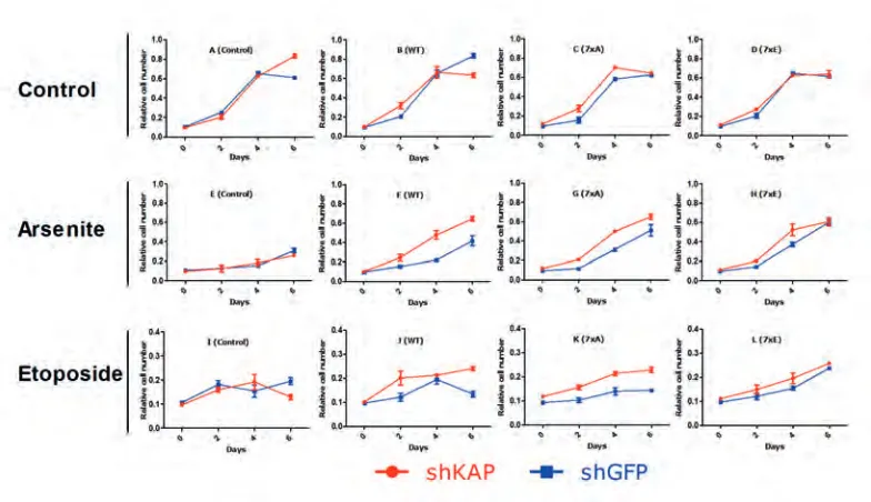
In order to assess the influence of KAP1 on cellular proliferation under both normal and cell stressed conditions we infected cell lines with short hairpin RNA’s (shRNA’s) to create knockdown of KAP1. shRNA targeting green fluorescent protein (shGFP) was used as control as U2OS cells do not express GFP. KAP1 and GFP knockdown was performed in BMI1 mutant (7xA and 7xE) and wild type overexpressing cells. Cells were subjected to different cell stressors and relative cell number was followed over time to create growth curves indicative of proliferation rate.
The loss of KAP1 in BMI1 mutants had no significant effect in control conditions of plain growth medium (figure 4 A-D). Loss of KAP1 in itself does not provide protection against arsenite induced cell stress, however, all BMI constructs in our model do (figure 4 F-H). In addition, loss of KAP1 provides an additional proliferative advantage in combination with all BMI1 constructs; this suggests a cooperative effect under arsenite-stressed conditions. Plotting the same data in graphs combining the BMI1 mutants, BMI1 wild-type and control cell lines all with KAP1 knockdown suggested that loss of KAP1 reduces the differences that were found in cells that do indeed express KAP1 (figure 5). Etoposide affects shKAP and shGFP cells equally, suggesting KAP1 depletion does not provide protection (figure 4I). Under these conditions BMI1 overexpression does not provide substantial protection, as proliferation with or without BMI1 overexpression remains unaltered. BMI1-WT and 7xA in combination with shKAP however show a slight proliferative advantage.
RingFinger domain required for KAP1 mediated changes in ATF3 expression
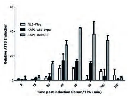
All cell lines show a transient induction of ATF3 post-serum/TPA stimulation (figure 6). shKAP1eV (NLS-Flag) cells showed an attenuated transcriptional response in comparison to cells expression shKAP1/KAP1-fl, as evidenced by a lower relative ATF3 induction. However, since ATF3 is not a KAP1 target (19), this response is expected to be indirect. This result is in concordance with its proposed repressive effects of KAP1 on BMI1. Loss of KAP1 would increase BMI1 levels, thereby increasing ATF3 gene repression by PRC1 and thereby decreasing its expression. Conversely, the overexpression of a KAP1 mutant lacking the RingFinger domain elicited a greater response than wild-type KAP1.
Figure 3. Phospho-mimic mutant BMI1 overexpression attenuates senescence by arsenite poisoning.
Figure 4. KAP1 knockdown increased BMI1 mutant overexpression induced changes in proliferation under arsenite and etoposide induced cell stress. Relative cell numbers indicate no change in proliferation in control situations between shKAP1 and shGFP cell lines in all BMI1 variants. KAP1 knockdown increased proliferation in BMI1 overexpressing cells under arsenite induced stress (F-G) and the 7xA BMI1 mutant proved to proliferate more rapidly under etoposide stress conditions when KAP1 is knocked down.
Figure 5. KAP1 knockdown in BMI1 mutant U2OS cells diminished differential effects between mutants. KAP1 knockdown was achieved with shKAP1 infection. The same cell lines were used as in figure 4 but the knock down of KAP1 proves to reduce the differences between BMI1 mutants we saw in proliferation under arsenite stimulation.
enhancement of ATF3 induction in the presence of a KAP1-RF mutant was surprising, as previous results suggested that the RingFinger domain is required for KAP1 mediated degradation and that KAP1 may control repressive action by PRC1 through inducing proteolytic degradation of BMI1. Combined, the above data suggests that KAP1 control initiation of transcription as well as silencing. Whether both properties involved KAP1-BMI1 interaction is currently not clear. It is currently also not possible to conclude whether KAP1 targets BMI1 and thereby PRC1-mediated repression, through RF-mediated E3-ligase activity, or whether it is the physical presence of KAP1, that through its RF domain recruits additional E3-ligase activity which targets BMI1 for degradation.
Figure 6. ATF3 expression increase following serum/TPA stimulation increased by loss of RingFinger domain of KAP1. The expression levels of PRC1 target gene ATF3 as measured by rtPCR and normalized to cyclophillin A are provided as relative induction compared to control situation (t=0). Double 3’UTR shKAP1 infected U2OS cells were infected with either NLS-Flag (control), full-length KAP1 or KAP1 delta-RingFinger viral vector to stably induce expression of these proteins (supplemental figure 5). Restoring KAP1 increases the transcriptional response measured as ATF3 induction. KAP1 DeltaRF shows a greater response compared to wild-type KAP1.
Chromatin association of KAP1 and BMI1 in response to mitogenic stimulation
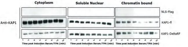
wild-type KAP1 and a mutant lacking the RingFinger domain (Flag-KAP1 DeltaRF). Cells infected with an empty vector construct (NLS-Flag) were used as controls. KAP1 and BMI1 protein were visualized by western blot in the different subcellular fractions.
Figure 7. Serum/TPA induced mitogenic stimulation changes subcellular localisation of KAP1. Protein lysates of cytoplasmic, soluble nuclear and chromatin bound fractions visualised are using western blot. KAP1 is removed from the chromatin fraction at 60 minutes post mitogenic stimulation for wild-type KAP1 and 45 minutes in the case of KAP1-DeltaRF. Chromatin localisation of KAP1 is restored around 1 hour later.
Due to unfortunate problems with the secondary antibodies targeting monoclonal primary antibodies in our lab, we were only able to visualise the expression of KAP1 (which is detected using a polyclonal primary antibody). Since the loading controls cannot be used to prove equal protein loading, we cannot be certain about the KAP1 levels in the different cellular compartments. Taking this into account we can however cautiously interpret the data at hand (figure 7). The findings suggest that KAP1-fl dissociates from chromatin around 60 minutes post-serum/TPA stimulation and is recruited to chromatin after 120 minutes. Remarkably, KAP1-DeltaRF dissociation dynamics are substantially altered: KAP1-DeltaRF dissociation is transient and recruitment of KAP1-DeltaRF is more profound compared to KAP1-fl. Taken together, RF-mutation clearly affects KAP-1 chromatin-binding dynamics and PCR1-mediated gene repression.
Discussion/Conclusion
BMI1 in arsenite-induced cell stressed conditions. Surprisingly, in cells exposed to selenite BMI1 overexpression seems, regardless of mutation, to decrease the rate of proliferation compared to the control cell line (figure 3C). We know that in response to DSB induced H2AX phosphorylation, BMI1 will ubiquitylate γ-H2AX to stimulate the correct DNA damage repair system (11). As selenite induces DSBs, BMI1 overexpression could be expected to rescue the cells by ensuring an adequate DSB repair response and prevent cell death, as was reported before (20), but this appears not to be the case in our experimental setting. Since phosphorylation status does not change the response BMI1 functioning alteration by phosphorylation events cannot be appointed as the cause of this effect. One explanation can be that the overexpression of BMI1 causes a DNA damage response signalling cascade that is so strong that it may push cells towards senescence (21). Combining KAP1 knock down with BMI1 overexpression proved to produce a significant increase in proliferation in arsenite induced cell stress in U2OS cells (figure 4). This synthetic effect implicates a functional interaction between KAP1 and BMI1 on proliferation under cell stress conditions. Our group has previously hypothesised that KAP1 targets BMI1 for degradation in cell stress conditions induced by arsenite and selenite. At first glance, our results fit with this explanation. If KAP1 is not present to inhibit BMI1, the expression of BMI1 is expected to increase. This has to be validated for each BMI1 type in our model. The fact that all three of the used BMI1 varieties show similar proliferative responses in the absence of KAP1 suggests that phosphorylation status of BMI1 is only of importance when KAP1 is present. While this is not conclusive proof, it points toward the notion that at least one of these seven amino acid residues is implicated in the BMI1/KAP1 interaction or other functional relations (e.g. ubiquitylation, degradation).
Although the results of the subcellular fractionation were inconclusive, we can state that the chromatin localisation of KAP1 is not dependent on its RingFinger domain (figure 7). A possible mechanism could be an auto-ubiquitylation event of KAP1, which would inhibit its function and attenuate co-localisation to chromatin following mitogenic stimulation. The proposed auto-phosphorylation would be abrogated in the RingFinger mutant of KAP1, providing a possible explanation for the observed increase and altered dynamics in chromatin localisation of KAP1-DeltaRF.
We have here demonstrated that the Polycomb protein BMI1 enhances resistance to arsenite induced– proliferative arrest. Furthermore we have provided further indication for a functional KAP1/BMI1 interaction both under cell stress conditions and mitogenic stimulation. While we found indications that phosphorylation status of BMI1 and the RingFinger domain of KAP1 are implicated in this interaction, we cannot provide conclusive evidence on the underlying molecular processes involved. Elucidating the mechanisms by which KAP1 modifies the function of BMI1 and what post-translational modifications of BMI1 are crucial for its function are compelling targets for future research.
Role of the student
J. van Haasteren was an undergraduate student in Biomedical Science working under the supervision of Dr Jan-Willem Voncken when the research in this report was performed. The topic and experiments were proposed by the supervisor. The design and planning of the experiments, the processing of the results as well formulation of the conclusions and the writing were done by the student.
References
1. Sparmann A, van Lohuizen M. Polycomb silencers control cell fate, development and cancer. Nature reviews Cancer. 2006;6(11):846-56.
2. Posfai E, Kunzmann R, Brochard V, Salvaing J, Cabuy E, Roloff TC, et al. Polycomb function during oogenesis is required for mouse embryonic development. Genes & development. 2012;26(9):920-32.
3. Surface LE, Thornton SR, Boyer LA. Polycomb group proteins set the stage for early lineage commitment. Cell stem cell. 2010;7(3):288-98.
4. Francis NJ, Kingston RE. Mechanisms of transcriptional memory. Nature Reviews Molecular Cell Biology. 2001;2(6):409-21.
5. Gunster MJ, Raaphorst FM, Hamer KM, den Blaauwen JL, Fieret E, Meijer CJ, et al. Differential expression of human Polycomb group proteins in various tissues and cell types. Journal of cellular biochemistry Supplement. 2001;Suppl 36:129-43.
6. Simon JA, Kingston RE. Mechanisms of polycomb gene silencing: knowns and unknowns. Nature reviews Molecular cell biology. 2009;10(10):697-708.
8. Wang H, Wang L, Erdjument-Bromage H, Vidal M, Tempst P, Jones RS, et al. Role of histone H2A ubiquitination in Polycomb silencing. Nature. 2004;431(7010):873-8.
9. Cao R, Tsukada Y, Zhang Y. Role of Bmi-1 and Ring1A in H2A ubiquitylation and Hox gene silencing. Molecular cell. 2005;20(6):845-54.
10. Alchanati I, Teicher C, Cohen G, Shemesh V, Barr HM, Nakache P, et al. The E3 Ubiquitin-Ligase Bmi1/Ring1A Controls the Proteasomal Degradation of Top2- Cleavage Complex–A Potentially New Drug Target. PloS one. 2009;4(12):e8104.
11. Ismail IH, Andrin C, McDonald D, Hendzel MJ. BMI1-mediated histone ubiquitylation promotes DNA double-strand break repair. The Journal of Cell Biology. 2010;191(1):45-60.
12. Vonlanthen S, Heighway J, Altermatt H, Gugger M, Kappeler A, Borner M, et al. The bmi-1 oncoprotein is differentially expressed in non-small cell lung cancer and correlates with INK4A-ARF locus expression. British journal of cancer. 2001;84(10):1372.
13. Kim JH, Yoon SY, Kim C-N, Joo JH, Moon SK, Choe IS, et al. The Bmi-1 oncoprotein is overexpressed in human colorectal cancer and correlates with the reduced p16INK4a/p14ARF proteins. Cancer letters. 2004;203(2):217-24. 14. Niessen HE, Demmers JA, Voncken JW. Talking to chromatin: post-translational modulation of polycomb group
function. Epigenetics & chromatin. 2009;2(1):10.
15. Voncken JW, Niessen H, Neufeld B, Rennefahrt U, Dahlmans V, Kubben N, et al. MAPKAP kinase 3pK phosphorylates and regulates chromatin association of the polycomb group protein Bmi1. The Journal of biological chemistry. 2005;280(7):5178-87.
16. Yadav AK, Sahasrabuddhe AA, Dimri M, Bommi PV, Sainger R, Dimri GP. Research Deletion analysis of BMI1 oncoprotein identifies its negative regulatory domain. Molecular cancer. 2010;9.
17. Spivakov M, Fisher AG. Epigenetic signatures of stem-cell identity. Nature Reviews Genetics. 2007;8(4):263-71. 18. Bracken AP, Dietrich N, Pasini D, Hansen KH, Helin K. Genome-wide mapping of Polycomb target genes unravels
their roles in cell fate transitions. Genes & development. 2006;20(9):1123-36.
19. O’Geen H, Squazzo SL, Iyengar S, Blahnik K, Rinn JL, Chang HY, et al. Genome-wide analysis of KAP1 binding suggests autoregulation of KRAB-ZNFs. PLoS genetics. 2007;3(6):e89.
20. Lu J, Kaeck M, Jiang C, Wilson AC, Thompson HJ. Selenite induction of DNA strand breaks and apoptosis in mouse leukemic L1210 cells. Biochemical pharmacology. 1994;47(9):1531-5.
21. d’Adda di Fagagna F. Living on a break: cellular senescence as a DNA-damage response. Nature reviews Cancer. 2008;8(7):512-22.
22. Burden DA, Kingma PS, Froelich-Ammon SJ, Bjornsti M-A, Patchan MW, Thompson RB, et al. Topoisomerase II· etoposide interactions direct the formation of drug-induced enzyme-DNA cleavage complexes. Journal of Biological Chemistry. 1996;271(46):29238-44.