1112
Renewable, Reagentless Glucose Sensor Based on a Redox Modified
Enzyme and Carbon-Silica Composite
Srinivasan Surnputh and Ovadiu Lev*
Division of Environmental Sciences, School of Applied Science, The Hebrew University of Jerusalem, Jerusalem 9 1904, Israel
Received: March 14, 1996 Final version: May 9, 1996
Abstract
A new type of sol-gel derived, ferrocene derivatized glucose oxidase based amperometric biosensor is introduced. The electrode consists of graphite powder impregnated with a ferrocene modified enzyme imbedded in an organically modified silicate network. In this assembly, graphite powder provides percolation conductivity, the organic modification of the silicate ensures that the whole bulk of the material is not wetted by the analyte, thus providing a thin reactive layer. The ferrocene which is covalently attached to the enzyme acts as a mediator for the enzymatic reaction, thereby comrnunicating with the conducting graphite in the matrix. The electrode can be reproducibly renewed by mechanical polishing. Thick film biosensors, where a drop of the analyte is sufficient for the sensing process is also demonstrated with this material.
Keywords: Carbon composite, Sol-gel materials, Biosensor, Glucose
1. Introduction
Efficient electron transfer communication between the con- ducting surface and the enzyme active centers is a prerequisite for constructing amperometric biosensors for practical uses. In glucose sensors based on glucose oxidase, direct electron transfer between the active centers, GOx(FAD)/GOx(FADH), and the electrode surface does not occur to any measurable extent. The common indirect method of quantifying the analyte is based on the reaction involving oxygen as the electron acceptor. Hydrogen peroxide formed is oxidized at the electrode surface, at high over potentials. Metallized carbon electrodes can be used to reduce the over potentials required for this reaction [l]. The drawback in using oxygen as the electron acceptor is that the signal depends on the oxygen tension which is highly undesirable, especially in the case of most biological liquids where the oxygen concentration is low. Use of several classes of mediators has been proposed to circumvent this problem [2-51. The most widely studied mediator is the ferrocene-ferricinium redox couple [2]. However, physically entrapped redox mediators tend to leach out during prolonged operation, which limits the operational life of the sensors and makes in vivo applications impossible. One possible answer to this problem is the chemical bonding of the mediator either to the enzyme itself or to the matrix that holds the biomolecules. This will ensure close proximity of the mediator to the enzyme active centers and will also lead to reagentless biosensors. This approach was demonstrated by Heller and co-workers [6-81 by covalently attaching the redox mediators to the biomolecules (enzymes) and using them in solution phase for the quantifica- tion of the analytes. Bartlett and co-workers [9] attached tetrathiafulvalene derivatives to glucose oxidase and showed that this derivatization resulted in a superior stability compared to the ferrocene monocarboxylic acid derivatized glucose oxidase. Recently, Willner and co-workers [ 101 achieved the modification of cofactors with ferrocene moieties, subsequently attaching them to the protein and using the resulting modified enzyme for the analysis of glucose. The research activities on redox modified biosensors concentrated rnainly on the mod- ification of the external surface of noble metal or graphite electrodes. It is desirable to immobilize the redox modified
enzymes within a conducting matrix to prevent leakage of the biomolecules into the analyte.
The advent of sol-gel technology and the subsequent discovery [ l l , 121 that biomolecules withstand the harsh preparation conditions, use of acid catalyst and alcoholic solvents, initiated a tremendous activity in the area of sol gel derived bioceramics [13-181. However, most of the published literature concentrate on optical biosensors [I 5, 17, 181. Recently, our group has introduced carbon ceramic electrodes (CCE) based on the combination of sol gel derived, organically modified silicate and graphite [ 19-21]. In these constructions, graphite is impregnated into an organically modified silicate matrix yielding a conducting composite xerogel. This matrix, when appropriately prepared, could retain the activity of biomolecules for a long time [22]. These matrices can be renewed by a simple polishing step, thus providing a new porous, bioreactive layer. Previous work related to renewable biosensors were carried out mainly by using the carbon-epoxy and carbon paste matrices [23-251. In this article we introduce a new type of reagentless, polishable, glucose biosensor by combining the advantages of the sol-gel bioceramics and the mediator derivatized enzyme. This class of reagentless biosen- sors can also be produced as thick film, disposable sensors amenable for mass production.
2. Experimental
Methyltrimethoxysilane (MTMOS) was purchased from ABCR Inc. (Karlsruhe, Germany). High purity graphite powder was the product of Bay Carbon, Inc. (pp 300, Bay City, MI). Glucose oxidase (EC 1.1.3.4. Type VII-S from
Aspergillus niger) was purchased from Sigma. Ferrocene carboxylic acid, Na-HEPES [sodium 4 (2-hydroxyethyl)-l- piperazineethane sulfonate], DEC 1-(3-dimethyl aminopropyl) 3-ethyl carbodiimide hydrochloride and urea were purchased from Aldrich. Sephadex G-15 was from Pharmacia fine chemicals. Analytical grade reagents and triple distilled water (resistivity greater than 20 Mf2 cm) were used unless otherwise specified.
Glucose Sensor Based on a Redox Modified Enzyme and Carbon-Silica Composite 1113
An EG&G PARC Model 273 potentiostat-galvanostat in conjunction with a Watanabe WX 4421 x-y recorder was used for the voltammetric studies. A three electrode cell with a calomel reference electrode (SCE) and a platinum foil counter electrode was used. All the measurements were carried out at approximately 25 "C. BET surface area measure- ment was carried out using a Micromeritics Gemini I1 2370 surface area analyzer with nitrogen as the adsorbent. Water contact angle measurements were carried out using a telescope and a goniometer (Rame-Hart Inc., NRL, C.A, Model No. 100-00230). Atomic absorption measurements were carried out using a Perkin Elmer 4000 atomic absorption spectrophotometer.
2.1. Preparation of the Derivatized Enzyme
The derivatization of glucose oxidase was carried out according to the procedure of Degani and Heller [6, 71. 80mg of ferrocenecarboxylic acid was dissolved in 4mL of Na- HEPES buffer and the pH was adjusted to be between 7.2 and 7.3. DEC (100mg) was added followed by urea (810mg). Subsequently, the pH of the solution was adjusted to 7.2 and then 60mg of glucose oxidase was added. The solution was maintained on an ice bath for about 24 h. The turbid mixture was then centrifuged and the clear liquid passed through a 0.2 pm membrane filter. Subsequently, the modified enzyme was separated either by passing the liquid through a sephadex column or using a dialysis membrane. The separated enzyme solution was then lyophilized to get a powder of ferrocene attached glucose oxidase.
2.2. Preparation of the Modified Electrodes
The electrodes were prepared by a two step preparation protocol as follows: First, modified enzyme loaded carbon powder was prepared by dissolving 25 mg of the lyophilized enzyme in distilled water (4°C) and immediately mixing thoroughly with 100mg of graphite. This mixture was kept in a desiccator at 4 "C in order to evaporate water. The silica sol used for making the electrodes was prepared by mixing 0.8 mL of methyltrimethoxysilane, 0.5 mL of water and 0.1 mL of 0.1M HCI with sonication for 5min. The mixture was kept in a refrigerator overnight before use. A suitable amount (M 0.1 g) of enzyme impregnated graphite powder was mixed with a required amount of silica sol (0.4-0.6 mL) and molded into capillaries of 1-3 mm diameter. Electrical contacts were made by inserting a copper wire into the gel, during drying. The gelation and drying were completed in three to four days, at ambient temperatures or in about a week at 4 "C. The two step preparation procedure is necessary to avoid the denaturation of the modified enzyme, by consuming most of the MTMOS prior to mixing the silica sol with the enzyme loaded graphite powder. The acid concentration is so optimized not to denature the enzyme but still yield a reasonable catalysis for the poly- condensation of the silicate backbone. The electrode surface was polished with 400 and 600 grit polishing papers and washed thoroughly with deionized water before use. The electrodes were stored in dry conditions at 4 "C.
3. Results and Discussion
The lyophilized, ferrocene attached glucose oxidase enzyme was brownish yellow in color. This powder was found to be
stable for months if stored in a freezer. The number of ferrocene units attached to glucose oxidase was found to be 12 f 2, based on the iron content, determined from the atomic absorption spectrophotometric studies.
The carbon ceramic electrodes with the ferrocene modified enzyme were black in color, porous, rigid and the outer dimension did not shrink upon gelation and drying. The good adhesion of the CCE material to the supporting glass capillaries was maintained without any additional treatment. The BET surface area measured using nitrogen as the adsorbent, was
M 10 m2/g. The adsorption-desorption isotherms exhibited open
hysteresis loops indicating nonequilibrium conditions and microporous structure [26]. The bulk density was calculated to be 0.94 while the skeletal density was 1.60. The water contact angle measured on a film of the CCE modified with the derivatized enzyme was 65 degrees. This value should be taken cautiously due to the rough surface of the CCEs. However, it still shows that the material was hydrophobic in nature. The CCEs prepared using a tetramethoxysilane precursor was hydrophilic and got completely wetted in aqueous solutions. The present material combines the hydrophobic nature of the organically modified silicate matrix and the hydrophilic nature of the enzyme to yield a material where the wetted area is a thin porous section of the electrode [20, 271.
3.1. Cyclic Voltammetric Response
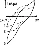
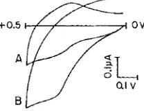
The electrochemical experiments were carried out in a deaerated 0.15M phosphate buffer solution of pH 5.8. Figure 1 shows cyclic voltammograms of ferrocene modified enzyme in the deaerated phosphate buffer, on a glassy carbon electrode. The redox reactions of the attached ferrocene units were clearly seen in the absence of glucose. Once glucose was added, the oxidation currents increased considerably showing the catalytic response of the derivatized enzyme in accordance with the results of Degani and Heller [6, 71. Figure 2 shows the cyclic voltammetric response of the CCE modified with the derivatized enzyme. The oxidation of ferrocene moiety was found to occur at N 0.340V (vs. SCE). Though the background currents were
large, the catalytic currents were clearly seen on the addition of glucose (Fig. 2). The calibration plot at 0.4V (Fig. 3) shows linearity up to ca. 8 mM and the dynamic response spans over a range of 0 to 35 mM glucose. The Lineweaver-Burk type plot of reciprocals of current response and the concentration of glucose yielded a KM value of 37mM, which is close to the value reported for the native enzyme in the solution phase [28, 291.
The agreement of the activity coefficients of the ferrocene modified glucose oxidase in the CCE and that of the native
Fig. 1. Cyclic voltammograms in phosphate buffer, pH 5.8, on a glassy carbon electrode in the base electrolyte (l), in the presence of 3.5mg of ferrocene derivatized glucose oxidase (Z), and in the presence of 3.5 mg of derivatized enzyme and 6 mM glucose (3). Scan rate 10mV/s. Potentials referred to SCE.
[image:2.594.387.468.575.669.2]S. Sampath, 0 . Lev
1114
f0.5
A
8
Fig. 2. Cyclic voltammograms of the ferrocene derivatized glucose oxidase modified CCE, in phosphate buffer, p t i 5.8, in the absence of glucose (A) and in the presence of 6m M glucose (B). Scan rate 10 mV/s. Potentials referred to SCE.
enzyme in the solution phase leads to the conclusion that the microenvironment of the enzyme active site and subsequently the intrinsic characteristics of the enzyme were not altered by this derivatization and immobilization in the organically modified silicate matrix.
The steady state response of the electrode at 0.4 V (vs. SCE), to varying glucose levels in the solution is given in Figure 4. The response time of the sensor was rather fast, of the order of 20 s. The cyclic voltammograms and the steady state responses were not affected by stirring the solution. This points onto the fact that the limiting step is the diffusion of the analyte inside the porous matrix coupled with enzyme kinetics and not external diffusion in the solution.
[image:3.594.313.540.34.131.2]3.2. Influence of Oxygen and Other Interferences
Figure 5 shows the calibration plots obtained at 0.4V (vs. SCE), in the presence and in the absence of oxygen in the electrolyte. The difference between the two calibration plots was, however, small; the response in presence of oxygen was somewhat lower than in the presence of argon. Oxygen trapped inside the porous matrix of the carbon ceramic electrode was found to be responsible for this observation. In addition to the ferricinium ions formed at the electrode, oxygen also acts as a competitive electron acceptor. The over potential applied was not large enough to oxidize Hz02 and these species slowly diffused into the solution without contributing to the electrode signal. When argon was passed through the porous electrode from the top, the response became equal irrespective of whether the solution was saturated with oxygen or not. As expected, the derivatized glucose oxidase modified enzyme CCEs did not reduce the interferences of oxidizable compounds such as ascorbic acid and acetaminophen.
._ __ 0.5
0.4 Q *- =0.3
9
3 0.2 0
0.1
G
I
+---.I
0 5 10 15 20 25 30 35Concentration of Glucose, rnM
Fig. 3. Calibration plot for glucose based on the cyclic voltammetric current response at 0.4V (vs. SCE). Scan rate 10 mV/s. The response is corrected for background currents.
20 sec
*
Fig. 4. Steady state response of the CCE modified with the derivatized enzyme, at a bias of 0.4V (vs. SCE). The arrows indicate addition of 2.5mM of glucose each.
3.3. Renewal Repeatability
The CCEs were brittle and the porous structure did not clog after polishing. The organically modified silicate matrix ensured that the bulk of the electrode was not exposed to the analyte. Hence, the electrode surface can be renewed by mechanical polishing. The cyclic voltammetric background currents were found to be almost the same with a relative standard deviation of
<
5 YO for five consecutively renewed surfaces. The renewal repeatability tests with glucose were carried out in the steady state mode at 0.4V (vs. SCE). Different surfaces were exposed, after mechanical polishing, to a 5 mM glucose solution and the response was followed. The relative standard deviation for the amperometric signal using six consecutive polished surfaces of the same electrode was found to be ca. 5 % . The relative standard deviation for the inter electrode repeatability of seven electrodes from the same batch worked out to be<
8 YO.3.4. Thick Film Sensors
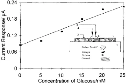
Thick film disposable sensors can easily be mass produced [30]. Leaching of mediators from the thick film disposable sensors constitutes one of the major drawbacks that limits the usefulness of such materials. The preparation conditions employed in the present studies and the resulting material promise a new way of preparation of disposable biosensors. The studies were carried out in a single drop of the electrolyte as shown in Figure 6. Thick films of the derivatized enzyme modified CCEs were prepared by coating the material on a microscopic glass slide. The film thickness was approximately
0 ~~ ~ ~~
10 20 30
Y
0Conc. of Glucose/mM
Fig. 5. Steady state calibration plots for the CCE glucose sensor at 0.4V (vs. SCE) in phosphate buffer, pH 5.8; Electrolyte deaerated with argon (A) and oxygen saturated (B).
[image:3.594.114.218.37.117.2] [image:3.594.320.527.534.688.2] [image:3.594.62.274.560.689.2]Glucose Sensor Based on a Redox Modified Enzyme and Carbon-Silica ComDosite
~
1115
0.24
Q,
2 0.18
ln c
0
Q
0.12
rY
c c
0.06
5
0
0 5 10 15 20 25
Concentration of Glucose/mM
Fig. 6. Calibration plot for glucose a t 0.4V (vs. SCE) using the ferrocene derivatized glucose oxidase modified C C E in the thick film form. Cyclic voltammograms taken in a single d r o p of the analyte of volume 15 pL. Scan rate 10mV/s. Inset: Scheme of the single drop measurement, 1) glass slide; 2) enzyme modified CCE; 3) contact for working electrode; 4) reference electrode; 5) counter electrode and 6) the analyte drop
1 mm. Electrical contacts were made using silver paint far away from the place of the electrolyte drop. Cyclic voltammetric studies were conducted in a 1 5 p L drop of the analyte. A platinum wire and the sharp tip of a calomel electrode were inserted into the drop with the help of a microscope. The measurements were carried out after five minutes of the application of a drop of 15pL. After one measurement, the film was thoroughly washed with distilled water and dried at ambient conditions. Again a I5 p L drop of a buffer containing a known amount of glucose was placed at the same point and the measurements carried out. Figure 6 shows the calibration plot at 0.4 V, obtained from the cyclic voltammograms in this mode. It is to be emphasized that the measurements using a single drop of the electrolyte can be carried out up to about 20min after the drop was applied. The signal instability observed after this time was associated with evaporation of the aqueous medium rather than the stability of the drop itself. The drop shape was visibly stable for at least 25 min, depending on the hydrophobicity of the matrix.
3.5. Stability
The carbon ceramic electrodes were found to encapsulate the biomolecules quite efficiently and no leaching from the rod or thick film type electrodes was seen even after immersing the electrode for 24h in aqueous solutions. This constitutes an advantage over the previously reported coimmobilization of ferrocene and glucose oxidase in the CCEs which exhibited a certain degree of reagent leaching [20]. This is especially important in the case of thick films since leaching of reagents or enzymes is not guaranteed by current technology. The stability of the electrodes was followed by two different methods; (1) by storing them at room temperature ( z 22 "C) and (2) by storing them in a refrigerator under dry conditions. In both cases, the sensors were periodically exposed to a 5 m M glucose solution and the response followed at different time intervals. Figure 7 shows the relative response of the sensors as a function of storage time. The stability tests were initiated seven days after the preparation of the electrodes, to allow complete drying and stabilization. It was found that the sensor's response continuously decreased with time,if stored at room temperature. On the other hand, refrigerated sensors were found to be stable
17 27 37
Timel Days
I\ A
t-- -I
47 57
Fig. 7. Stability of the CCEs modified with the derivatixed enzyme. Sensor stored at 4 ° C ( A ) and a1 N 22°C (B), room temperature.
for about four weeks after a small initial signal lose. These stability studies are still in progress.
4. Conclusions
The carbon ceramic electrode combined with the ferrocene derivatized glucose oxidase results in a reagentless glucose sensor with an active inner reaction layer. Thick films of the sensor can be easily prepared and the sensing step can be accomplished using a single drop of analyte. This, along with the fact that the signal is independent of oxygen, makes this material an attractive candidate for disposable biosensors.
5. Acknowledgements
We gratefully acknowledge the financial support of GBF, Gesellschaft Fuer Biotechnologische Forschung, M BH, Braunschweig, Germany and the Ministry of Science and Arts, Israel.
6. References
[l] J. Wang, J. Liu, L. Chen, F. Lu, Anal. Chem, 1994, 66, 3600.
[2] A.E.G. Cass, G . Davis, G.D. Francis, H.A.O. Hill, W.J. Aston, J.1. Higgins, E.V. Plotkin, L.D. Scott, A.P.F. Turner, Anal. Chem. 1984,56,
661.
[3] J.T. Kulys, N.K. Cenas, Biochim. Biophys. Acta 1983, 744, 57.
[4] W.J. Albery, P.N. Bartlett, D.H. Craston, J . Electrounul. Chcm. 1985, 194, 223.
[5] J.T. Kulys, T. Buch-Rasmussen. K . Bechgaard. V. Razutnas, J. Kazlanskaite, J. Marcinkeviciene, J.B. Christensen, H.E. Hansen, .I. Mol. Catal. 1994, 91, 407.
[6] Y. Degani, A. Heller, J . Phys. Chem. 1987, Y I , 1285. [7] Y. Degani, A. Heller, J . Am. Chem. Soc. 1988, 110, 2615.
[S] A. Heller, Acc. Chem. Res. 1990, 23, 128.
[9] C. Santamaria, P.N. Bartlett, D.J. Caruana, J. Kilburn, R. Parg, Abstracts of "Electrochem 'Y5", IO-141h Sept., University of Wales, Bangor, UK, SCI, London 1995.
A. Riklin, E. Katz, 1. Willner, A. Stocker, A.F. Buckmann, Nuturc 1995,
376, 612.
S. Brdun, S. Rappoport, R. Zusman, D. Avnir, M . Ottolenghi, Mutrr. Lett. 1990, 10, I .
S. Braun, S. Rappoport, R. Zusman. D. Avnir, M. Ottolenghi, in Biotechnology : Bridging Research and Applictrticins (Eds: D. Kamely, A .
Chakrabarty, S.E. Kornguth) Kluwer Academic, Boston 1990, p. 205. D. Avnir, S. Braun, 0. Lev, M. Ottolenghi, Chem. Muter. 1994, 6, 1605.
S. Braun, S. Shtelzer, S . Rappoport, D. Avnir, M. Ottolenghi, J . Non- Cryst. Solid~y 1992, 147P148, 139.
[image:4.594.324.538.26.172.2] [image:4.594.59.267.33.170.2]1116 S . Sampath, 0. Lev
[I51 0. Lev, M. Tsionsky, L. Rabinovich, V. Glezer, S. Sampath, I.
[ 161 L.M. Ellerby, C.R. Nishida, F. Nishida, S.A. Yamanaka, B. Dunn, J.S.
[I71 B.C. Dave, B. Dunn, J.S. Valentine, J . I . Zink, Anal. Chem. 1994, 66,
[I81 S. Shtelzer, S. Braun, Biotechnol. A p p l . Biochem. 1994, 19, 293.
[I91 M. Tsionsky, G. Gun, V. Glezer, 0 . Lev, Anal. Chem. 1994, 66, 1747. [20] I. Pankratov, 0. Lev, J . Electroanal. Chem. 1995, 3Y3, 3 5 .
[21] M. Tsionsky, 0. Lev, Anal. Chem. 1995, 67, 2409.
[22] S . Sampath, J. Gun, I. Pankratov, 0. Lev, 1. Sol-Gel Sci. Tech., Special
Pankratov, J. Gun, Anal. Chem. 1995, 67, 22A.
Valentine, Z.1. Zink, Science 1992, 255, 1 113.
1120A.
Issue on Bioceramics, (Eds. D. Avnir, S. Braun) 1996, 7, 123.
[23] J. Wang, K. Varnghese, Anal. Chem. 1990,62, 318.
[24] J . Wang, M.S. Lin, Anal. Chem. 1988, 60, 1545. [25] L. Gorton, Electroanalvsis. 1995, 7, 23.
[26] Y. Polevaya, J. Samuel, M. Ottolenghi, D. Avnir, J . Sol-Gel Sci. Tech.
[27] S. Sampath, 0. Lev, Anal. Chem. 1996, 68, 20 15.
[28] B.E.P. Swoboda, V. Massey, J . Biol. Chem. 1965, 240,2209.
[29] Q.H. Gibson, B.E.P. Swoboda, V. Massey, J . Biol. Chem. 1964, 239,
[30] M. Aharez-Icazd, U. Bilitewski, Anal. Chem. 1993, 65, 52SA.
1995, 5, 65.
3927.