Volume 14, Iss
ue 4, F
a
ll 2015
Editorial in Charge
Hossein Pakdaman, M.D.
Professor of Neurology, Shahid Beheshti
University of Medical Sciences,
Tehran, Iran
Editor-in-Chief
Shahriar Nafissi, M.D.
Associate Professor of Neurology,
Neurology Department, Tehran University of
Medical Sciences, Tehran, Iran
Deputy Editor
Farzad Fatehi, M.D.
Assistant Professor of Neurology, Neurology Department, Tehran University of
Medical Sciences, Tehran, Iran
Section Editors
Headache: Mansooreh Togha, M.D.,
Tehran
University of Medical Sciences, Tehran, Iran
Multiple
Sclerosis:
Mohammad
Ali
Sahraian, M.D., Neurology Department, Tehran
University of Medical Sciences, Tehran, Iran
Stroke: Afshin Borhanin Haghighi, M.D.,
Shiraz University of Medical Sciences, Shiraz,
Iran
Movement Disorders: Mohammad Rohani,
M.D., Iran University of Medical Sciences,
Tehran, Iran
Associate Editors
Shahin
Akhondzadeh,
Pharm.D.,
Ph.D,
Tehran University of Medical Sciences,
Tehran, Iran
Majid Ghafarpour, M.D., Tehran University of
Medical Sciences, Tehran, Iran
Massoud Nabavi, M.D., Shahed University of
Medical Sciences, Tehran, Iran
Scientific Assistant Editor
Ali Amini-Harandi, M.D., Shahid Beheshti University of Medical Sciences, Tehran, Iran
Editorial Board
Shahram Attarian, M.D., Centre de Référence
des Maladies Neuromusculaires et de la SLA,
France
Mahmoud R. Azarpazhooh, M.D., Mashhad
University of Medical Sciences, Mashhad, Iran
Keivan Basiri, M.D., Isfahan University of
Medical Sciences, Isfahan, Iran
Ahmad R. Dehpour, Pharm.D., Ph.D., Tehran
University of Medical Sciences, Tehran, Iran
Masoud Etemadifar, M.D., Isfahan University
of Medical Sciences, Isfahan, Iran
Kavian
Ghandehari,
M.D.,
Mashhad
University of Medical Sciences, Mashhad, Iran
Kurosh Gharagozli, M.D., Shahid Beheshti
University of Medical Sciences, Tehran, Iran
Mohammad H. Harirchian, M.D., Tehran
University of Medical Sciences, Tehran, Iran
Payam Kabiri, M.D., Ph.D., Tehran University
of Medical Sciences, Tehran, Iran
Hossein Kalani, M.D., Shahid Beheshti
University of Medical Sciences, Tehran, Iran
Jamshid Lotfi, M.D., Tehran University of
Medical Sciences, Tehran, Iran
Alireza Minagar, M.D., Louisiana State
University Health Sciences Center, USA
Ali Moghtaderi, M.D., Zahedan University of
Medical Sciences, Zahedan, Iran
Mahmood Motamedi, M.D., Tehran University
of Medical Sciences, Tehran, Iran
Alireza Nikseresht, M.D., Shiraz University of
Medical Sciences, Shiraz, Iran
Abdolmohamad M. Rostami, M.D., Thomas
Jefferson University Hospitals, USA
Mohammad
Saadatnia,
M.D.,
Isfahan
University of Medical Sciences, Isfahan, Iran
Mohammad K. Salajegheh, M.D., Brigham and
Women's Hospital and Harvard Medical School,
USA
Gholam A. Shahidi, M.D., Tehran University
of Medical Sciences, Tehran, Iran
Vahid
Shaygannejad,
M.D.,
Isfahan
University of Medical Sciences, Isfahan, Iran
Akbar Soltanzadeh, M.D., Tehran University
of Medical Sciences, Tehran, Iran
Amir A. Zamani, M.D., Brigham and Women's
Hospital and Harvard Medical School, USA
Babak Zamani, M.D., Tehran University of
Medical Sciences, Tehran, Iran
Secretary: Samaneh Bahraminejad, BSc
Email: [email protected]
http://ijnl.tums.ac.ir
Copy Edit, Layout Edit, Proof Reading and Design: Farzanegan Radandish Co. Postal Code: 81465-1798, Isfahan, Iran; Telefax: +98 311 6686302
www.farzaneganco.ir; Email: [email protected]
Indexed in
•PubMed, •PubMed Central, •Academic Keys,
•Cite Factor (Directory Indexing of International Research Journals),
•Directory of Open Access Journals (DOAJ), •Directory of Research Journal Indexing (DRJI), •Ebsco,
•InfoBase Index,
•Islamic World Science Citation Center (ISC), •LocatorPlus,
•Scientific Information Database (SID), •Ulrichsweb Global Serials Directory, •Universal Impact Factor,
•WorldCat
Iranian Journal of Neurology © 2015
II Iran J Neurol 2015; 14(4)
INFORMATION FOR AUTHORS
Aim and Scope
The Iranian Journal of Neurology is dedicated to the
Iranian Neurological Association. The journal is a
peer-reviewed journal published quarterly and publishes
neurological experiences in basic or clinical fields in
English Language. The Iranian Journal of Neurology aims
to publish manuscripts of a high scientific quality
representing original clinical, diagnostic or experimental
works or observations in neurological sciences. Papers in
English are welcomed, particularly those which bring
novel information and researches in clinical or basic fields
from the neurological disorders. All received manuscripts
coving the scope of the journal will be evaluated by
properly competent referees.
Submission
Cover Letter:
Submissions should be accompanied by a cover letter
including a declaration by the first author on behalf of the
others to the effect that
(1) The paper has not been published to date (except
for abstracts of conference materials).
(2) The paper has not been accepted for publication
elsewhere.
(3) All persons listed as the authors have read it and
approved it for publication. The cover letters should be
submitted in section "Comments for the Editor".
Articles must be written in accurate scientific English
appropriate for publication. The articles are subject to
review and editing; however, the authors are responsible
for the correctness the manuscript's English language.
The articles must be submitted only online:
ijnl.tums.ac.ir
Policies
The Editorial Board reserves the right to reject a paper
without seeking reviewers’ opinion provied the content or
the form of the paper does not meet minimum acceptance
criteria or if the subject of the paper is beyond the aims
and scope of the journal.
Everyone listed as the author of a paper is responsible
for the reliability and completeness of data presented in
the paper.
Do not submit papers that copy fully or partially
previously published papers.
Indicate that this submission is ready to be considered
by this journal by checking off the following:
•
The submission has not been previously published,
nor is it before another journal for consideration (or an
explanation has been provided in Comments to the Editor).
•
The submission file is in Microsoft Word document
file format.
•
Where available, URLs for the references have
been provided.
•
The text is double-spaced; uses an Arial 12-point
font; and all illustrations, figures, and tables are placed
within the text at the appropriate points, rather than at the
end.
•
The text adheres to the stylistic and bibliographic
requirements outlined in the Author Guidelines, which is
found in About the Journal.
If the Editorial Board is not notified in advance and the
paper is found to have been copied during editorial work,
the paper shall be rejected.
We expect that all studies reported in the journal
conform to the requirements of the Declaration of Helsinki
(1989). Information on the consent of a relevant ethics
committee to perform the trial and the informed consent of
the patients to participate in the trial should be given in the
Material and methods section of each paper in which
diagnostic or therapeutic intervention does not follow from
the standard procedure. Authors of case reports must not
disclose personal data of patients described.
Manuscripts
The journal publishes:
•
Original Article
•
Review Article
•
Case Report
•
Short Communication
•
Clinical Notes
•
Editorial
•
Letters to Editor
•
Neurological Images
•
Neurological Videos
•
Iranian Neurological Events
•
Clinical Quiz
Details
Original and review papers: The maximum length of
original and review papers (including tables and figures
materials) is 3000 words.
Case reports: Should not be longer than 1200 words,
while letters to the Editor, reports and critical reviews
should not exceed 800 words.
Short communications: The maximum word number of
short communications should be below 1200 words with
maximum one table or figure and 10 references. The
manuscript should be structured including introduction,
materials and methods, results, discussion, and conclusion
with a structured abstracts as original articles.
Iranian Journal of Neurology © 2015
Iran J Neurol 2015; 14(4) III
Neurological images or videos: Interesting cases as
neurological images or videos are welcome. They should
be maximally 400 words with legends without abstract and
unstructured. The videos should be uploaded as
supplementary files.
Letter to the Editor: May concern short scientific reports
and comments. The maximum number of words should be
below 800 words with maximum 5 references, no abstract,
no table or figure, and unstructured.
Clinical notes: Refer to important interesting observations
which are imperative for reminders in clinical practice.
The maximum number is 1000 words with maximum 5
references, 1 table and 1 figure with no abstract.
Iranian neurological events: Include the brief description
of major regional events (congresses or seminar)
implemented in Iran.
Structure of Articles
•
Manuscripts should be submitted in 12 points, Arial
font, with double line spacing and sufficient margins of
2.5 cm.
•
The text should not be formatted.
•
Each section of the paper should begin on a new page
The manuscript must include:
•
Page 1: Title Page
•
Page 2: Abstract and Key Words
•
Page 3 and subsequent pages: manuscript body
including Introduction, Materials and Methods, Results,
Discussion, Conclusion, References, Tables, Figures
1. Title page:
Title page should contain paper title, full names of
authors, authors’ place of work, full name and address of
the corresponding author (including e-mail address and
telephone number), given in that order.
2. Abstract page:
•
The length of the abstract should be at least 200
and not more than 250 words for original papers and not
more than 150 words for review papers and case reports.
Abstracts of original papers should be structured to
include the background, methods, results and conclusion.
•
Below the abstract authors should provide between
three and six keywords conforming to Medical Subject
Headings (Index Medicus).
3. Page three and subsequent pages of the original paper
and short communication should include the text arranged
in the following order (for other mansucript type, see
above):
1.
Introduction: The introduction should be as
concise as possible and introduce the context of the paper
to the reader; the paper should clearly state the research
hypothesis and the objective of the study.
2.
Materials and Methods: Description of the
studied population or material should be detailed and
include all information necessary to assess the reliability
of results obtained in the study and/or allow the
experiment to be repeated by other researchers; the section
related to statistical analysis should have information on
applied statistical tests and programs.
3.
Results: Present results directly related to the topic
of the paper only; tables and/or figures are recommended.
4.
Discussion
5.
Conclusions: These should be brief, follow directly
from results presented above and correspond to the aim of
the paper outlined in the introduction.
6.
Acknowledgements: Should comprise information
on sources of funding (grant numbers); acknowledgements
should concern those who made a significant contribution
to the paper, but who did not meet the criteria to be listed
as authors.
7.
References: References should be listed in the
order quoted in the paper. Please cite source and major
papers that offer interested readers an opportunity to
obtain more detailed information. Avoid citing review
papers and conference reports, if they are not the only
materials on a given topic.
References
In the paper references should be given in superscripts
with no space between the comma and the consecutive
number.
Authors are advised to carefully verify citation details.
Give names of first six authors; if there are more
authors, add “et al.“. Use Index Medicus abbreviations for
journal titles. Then mention the volume and the issue of
the journal.
The recommended style for journal references is as
follows:
[Reference number][Authors]. [Article title]. [Journal
Name] [Year of publication]; [volume](issue): [Pages
range].
For Journal Example:
1. Janghorbani M, Amini M, Willett WC, Mehdi Gouya
M, Delavari A, Alikhani S, et al. First nationwide survey
of prevalence of overweight, underweight, and abdominal
obesity in Iranian adults. Obesity (Silver Spring) 2007;
15(11): 2797-808.
For Books Example:
2. Ropper AH, Brown RJ. Adams and Victors principles
of neurology. 8
thed. New York, NY: McGraw Hill
Professional; 2005. p. 271.
Tables: Each table should be placed on a separate page.
Tables should be numbered with Arabic numerals in the
order in which they appear in the text. Authors should
indicate the position of tables in the paper. Titles and
headings of tables should be given in English. Information
given in tables should not be repeated in the body of the
text. Explanations concerning tables, e.g. full names of
abbreviations should be given in footers below tables and
should be consecutively marked: “*”,“**”,“***” etc.
Figures: Figures and photographs should be numbered
Iranian Journal of Neurology © 2015
IV Iran J Neurol 2015; 14(4)
Photographs sent electronically should be of the resolution
of 300 dpi and in the .tif or .jpg format. Figures and
photographs are placed in the paper in the form delivered,
so they must be prepared carefully. Please indicate where
they should be placed in the text.
Abbreviations should be always clarified when used for
the first time in the text (including the abstract).
Abbreviations should not be used in paper titles, unless in
exceptional circumstances.
Review process: All papers submitted for publication in
the journal are assessed by two independent reviewers
with the mutual anonymity rule as to the names of
reviewers and authors observed.
Plagiarism policy: According to the plagiarism policy of
Iranian Journal of Neurology, plagiarism is defined as a
paper which replicates another publication with as a
minimum 25% resemblance and devoid of citation.
In any time the evidence of plagiarism is detected, the
manuscript will be withdrawn and the author will be
sanctioned from publishing papers permanently.
Proofs: The proofs will be sent via email and must be
Iranian Journal of Neurology © 2015
Iran J Neurol 2015; 14(4) V
Table of Contents
Original Article(s)
Assessment of clinicopathologic features in patients with pituitary adenomas in
Northeast of Iran: A 13-year retrospective study
Kazem Anvari, Mahmoud Reza Kalantari, Fariborz Samini, Soodabeh Shahidsales, Mehdi
Seilanian-Toussi, Zakiyeh Ghorbanpour ………...…...………...…..185-189
Mutation analysis in exons 22 and 24 of SCN4A gene in Iranian patients with
non-dystrophic myotonia
Mohammad Mehdi Heidari, Mehri Khatami, Shahriar Nafissi, Faezeh Hesami-Zokai,
Afshin Khorrami ………...…….190-194
Effects of L-arginine pre-treatment in
1-methyl-4-phenyl-1,2,3,6-tetrahydropyridine-induced Parkinson’s diseases in Balb/c mice
Javad Hami, Mehran Hosseini, Sekineh Shahi, Nassim Lotfi, Abolfazl Talebi, Mohammad Afshar
………...195-203
A descriptive study of prevalence, clinical features and other findings of neuromyelitis
optica and neuromyelitis optica spectrum disorder in Khuzestan Province, Iran
Davood Kashipazha, Seyed Ehsan Mohammadianinejad, Nastaran Majdinasab, Mostafa Azizi,
Majid Jafari ………...…204-210
Policy interventions to improve rural retention among neurosurgeons in Iran: A
discrete choice experiment
Sima Rafiei, Mohammad Arab, Arash Rashidian, Mahmood Mahmoudi, Vafa Rahimi-Movaghar
……….211-218
Clinical Notes
Cortical tumor presenting with Parkinsonism
Mi-Song Choi, Bom Choi, Soo-Jin Cho, Joo Yong Kim, Ki-Han Kwon, Suk Yun Kang…..219-221
Letter(s) to Editor
Cerebellar infarction and aneurysmal subarachnoid hemorrhage: An unusual
presentation and rare complications of rhinocerebral mucormycosis
Payam Sasannejad, Ali Ghabeli-Juibary, Samira Aminzadeh, Nahid Olfati ………..…222-224
Complicated orolingual angioedema after recombinant tissue plasminogen activator
treatment in stroke patients under angiotensin converting enzyme inhibitor: Report of
two cases
Mohammad Reza Motamed, Fereshteh Nasiri, Seyed Mohammad Fereshtehnejad, Masoud
Mehrpour, Babak Zamani, Bahram Haghi-Ashtiani, Farzaneh Rohani ………..…225-227
Nasopharyngeal carcinoma presenting as Garcin’s syndrome: A rare case report
Shivani Patel, Apurva Patel, Monil Majmundar, Irappa Madabhavi, Ravi Shah,
Jay Soni………...…228-230
The prevalence of Martin-Gruber anastomosis in Iranian subjects by electrodiagnostic
criteria
Iranian Journal of Neurology © 2015 Corresponding Author: Soodabeh Shahidsales
Email: [email protected] Email: [email protected]
Original Paper
Iran J Neurol 2015; 14(4): 185-189
Assessment of clinicopathologic
features in patients with pituitary
adenomas in Northeast of Iran:
A 13-year retrospective study
Kazem Anvari
1, Mahmoud Reza Kalantari
2, Fariborz Samini
3, Soodabeh Shahidsales
1,
Mehdi Seilanian-Toussi
1, Zakiyeh Ghorbanpour
41 Cancer Research Center AND Department of Radiation Oncology, School of Medicine, Mashhad University of Medical Sciences, Mashhad, Iran
2 Department of Pathology, School of Medicine, Ghaem Hospital, Mashhad University of Medical Sciences, Mashhad, Iran
3Department of Neurosurgery, School of Medicine, Shahid Kamiab Hospital, Mashhad University of Medical Sciences, Mashhad,
Iran
4Department of Radiation Oncology, School of Medicine, Omid Hospital, Mashhad University of Medical Sciences, Mashhad, Iran
Keywords
Pituitary Adenoma, Functional Adenoma, Survival
Rate
Abstract
Background: Intracranial lesions of the pituitary
gland are common pituitary adenomas, accounting
for 6-10% of all symptomatic intracranial tumors. In
this
retrospective
study,
the
clinicopathologic
features and survival rate of pituitary adenomas
were evaluated.
Methods: The present retrospective study was
conducted on 83 patients with pituitary adenomas,
referring to radiation oncology departments of Ghaem
and Omid Hospitals, Mashhad, Iran, over a period of
13 years (1999-2012). Data obtained from clinical
records including clinical features, type of surgery (if
performed), treatment modality, overall survival rate,
and progression-free survival rate were analyzed.
Results:
Eighty-three patients including 44 males
(53%) and 39 females (47%) participated in this
study. The median age was 40 years (age range:
10-69 years). Chiasm compression was reported in
62 patients (74.4%), and 45.78% of the subjects
suffered from headaches. Functional and
non-functional adenomas were reported in 44 (53.01%)
and 39 (46.99%) patients, respectively. In cases with
functional and non-functional adenomas, the disease
was controlled in 95 and 84.5% of the subjects for 3
years,
respectively.
Furthermore,
1- and 3-year survival rates for functional adenoma
were 84.6 and 23%, respectively; the corresponding
values were 90.9 and 22.7% in non-functional
adenomas, respectively
.
Conclusion:
In this study, a significant correlation
between headache severity and type of adenoma
was observed. So, application of surgery and
radiotherapy together could be a highly effective
approach for treating functional adenomas, although
it is less efficient for the non-functional type.
Introduction
Intracranial lesions of pituitary gland are common
pituitary adenomas, accounting for 6-10% of all
symptomatic
intracranial
tumors.
1-3Pituitary
adenomas are defined as abnormal growth of
tumors in pituitary glands (benign adenomas,
Iranian Journal
of Neurology
186 Iran J Neurol 2015; 14(4) Anvari et al.
invasive adenomas, and carcinomas).
4,5Recent
studies have shown that invasive adenomas may
approximately affect 1 in 1000 people of the general
population.
6The most frequent pituitary adenomas are
microadenomas with an estimated incidence of
16.7%. Pituitary adenomas are also categorized as
active-functioning and non-functioning adenomas;
two-thirds of clinically diagnosed lesions are
functional adenomas.
7Symptoms of pituitary disorders are often
non-specific and may differ given the effects of
space-occupying lesions, increased hormonal release or
both.
8,9Among patients with pituitary adenomas,
different types of headaches such as chronic,
episodic migraines and unilateral headaches
including primary stabbing headache, short-lasting
unilateral neuralgiform headache, cluster headache,
and hemicrania continua are common.
10-13Pituitary
adenomas are also associated with psychiatric
disorders including hostility, anxiety, apathy,
depression, emotional instability, and irritability.
14,15Although the treatment of pituitary adenoma
depends on the size and type of tumor, surgery is
the common treatment modality. Transsphenoidal
adenomectomy is a method for tumor removal,
though recently, endoscopic surgery has been
commonly applied.
16Due to the importance of
pituitary disorders and insufficient research in this
field, this study aimed to assess the clinicopathologic
features and treatment outcomes of patients with
pituitary adenomas over a 13-year period in the
Northeast of Iran.
Materials and Methods
We studied the clinicopathologic features of all
patients, presenting with pituitary adenomas. The
subjects had referred to the departments of radiation
oncology at Omid and Ghaem hospitals in years
1999-2012.
The sample size included 83 patients, according to
inclusion criteria. Since all eligible subjects were
recruited within a specific time span, use of a formula
for calculating the sample size was not necessary. The
inclusion criteria were as follows: (1) Pathological
evidence
of pituitary adenoma; (2) essential
information including age, gender, treatment modality,
and type of surgery and medical records; and (3)
undergoing medically proposed treatments. The
exclusion criterion was unfinished complementary
treatment (the recommended treatment).
In this retrospective, cohort study, all patients’
medical records were collected and examined after
review and pathological assessment for the selection
of definitive or complementary treatments. Data
obtained from the clinical records such as clinical
signs, type of surgery (if performed), treatment
modality, overall survival rate, and progression-free
survival were examined; in addition, previous
medical histories and clinical variables were
recorded. We contacted the patients in case the data
needed to be corrected or completed.
Patients with pituitary adenomas, diagnosed via
pathological assessment were included in this study
after meeting the inclusion criteria. All patients’
records
were
included
in
the
predesigned
questionnaires.
Data obtained from patients’ records and
recorded calls were analyzed by SPSS software
(version 16, SPSS Inc., Chicago, IL, USA). For
descriptive data, statistical indices, tables, and
diagrams were used. Conventional methods of
survival analysis including Kaplan–Meier and Cox
regression were employed in order to study the
effects of variables on disease-free survival rate.
Since no medical interventions were performed
in this study and the patients’ records were kept
confidential, no written consents were obtained.
Results
Eighty-three patients including 44 males (53%) and
39 females (47%) participated in this study. 4, 12, 24
and 19 patients were within the age range of 10-20,
20-29, 30-39, and 40-49 years, respectively; in
addition, 19 patients were 50-59 years old, and five
subjects were within the age range of 60-69 years.
The median age was 40 years (age range: 10-69
years), and the majority of the subjects (28.9%) were
30-39 years old.
Chiasm compression was reported in 62 patients
(74.4%) and 45.78% of the subjects suffered from
headaches. Functional and non-functional adenomas
were observed in 44 (53.01%) and 39 (46.99%)
patients, respectively.
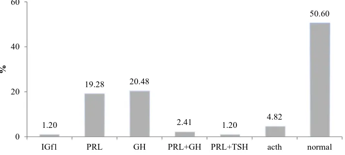
Prolactin (PRL) and growth hormones (GH) were
the most secreted hormones (19.3% and 20.5%,
respectively). Of all patients, one showed an increased
insulin-like growth factor-1 (IGF-1) level, 16
experienced prolactin elevation and 17 cases had
elevated
growth
hormone;
in
addition,
adrenocorticotropic hormone (ACTH) level increased
in 4 cases (Figure 1).
Galactorrhea and acromegaly were observed in
6% and 22% of the patients, respectively, and
Cushing’s syndrome was reported in 4.82% of the
subjects; this disease recurred in 25 patients (30%).
Assessment of clinicopathologic features Iran J Neurol 2015; 14(4) 187
suffered from non-functional adenomas, including
22 males (50%) and 17 females (43.6%). According to
the findings of the present study, no significant
association was found between adenoma type and
gender (P = 0.540).
Among 44 patients with functional adenomas, 25
patients aged < 40 years (56.8%) and 19 cases were > 40
years (43.2%). Furthermore, in patients with
non-functional adenomas, 17 and 22 individuals were < 40
and > 40 years of age, respectively (43.6% and 56.4%,
respectively). There was no significant association
between age and type of adenoma (P = 0.229).
Chiasm compression, accompanied by adenoma,
was assessed in patients with functional and
non-functional adenomas. Signs of compression were
reported in 31 patients with functional adenomas
(70.5%) and 31 subjects with non-functional
adenomas (79.5%); no significant relationship was
observed between chiasm compression and tumor
type (P = 0.345).
The headache was reported in 13 individuals
with functional adenoma (29.5%) and 25 patients
with non-functional adenoma (64.1%). The findings
showed that headache was prevalent among
patients with non-functional adenomas; therefore,
there was a significant association between adenoma
type and headache (P = 0.001, r = −0.346).
Adjuvant radiotherapy, as a complementary
treatment, was applied for 60 patients (72.3%).
Radiotherapy was also performed for 22 subjects
(26.5%) after surgical failure; in addition, the mean
duration of follow-up was 16 months (6-120).
Active disease was reported in only one patient
(2.3%) with functional adenoma. Among patients
with non-functional adenomas, five cases (12.8%)
showed signs of active disease; in both groups, the
disease was controlled satisfactorily (P = 0.064). In
the evaluation of the relationship between tumor
type and disease activity, no significant difference
was observed between patients undergoing primary
radiotherapy
and
those
receiving
adjuvant
radiotherapy (P = 0.534).
Out of 27 patients with non-functional adenoma,
24 individuals (88.9%) had non-active disease and 3
patients (11.1%) showed signs of active disease. As
to the findings, there was a significant relationship
between the type of adenoma and disease control; in
patients with functional adenoma, disease control
was significantly higher (P = 0.004).
Analysis of overall survival rate
The mean follow-up duration was 30 months, with a
median of 16 months (range: 6-125). We compared the
overall survival rate of patients, based on the type of
adenoma. Three-year survival rate was 95% in
patients with functional adenomas and 84.5% in
patients with non-functional adenomas. According to
these results, there was no correlation between
patients’ overall survival rate and adenoma type
(Table 1).
Figure 1. Increased secreted hormones
IGF1: Insulin-like growth factor-1; PRL: Prolactin; GH: Growth hormone; TSH: Thyroid stimulating hormone
Table 1. Relationship between type of adenoma and mean ± SD of overall survival rate
Type of adenoma
Number
Mean ± SD
Functional
44
31.659 ± 29.953
Non-functional
39
30.282 ± 28.799
Total
83
31.012 ± 29.246
SD: Standard deviation
1.20
19.28 20.48
2.41 1.20 4.82
50.60
0 20 40 60
IGf1 PRL GH PRL+GH PRL+TSH acth normal
188 Iran J Neurol 2015; 14(4) Anvari et al.
Discussion
Pituitary adenomas account for 10-15% of primary
intracranial neoplasms. In the current study, we
examined the clinicopathologic features of 83
patients, presenting with pituitary adenomas during
13 years. The most common signs were vision
impairment
and
chiasm
compression;
also,
galactorrhea was more prevalent than Cushing’s
disease or acromegaly. Functional adenoma was
observed in more than half of the patients.
Three-year survival rate was 95% for functional adenoma
and 84.5% for non-functional adenoma, although no
association was found between survival rate and
type of adenoma.
Rim et al.
17evaluated patients with pituitary
adenoma, using external beam radiation therapy
(EBRT). Correspondingly, it was revealed that
headache, vision impairment, and hormonal
disorders were the most common signs. In the
mentioned study, EBRT was considered to be an
effective method for controlling the pressure effects
of non-functional adenoma; similarly.
Furthermore, Shao and Li
18in a similar study
reported that headache and hormonal disorders
were more prevalent among patients with pituitary
adenomas; however, lower levels of hormones were
observed, compared to our findings; this may be due
to differences in the studied populations.
In the current study, 74.4% of the studied patients
suffered from vision impairment. Another similar
study performed in Egypt showed that 57% of
patients had optical disorders, which is different
from the findings of the present study; the mean
follow-up duration was 44 months in El-Shehaby et
al’s.
19study, while it was 31 months in our study.
In the present study, adenoma type was not
significantly associated with gender, age or signs of
chiasm
compression,
though
a
significant
relationship was observed between adenoma type
and headache severity; this could be due to
hormonal activity in patients with functional
adenoma. Furthermore, prolactin, IGF-1, growth
hormone and ACTH levels were high in some
patients in the present study.
In our study, there was a significant relationship
between adenoma type and disease activity in patients
undergoing complementary adjuvant radiotherapy.
Becker et al.
20in a review study reported that
non-functional adenomas are more significantly affected by
radiotherapy, compared to functional adenomas
(80-90%), while our findings showed that functional
adenomas can be better controlled.
Rim et al.
17in another study showed that external
radiotherapy plays a critical role in the recurrence of
non-functional adenomas; correspondingly, as it is
presented in this study, use of radiotherapy is
recommended for controlling adenoma, although
with a different impact.
Mecca et al.
21studied the efficacy of external
conventional radiotherapy (CRT) in short- and
long-term control of acromegaly; they indicated the
long-term effects of CRT on active acromegaly.
Similar to our study, active adenoma was
controlled in several cases.
As the results indicated, 3-year survival rate was
95% for functional adenoma and 84.5% for
non-functional adenoma; therefore, there was no
relationship between survival rate and adenoma type.
Also, in our study, 1- and 3-year survival rates
for functional adenoma were 84.6 and 23%,
respectively; however, regarding the non-functional
type, these values were 90.9 and 22.7%, respectively.
Rim et al.
17reported 10-year control rates to be 96%
and 66% for functional and non-functional
adenomas, respectively.
We also showed that survival in functional
adenoma was more than that observed in
non-functional tumors. Puataweepong et al.
22and Wilson
et al.
23reported the 5 years survival rate to be 91%
and 87%, respectively. Zargar et al.
24studied the
clinical and endocrine aspects of pituitary tumors.
As they indicated, in an endocrine center, functional
pituitary tumors were more prevalent than
non-functioning tumors; similarly, we showed that
functional adenomas are more frequent than the
non-functional type.
Limitations
Unfortunately, in the present study, the exact time of
disease recurrence was not recorded and many
patients did not refer for follow-up sessions after the
initial treatment.
Conclusion
In the present study, type of adenoma was not
associated with age, gender or signs of chiasm
compression, although there was a significant
association between headache and type of
adenoma.
Application
of
surgery
and
radiotherapy together could be a highly effective
approach for treating functional adenoma,
although it is less efficient for the non-functional
type. It is suggested that further research with
different methods be performed on larger
populations to obtain better outcomes.
Conflict of Interests
The authors declare no conflict of interest in this study.
Acknowledgments
Assessment of clinicopathologic features Iran J Neurol 2015; 14(4) 189
Ghorbanpour and was supported by the Research
Deputy of Mashhad University of Medical Sciences. The
authors would like to thank the vice chancellor for his
assistance and the Research Committee for their
support.
How to cite this article: Anvari K, Kalantari MR,
Samini F, Shahidsales S, Seilanian-Toussi M,
Ghorbanpour Z. Assessment of clinicopathologic
features in patients with pituitary adenomas in
Northeast of Iran: A 13-year retrospective study. Iran
J Neurol 2015; 14(4): 185-89.
References
1. Melmed Sh. Pathogenesis of pituitary tumors. Nature Reviews Endocrinology 2011; 7: 257-66.
2. Mete O, Asa SL. Clinicopathological correlations in pituitary adenomas. Brain Pathol 2012; 22(4): 443-53.
3. Zada G, Woodmansee WW, Ramkissoon S, Amadio J, Nose V, Laws ER. Atypical pituitary adenomas: incidence, clinical characteristics, and implications. J Neurosurg 2011; 114(2): 336-44.
4. Ezzat S, Asa SL, Couldwell WT, Barr CE, Dodge WE, Vance ML, et al. The prevalence of pituitary adenomas: a systematic review. Cancer 2004; 101(3): 613-9.
5. Ironside JW. Best Practice No 172: pituitary gland pathology. J Clin Pathol 2003; 56(8): 561-8. 6. Daly AF, Rixhon M, Adam C, Dempegioti
A, Tichomirowa MA, Beckers A. High prevalence of pituitary adenomas: a cross-sectional study in the province of Liege, Belgium. J Clin Endocrinol Metab 2006; 91(12): 4769-75.
7. Asa SL. Practical pituitary pathology: what does the pathologist need to know? Arch Pathol Lab Med 2008; 132(8): 1231-40. 8. Chahal HS, Stals K, Unterlander M, Balding
DJ, Thomas MG, Kumar AV, et al. AIP mutation in pituitary adenomas in the 18th
century and today. N Engl J Med 2011; 364(1): 43-50.
9. Mezosi E, Nemes O. Treatment of pituitary adenomas. Orv Hetil 2009; 150(39): 1803-10.
10. Levy MJ, Matharu MS, Meeran K, Powell M, Goadsby PJ. The clinical characteristics of headache in patients with pituitary
tumours. Brain 2005; 128(Pt 8): 1921-30. 11. Matharu MS, Levy MJ, Merry RT, Goadsby
PJ. SUNCT syndrome secondary to prolactinoma. J Neurol Neurosurg Psychiatry 2003; 74(11): 1590-2.
12. Milos P, Havelius U, Hindfelt B. Clusterlike headache in a patient with a pituitary adenoma. With a review of the literature. Headache 1996; 36(3): 184-8.
13. Levy MJ, Matharu MS, Goadsby PJ. Prolactinomas, dopamine agonists and headache: two case reports. Eur J Neurol 2003; 10(2): 169-73.
14. Sievers C, Ising M, Pfister H, Dimopoulou C, Schneider HJ, Roemmler J, et al. Personality in patients with pituitary adenomas is characterized by increased anxiety-related traits: comparison of 70 acromegalic patients with patients with non-functioning pituitary adenomas and age- and gender-matched controls. Eur J Endocrinol 2009; 160(3): 367-73.
15. Weitzner MA, Kanfer S, Booth-Jones M. Apathy and pituitary disease: it has nothing to do with depression. J Neuropsychiatry Clin Neurosci 2005; 17(2): 159-66. 16. American Cancer Society. Surgery for
pituitary tumors [Online]. 2008 [cited 2014 Aug 5]; Available from: URL:
http://www.cancer.org/cancer/pituitarytumo rs/detailedguide/pituitary-tumors-treating-surgery
17. Rim CH, Yang DS, Park YJ, Yoon WS, Lee JA, Kim CY. Radiotherapy for pituitary adenomas: long-term outcome and complications. Radiat Oncol J 2011; 29(3): 156-63.
18. Shao S, Li X. Clinical features and analysis in
1385 Chinese patients with pituitary adenomas. J Neurosurg Sci 2013; 57(3): 267-75. 19. El-Shehaby AM, Reda WA, Tawadros SR,
Abdel Karim KM. Low-dose Gamma Knife surgery for nonfunctioning pituitary adenomas. J Neurosurg 2012; 117(Suppl): 84-8. 20. Becker G, Kocher M, Kortmann RD,
Paulsen F, Jeremic B, Muller RP, et al. Radiation therapy in the multimodal treatment approach of pituitary adenoma. Strahlenther Onkol 2002; 178(4): 173-86.
21. Mecca N, Barbarulo F, Mercuri V, D'Amico T, Minniti G, Tamburrano G, et al. Efficacy of external conventional radiotherapy in short and long term control of acromegaly; a comparison with stereotactic irradiation. Clin Ter 2009; 160(4): 277-82.
22. Puataweepong P, Dhanachai M, Dangprasert S, Laothamatas J, Theerapancharoen V, Yongvithisatid P. Comparison of conventional external radiotherapy and stereotactic radiotherapy in the treatment of pituitary adenoma. J Med Assoc Thai 2009; 92(3): 382-9.
23. Wilson PJ, De-Loyde KJ, Williams JR, Smee RI. A single centre's experience of stereotactic radiosurgery and radiotherapy for non-functioning pituitary adenomas with the Linear Accelerator (Linac). J Clin Neurosci 2012; 19(3): 370-4.
Iranian Journal of Neurology © 2015 Corresponding Author: Mohammad Mehdi Heidari
Email: [email protected] Email:[email protected]
Received: 24 Nov 2014 Accepted: 31 Jul 2015
Original Paper
Iran J Neurol 2015; 14(4): 190-194
Mutation analysis in exons 22 and 24
of SCN4A gene in Iranian patients with
non-dystrophic myotonia
Mohammad Mehdi Heidari
1, Mehri Khatami
1, Shahriar Nafissi
2, Faezeh Hesami-Zokai
1,
Afshin Khorrami
11 Department of Biology, School of Science, Yazd University, Yazd, Iran
2 Department of Neurology, School of Medicine, Tehran University of Medical Sciences, Tehran, Iran
Keywords
Nondystrophic Myotonia, Mutation, SCN4A, Polymerase
Chain
Reaction-Single
Strand
Conformational
Polymorphism
Abstract
Background:
Non-dystrophic
myotonias
are
a
heterogeneous set of skeletal, muscular channelopathies,
which have been associated with point mutations within
sodium channel α-subunit (SCN4A) gene. Because exons
22 and 24 of SCN4A gene are recognized as hot spots for
this disease, the purpose of the study is to identify
mutation in exons 22 and 24 of SCN4A gene in Iranian
non-dystrophic myotonias patients.
Methods:
In this study, 28 Iranian patients with
non-dystrophic myotonia analyzed for the mutation scanning
in exons 22 and 24 of SCN4A gene by polymerase chain
reaction-single strand conformational polymorphism
(PCR-SSCP) and sequencing.
Results:
We found 29073G>C substitution in SCN4A
gene in one case and 31506A>G substitution in seven
cases. The 29073G>C substitution causes a missense
mutation G1306A, located in the conserved cytoplasmic
loop connecting repeat III and IV of the SCN4A channel
but, 31506A>G substitution do not alter amino acid in
SCN4A protein
.
Conclusion:
G1306A residue is located in functionally
important protein region. In “hinged-lid model” for Na
+channel inactivation in which glycines
1306act as the
hinge of the lid occluding the channel pore. Mutation
in this region slowed fast inactivation. Therefore, it
might be a pathogenic mutation. The causal
relationship of this mutation with the disease is an
object for further discussion
.
Introduction
Muscle channelopathies (the inherited muscle ion
channel diseases) are rare disorders of the skeletal
muscle.
Non-dystrophic
myotonias
are
a
heterogeneous
set
of
skeletal,
muscular
channelopathies, which have been associated with
specific point mutations within sodium channel
α-subunit (SCN4A) or Cl–channel (CLCN1) genes.
1,2The prevalence of non-dystrophic myotonia has
been estimated to be ~1 in 100,000 in the
worldwide.
3Voltage-gated sodium channels are
prominent transmembrane proteins in excitable
tissues and are responsible for the rising phase of the
action potential in the membranes of neurons and
most electronically excitable cells.
1,4The skeletal muscle sodium channel comprises a
principal pore-forming and voltage sensing subunit
(the alpha subunit), which is associated with an
Iranian Journal
Nondystrophic myotonia Iran J Neurol 2015; 14(4) 191
accessory beta-1 subunit. The beta-1 subunit has not
been reported to be linked to any disease. It’s alpha is
encoded by the SCN4A gene, which is located on
chromosome 17q23-25, comprises 24 exons with a 5.5
kb open reading frame, is associated with various
neuromuscular disorders.
5The alpha subunit consists
of four homologous domains, and each domain
possesses six hydrophobic putative transmembrane
segments (S1–S6).
4Conserved sequences in these
channels promote specific functions.
6,7SCN4A mutations produce several clinically
distinct
skeletal
muscle
disorders
including
hyperkalemic periodic paralysis, paramyotonia
congenita,
potassium-aggravated
myotonia,
hypokalemic periodic paralysis, and congenital
myasthenic syndrome.
5The similarities between sodium channel
myotonia and myotonia congenita can lead to
difficulty in prioritizing genetic testing. Clinical
history and examination considered in conjunction
with electromyogram findings can improve the
ability to distinguish between the two and guide
genetic analysis, but in some cases screening of
SCN4A genes will be required.
8More than 40 mutations have been reported in
SCN4A gene, but exons 22 and 24 of SCN4A gene
are recognized as hot spots for myothonia,
9and
there is no study investigating on Iranian patients
with non-dystrophic myotonia, so the aim of this
study was to screen this hotspot exon of SCN4A
gene in Iranian patients with non-dystrophic
myotonia by polymerase chain reaction-single
strand conformational polymorphism (PCR-SSCP)
and sequencing.
Materials and Methods
Twenty-eight Iranian patients with non-dystrophic
myotonia were included in the present study
(Table 1). The control group comprised 30 healthy
controls that matched for age, sex, and ethnicity.
Control subjects had no signs of the neuromuscular
disease when enrolled in the study. All of the
patients and the control group were informed of the
aims of the study and gave their informed consents
for the genetic analysis. Patients were referred for
assessment by consultant neurologists in Iran.
DNA was obtained directly from peripheral
blood samples by chloroform extraction and ethanol
precipitation. Samples of genomic DNA were
amplified by the polymerase chain reaction (PCR)
with specific primers. The experimental conditions
were optimized for each pair of primers.
The following primer pairs were designed to
amplify the exon 22 of the SCN4A gene and the exon
24 of SCN4A gene (Table 2). Primers were designed
by Primer Design Software (Primer Premier 5.0;
Premier Biosoft Inc., Canada), and their secondary
structure was examined with Gene Runner version
3.05 (Hastings Software Inc. Hastings, NY, USA,
http://www.generunner.com).
PCR was performed in a total volume of 25 µl
containing 100 ng of template DNA, 10 pmol of each
primer and 1× PCR Master Mix (Yekta Tajhiz Azma,
Tehran, Iran). The PCR was performed based on the
following conditions: initial denaturation at 94° C for 2
minutes; followed by 35 cycles including denaturation
at 94 °C for 35 seconds, annealing at 64 °C (exon 22)
and 62 °C (exon 24) for 50 seconds, and extension at
72 °C for 30 seconds; and a final extension at 72 °C for 5
minutes followed by a final extension for 5 minutes.
The PCR products were electrophoresed on an
ethidium bromide-stained 2% agarose gel.
The amplified PCR products were analyzed using
SSCP analysis.
106 µl of the amplified samples were
diluted with 6 µl of SSCP loading buffer dye,
denatured at 94° C for 3 minutes and then kept on ice
for 5 minutes until loaded onto 8% polyacrylamide
gels. Gels were run at 120 V for 12 hours in a buffer
containing TBE ×0.5 (pH = 8.3). After electrophoresis;
the gels were stained by silver nitrate.
Table 1. Clinical data for non-dystrophic myotonia patients
Group
Sex
Number
Age
Age of onset
Effected of family
Patient
Male
14
36 ± 12
9 ± 5
16 famillial
Female
14
12 sporadic
Healthy control
Male
17
34 ± 14
-
-
Female
13
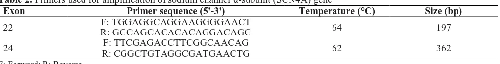
Table 2. Primers used for amplification of sodium channel
α
-subunit (SCN4A) gene
Exon
Primer sequence (5'-3')
Temperature (°C)
Size (bp)
22
F: TGGAGGCAGGAAGGGGAACT
R: GGCAGCACACACAGGACAGG
64
197
24
F: TTCGAGACCTTCGGCAACAG
R: CGGCTGTAGGCGATGAACTG
62
362
192 Iran J Neurol 2015; 14(4) Heidari et al.
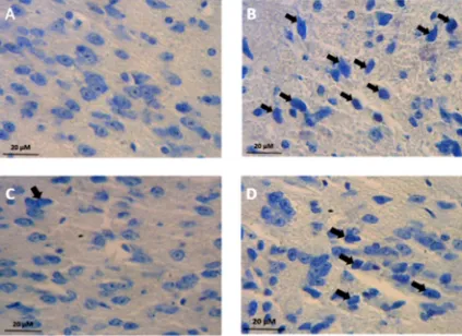
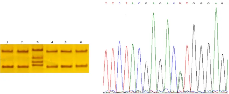
Figure 1. (A) Single strand conformational polymorphism gel electrophoresis of exon 22 of the
SCN4A gene. Line 1 shows different pattern banding regard to lines 2, 3, 4, 5, 6, and 7. Line 7
is normal control. (B) Chromatogram of sample 7 (without mutation) and (C) Chromatogram of
sample 1 (without 29073G>C mutation).
Figure 2. Single-strand conformational polymorphism results. Line 3 shows shift bands in a
patient with a heterozygous variation. Lines 1, 2, 4, and 5 show patients without any variations
and line 6 is a control sample. Sequence analysis of the genomic DNA of the patient revealed
the heterozygous transition of an A to G at position 31506 in exon 24 SCN4A genes in a patient
with non-dystrophic myotonia (left)
PCR products of samples with various band
patterns in SSCP gel were sent to a commercial
agency (Macrogene Seoul, South Korea) for
sequencing. The online multiple sequence alignment
software;
ClustalW2
(http://www.ebi.ac.uk/
tools/msa/ clustalw2/) and Blast analysis was used
to find the percent homology of the sequences that
has been obtained in the study and with all other
sequences of the other species.
Levels of the quantitative variables are presented
as a mean ± standard deviation. Student’s t-test was
used for comparison of continuous variables;
Fisher’s exact test was used for comparison of
categorical variables. The GraphPad Prism software
(version 3.00, GraphPad Software, La Jolla, CA,
USA) was used for statistical analysis, with P < 0.05
considered indicative of statistical significance.
Results
The mobility of single-stranded DNA fragments in
SSCP gel was conducted on 28 patients and 30
healthy controls. Mean age was 36 ± 12 and 34 ± 14
years for patients and controls, respectively. The
screening of exon 22 of SCN4A gene led to the
identification of one mutation in one out of 28
patients. DNA sequencing revealed 29073G>C
variant (Figure 1). This variant causes a missense
mutation G1306A (Glycine to Alanine).
Nondystrophic myotonia Iran J Neurol 2015; 14(4) 193
previously reported in SCN4A gene (Figure 2).
The results of multiple sequence alignment with
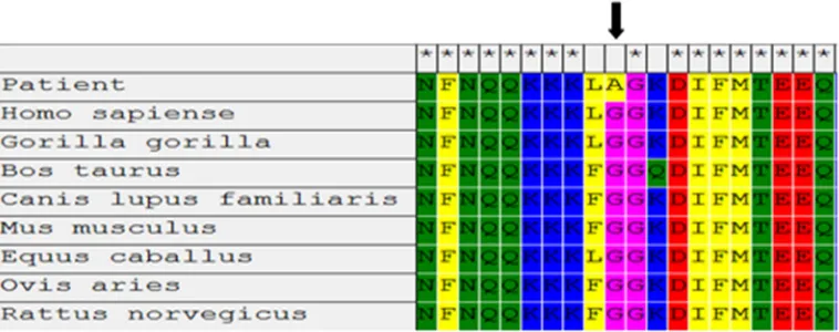
various species showed that G1306 is conserved during
evolution (Figure 3).
Discussion
The clinical signs and electrophysiological indicators
are used to prioritize genetic testing in the
non-dystrophic myotonias but, the detection of sodium
channel
myotonia
from
dominant
myotonia
congenital is difficult. Hence, it requires that a
proportion of patients to the screen of both SCN4A
and CLCN-1 genes.
11Lerche et al. found three point
mutations at the same nucleotide position 29073 of
the SCN4A gene in three families with a form of
myotonia. These mutations change glycine 1306 to
glutamic acid, valine, or alanine in SCN4A protein.
12Furthermore, McClatchey et al. found one of the
three substitutions at location 1306
(glycine-to-valine) in a family characterized by chronic
myotonia.
13Vicart et al. demonstrated that the most
common sodium channel myotonia mutations are
V1589M and G1306 position.
14Matthews et al. studied the clinical and genetic
features a long cohort of UK patients with
non-dystrophic myotonia and they demonstrated that 3
of their patients had mutations (G1306A, G1306E)
that previously described and identified two novel
mutations (R1448L, L1436P) in SCN4A gene.
9Here, we found one homozygous missense
mutation G1306A (Glycine to Alanine) in SCN4A gene
in one patient with mild painful myotonia. Multiple
sequence alignment with various species showed that
G1306 is conserved during evolution (Figure 3). Data of
Polyphen-2 software (with 0.54 score) predicted this
mutation is a possible pathogen. This substitution has
been
previously
predicted
in
the
conserved
cytoplasmic loop connecting repeat III and IV domains
of the sodium channel α-subunit could cause to
inactivation of gate of the sodium channel. The glycine
1306 confers a good flexibility of the hinge that could
restricted by side-chains of other amino acids.
12,15,16Second nucleotide variation, the heteroplasmic
31506 A>G polymorphism in exon 24 SCN4A gene,
which no alter amino acid sequences, has not been
previously described. This synonymous mutation is
located in a moderately conserved amino acid of the
C-terminal loop of SCN4A protein.
Study limitations
No accessibility to tissue samples from our patients
is the major limitation of our study. Another
limitation is the lack of classification of the patients
according to their clinical findings. Further studies
with larger cohorts of patients are warranted to
reveal the relationship of these nucleotide changes
with non-dystrophic myotonias.
Conclusion
Our mutational analysis confirms the role of single
nucleotide polymorphisms in SCN4A gene in
Iranian patients with non-dystrophic myotonias.
Hence, to find out and understand the nature of
pathogenesis and predisposition effects of these
variations on non-dystrophic myotonias, further
genetic, and functional studies are necessary.
Conflict of Interests
The authors declare no conflict of interest in this study.
Acknowledgments
The authors would like to thank Yazd University
Research Council, IUT Research Council and
Excellence in Sensors for financial support of this
research. We thank all the patients for providing
blood samples for the scientific research. The study
was approved by Yazd University Human Research
Ethics Committee.
Figure 3. Multiple sequence alignment of a part of exon 22 from various species. Glycine
194 Iran J Neurol 2015; 14(4) Heidari et al.
How to cite this article: Heidari MM, Khatami M, Nafissi Sh,
Hesami-Zokai F, Khorrami A. Mutation analysis in exons
22 and 24 of SCN4A gene in Iranian patients with
non-dystrophic myotonia. Iran J Neurol 2015; 14(4): 190-4.
References
1. Heatwole CR, Moxley RT. The nondystrophic myotonias. Neurotherapeutics 2007; 4(2): 238-51. 2. Lehmann-Horn F, Rudel R. Hereditary
nondystrophic myotonias and periodic paralyses. Curr Opin Neurol 1995; 8(5): 402-10.
3. Emery AE. Population frequencies of inherited neuromuscular diseases--a world survey. Neuromuscul Disord 1991; 1(1): 19-29.
4. Chino N, Noda Y, Oda N. Conduction study in human muscle fibers in situ-a useful technique for diagnosing myopathies. Electroencephalogr Clin Neurophysiol 1984; 58(6): 513-6.
5. Lee SC, Kim HS, Park YE, Choi YC, Park KH, Kim DS. Clinical Diversity of SCN4A-Mutation-Associated Skeletal Muscle Sodium Channelopathy. J Clin Neurol 2009; 5(4): 186-91.
6. Stuhmer W, Conti F, Suzuki H, Wang XD, Noda M, Yahagi N, et al. Structural parts involved in activation and inactivation of the sodium channel. Nature 1989; 339(6226): 597-603.
7. Bendahhou S, Cummins TR, Kwiecinski H, Waxman SG, Ptacek LJ. Characterization of a new sodium channel mutation at arginine 1448 associated with moderate Paramyotonia congenita in humans. J Physiol 1999; 518 (Pt 2): 337-44.
8. Matthews E, Fialho D, Tan SV, Venance SL, Cannon SC, Sternberg D, et al. The non-dystrophic myotonias: molecular pathogenesis, diagnosis and treatment. Brain 2010; 133(Pt 1): 9-22.
9. Matthews E, Tan SV, Fialho D, Sweeney MG, Sud R, Haworth A, et al. What causes paramyotonia in the United Kingdom? Common and new SCN4A mutations revealed. Neurology 2008; 70(1): 50-3. 10. Sambrook J, Russell DW. Molecular
Cloning: A Laboratory Manual. New York, NY: Cold Spring Harbor Laboratory; 2001. 11. Trip J, Drost G, Verbove DJ, van der Kooi
AJ, Kuks JB, Notermans NC, et al. In tandem analysis of CLCN1 and SCN4A greatly enhances mutation detection in families with non-dystrophic myotonia. Eur J Hum Genet 2008; 16(8): 921-9.
12. Lerche H, Heine R, Pika U, George AL,
Mitrovic N, Browatzki M, et al. Human sodium channel myotonia: slowed channel inactivation due to substitutions for a glycine within the III-IV linker. J Physiol 1993; 470: 13-22.
13. McClatchey AI, Van den Bergh P, Pericak-Vance MA, Raskind W, Verellen C, McKenna-Yasek D, et al. Temperature-sensitive mutations in the III-IV cytoplasmic loop region of the skeletal muscle sodium channel gene in paramyotonia congenita. Cell 1992; 68(4): 769-74.
14. Vicart S, Sternberg D, Fontaine B, Meola G. Human skeletal muscle sodium channelopathies. Neurol Sci 2005; 26(4): 194-202.
15. West JW, Patton DE, Scheuer T, Wang Y, Goldin AL, Catterall WA. A cluster of hydrophobic amino acid residues required for fast Na(+)-channel inactivation. Proc Natl Acad Sci U S A 1992; 89(22): 10910-4.
![Figure 2. Photomicrograph of coronal section from the mice substantia nigra (SN) sub-regions [SNc: Substantia Nigra pars compacta (Red); SNr: Substantia Nigra pars reticulata (Yellow)] illustrating 1-methyl-4-phenyl-1,2,3,6-tetrahydropyridine-induced dark neurons production in SNc sub-region where used for the stereological study; Regional boundaries were determined by cross-referencing with the atlases of Paxinos and Watson](https://thumb-us.123doks.com/thumbv2/123dok_us/8751871.1748804/21.595.46.281.274.493/photomicrograph-substantia-substantia-substantia-illustrating-tetrahydropyridine-stereological-referencing.webp)