Germination Involves Seed Compartment-Speci
fi
c
Expression and Activity of Pectin Methylesterases
1[OPEN]
Claudia Scheler2, Karin Weitbrecht2, Simon P. Pearce2, Anthony Hampstead, Annette Büttner-Mainik, Kieran J.D. Lee, Antje Voegele, Krystyna Oracz, Bas J.W. Dekkers, Xiaofeng Wang,
Andrew T.A. Wood, Leónie Bentsink, John R. King, J. Paul Knox, Michael J. Holdsworth3, Kerstin Müller3, and Gerhard Leubner-Metzger3*
Botany and Plant Physiology, Institute for Biology II, Faculty of Biology, University of Freiburg, D–79104 Freiburg, Germany (C.S., K.W., A.B.-M., K.O., G.L.-M.); Institute of Biochemical Plant Pathology, Helmholtz Zentrum München, Deutsches Forschungszentrum für Gesundheit und Umwelt, D–85764 Neuherberg, Germany (C.S.); Staatliches Weinbauinstitut Freiburg, D–79104 Freiburg, Germany (K.W.); Centre for Plant Integrative Biology (S.P.P., A.H., A.T.A.W., J.R.K., M.J.H.) and Division of Plant and Crop Science (S.P.P., M.J.H., K.M.), School of Biosciences, University of Nottingham, Sutton Bonington Campus, Sutton Bonington, Leicestershire LE12 5RD, United Kingdom; School of Mathematical Sciences, University of Nottingham, University Park, Nottingham NG7 2RD, United Kingdom (S.P.P., A.H., A.T.A.W., J.R.K.); Agroscope, Institute for Plant Production Sciences, Seed Quality, CH–8046 Zurich, Switzerland (A.B.-M.); Centre for Plant Sciences, Faculty of Biological Sciences, University of Leeds, Leeds LS2 9JT, United Kingdom (K.J.D.L., J.P.K.);
National Institute for Health Research Trainees Coordinating Centre, Leeds Innovation Centre, Leeds LS2 9DF, United Kingdom (K.J.D.L.); School of Biological Sciences, Plant Molecular Science and Centre for Systems and Synthetic Biology, Royal Holloway, University of London, Egham, Surrey TW20 0EX, United Kingdom (A.V., G.L.-M.); Department of Plant Physiology, Warsaw University of Life Sciences, 02–776, Warsaw, Poland (K.O.); Wageningen Seed Laboratory, Laboratory of Plant Physiology, Wageningen University and Research Centre, NL–6708 PB Wageningen, The Netherlands (B.J.W.D., L.B.); College of Life Sciences, South China Agricultural University, Guangzhou 510642, China (X.W.); and Laboratory of Growth Regulators, Faculty of Science, Palacký University and Institute of Experimental Botany, CZ–783 71 Olomouc, Czech Republic (G.L.-M.)
Pectin methylesterase (PME) controls the methylesterification status of pectins and thereby determines the biophysical properties of plant cell walls, which are important for tissue growth and weakening processes. We demonstrate here that tissue-specific and spatiotemporal alterations in cell wall pectin methylesterification occur during the germination of garden cress (Lepidium sativum). These cell wall changes are associated with characteristic expression patterns of PME genes and resultant enzyme activities in the key seed compartments CAP (micropylar endosperm) and RAD (radicle plus lower hypocotyl). Transcriptome and quantitative real-time reverse transcription-polymerase chain reaction analysis as well as PME enzyme activity measurements of separated seed compartments, including CAP and RAD, revealed distinct phases during germination. These were associated with hormonal and compartment-specific regulation of PME group 1, PME group 2, and PME inhibitor transcript expression and total PME activity. The regulatory patterns indicated a role for PME activity in testa rupture (TR). Consistent with a role for cell wall pectin methylesterification in TR, treatment of seeds with PME resulted in enhanced testa permeability and promoted TR. Mathematical modeling of transcript expression changes in germinating garden cress and Arabidopsis (Arabidopsis thaliana) seeds suggested that group 2 PMEs make a major contribution to the overall PME activity rather than acting as PME inhibitors. It is concluded that regulated changes in the degree of pectin methylesterification through CAP- and RAD-specific PME and PME inhibitor expression play a crucial role during Brassicaceae seed germination.
Mature seeds of members of the Brassicaceae family such as Arabidopsis (Arabidopsis thaliana) and garden cress (Lepidium sativum) are endospermic (i.e. the embryo is surrounded by a thin living cell layer, the endosperm, and a dead outer layer, the testa). Many angiosperm seeds, including those of garden cress and Arabidopsis, germinate in a two-step process: after the initial phase of water uptake by the dry seeds (imbibition), testa rupture (TR) occurs and is subsequently followed by endosperm rupture (ER), which marks the completion of germina-tion (Liu et al., 2005; Müller et al., 2006). Germinagermina-tion is
controlled by two opposing forces, the increasing growth potential of the radicle and the resistance of the testa and endosperm tissues covering it (Bewley, 1997; Schopfer, 2006; Linkies and Leubner-Metzger 2012). After TR, en-dosperm resistance decreases through tissue softening, a process called endosperm weakening (Müller et al., 2006; Linkies et al., 2009). Both radicle elongation and endosperm weakening require cell wall modifications (Schopfer, 2006; Müller et al., 2009; Morris et al., 2011).
Plant cell walls are the main determinants for the shape and biomechanical properties of plant tissues,
organs, and even the whole plant body. They control turgor-driven water uptake to allow cell growth through changes in extensibility, which depend on wall compo-sition and the interaction between their components (Thompson, 2005; Cosgrove and Jarvis, 2012; Yoshida et al., 2014). One of the most abundant groups of poly-saccharides in primary cell walls is pectins. Pectins are complex polysaccharides that are characterized bya -1,4-linked galacturonic acid (Willats et al., 2001; Mohnen, 2008; Tan et al., 2013); they are present in the middle lamellae and are key polymers in cell separation processes. The most abundant plant cell wall pectin is homogalacturonan (HG). HG is a linear polymer of (1,4)-linked-a-D-galacturonic acid that can be modified by methylesterification at the C-6 carboxyl position to form methylesterified homogalacturonan (Me-HG; Wolf et al., 2009a). The degree of methylesterification is variable between developmental stages, tissues, and even regions of the wall of an individual cell and strongly affects the mechanical properties of cell walls (Braybrook et al., 2012). After synthesis in the endomembrane system, HG is secreted in a highly methylesterified form into the plant cell wall of growing cells (Mohnen, 2008).
Pectin methylesterases (PMEs; EC 3.1.1.11) catalyze the demethylesterification of HG (Wolf et al., 2009a). PMEs are ubiquitous cell wall-associated enzymes that are found in all higher plants as well as in some bacteria and fungi. PMEs can act linearly to deesterify stretches of Me-HG to give rise to blocks of free carboxyl groups that can be cross-linked by calcium ions. These calcium bridges influence cell wall porosity and may enhance the overallfirmness of tissues. PMEs also can act in a nonlinear fashion and deesterify only individual gal-acturonate residues or short stretches, which does not allow for calcium bridges to form, leading to a looser cell wall matrix structure. PME activity might promote the subsequent action of cell wall hydrolases such as endopolygalacturonases (Wakabayashi et al., 2000, 2003),
which contribute to cell wall weakening and/or cell separation (González-Carranza et al., 2007). PMEs have been shown to be involved in pectin remodeling at different developmental stages, such as pollen tube growth (Eckardt, 2005), root elongation and its reac-tion to soil aluminum concentrareac-tions (Yang et al., 2013), hypocotyl elongation (Pelletier et al., 2010), fruit ripening (Hyodo et al., 2013), and seed germination (Müller et al., 2013). PMEs are encoded by a large multigene family that has been classified into two groups (Wang et al., 2013): all PMEs have a conserved pectinesterase domain (Pfam01095), but only group 2 has in addition a PME inhibitory domain (Pfam04043). PME activity is regulated by pectin methylesterase inhibitor (PMEI) proteins (Giovane et al., 2004; Wolf et al., 2009a).
In our integrative study, we discovered that during the seed germination of garden cress, changes in the transcript abundance of specific PMEs and PMEIs are reflected in seed compartment-specific changes in PME activity and accompanied by spatiotemporal changes in cell wall pectin methylesterification. We mathemati-cally modeled the contribution of the different groups of PMEs and of PMEIs to the degree of methylesterification in the garden cress seed cell walls. Based on these mo-lecular, physiological, histochemical, and biophysical analyses, we propose that PME activity is involved in the germination process of garden cress and is differentially regulated in a spatial and temporal manner.
RESULTS
Seed Compartment-Specific Transcriptome Analysis of Garden Cress Germination
We utilized the fact that garden cress has a two-step germination process, with TR and ER separated by several hours, to conduct a dense spatiotemporal transcriptome analysis during the germination process (Fig. 1). We in-vestigated transcriptome changes in the seed compart-ments that are directly involved in ER, the micropylar endosperm (CAP; Fig. 1B) and the radicle with the lower part of the hypocotyl (RAD; embryo growth zone; Fig. 1B), at key time points during the germination process from early imbibition to the completion of ER (Fig. 1, A and B). At the time points when approximately 50% of the seed population had reached TR and ER, respectively, we divided the sampling population be-tween the seeds that had already undergone rupture (+TR or +ER) and those that had not (2TR or2ER), so that we could compare samples that would have un-dergone TR within the next 1 or 2 h with those that already had ruptured testas. We also sampled cotyle-dons (COTs) and nonmicropylar endosperms (NMEs) at three time points (Fig. 1). Seed compartment-specific gene expression analyses for garden cress have been performed successfully before (Linkies et al., 2009) using Complete Arabidopsis Transcriptome Microarray-spotted PCR-amplified gene-specific tag-based chips, but only for a small number of samples that allowed for a 1
This work was supported by the ERA-NET Plant Genomics vSEED project through the Deutsche Forschungsgemeinschaft (grant nos. DFG LE720/8 and DFG LE720/6 to G.L.-M.), the U.K. Biotech-nology and Biosciences Research Council (grant no. BB/G024898/1 to J.P.K. and grant no. BBG02488X1 to M.J.H. and J.R.K.), the Neth-erlands Organization for Scientific Research (grant no. 855.50.011 to L.B.), the Wissenschaftliche Gesellschaft Freiburg (to G.L.-M.), the Guangdong Natural Science Foundation (grant no. 07006658 to X.W.), a Marie Curie International Outgoing Fellowships for Career Development fellowship (to K.M.), an Alexander von Humboldt Foundation research fellowship (to K.O.), and the Royal Society and the Wolfson Foundation (to J.R.K.).
2These authors contributed equally to the article. 3These authors contributed equally to the article. * Address correspondence to [email protected]. The author responsible for distribution of materials integral to the
findings presented in this article in accordance with the policy de-scribed in the Instructions for Authors (www.plantphysiol.org) is: Gerhard Leubner-Metzger ([email protected]).‘The Seed Biology Place’www.seedbiology.eu.
[OPEN]
limited analysis of the garden cress germination process. Our sampling concept in this work with garden cress (Fig. 1) led to a time course with sufficient spatiotemporal resolution to investigate the distinct phases of germina-tion and make physiologically relevant comparisons.
We performed microarrays by hybridizing garden cress RNA to Affymetrix ATH1 microarrays designed for Arabidopsis. Heterologous microarrays on Arabidopsis ATH1 chips have been employed successfully by several groups to elucidate transcriptome changes in unsequenced Brassicaceae species or in species without commercially available microarrays (Hammond et al., 2006; Slotte et al.,
[image:3.585.79.496.70.496.2]2007). To further improve the method for garden cress, we developed a sophisticated masking approach that allowed us to extract a maximum of information from the arrays (see“Materials and Methods”). Using the masking method presented here, with a false discovery rate of 0.01, 36.6% of probes and 65.1% of probe sets were retained, leading to 5,793 genes identified as being differentially expressed between 1 and 16 h after sowing in the CAP, and 6,098 genes in the RAD. Conversely, using the method of Hammond et al. (2005), the maximal number of differen-tially expressed genes was 1,712 at a cutoff of 100. Thus, our method retained a much larger number of differentially Figure 1. Spatiotemporal transcriptome analysis of garden cress ‘FR14’ seed compartments during germination. A, The kinetics
of garden cress TR and ER at 24˚C in continuous light. Arrows indicate sampling time points for RNA extraction at which the seeds were dissected into CAP, RAD, COT, and NME, as indicated. Note that at 0 h (dry seed stage), CAP plus RAD as well as NME plus COT were sampled together. Mean values6SEofn= 4 plates each with 100 seeds are presented for TR and ER. B,
expressed genes that could then be used for further anal-ysis. Therefore, normalized expression values for garden cress were obtained for 13,895 transcripts (Supplemental Data Set S1), with garden cress gene transcripts referring to the putative Arabidopsis orthologs defined by having an Arabidopsis Genome Initiative identifier such as At1g62380. Supplemental Data Sets S2 and S3 provide the normalized log2values for the mean andSDexpression results, respectively.
Seed Compartment-Specific Differentially Regulated Garden Cress Transcriptome during Germination Shows Overrrepresentation of Cell Wall-Related Transcripts at the TR Stage Transition
A principal component analysis (PCA) shows the gen-eral distribution of values calculated into eigenvectors and makes it possible to identify the eigenvectors with the greatest influence on sample variance. In our arrays, the eigenvectors with the biggest influence appear to corre-spond to time and seed compartment (Fig. 1C). In dry seeds and at 1 h after sowing, RAD and CAP samples still cluster together, separating from 3 h after sowing. In the RAD, distinct clusters formed at the phase transition time point at 7 h, where we segregated for2TR and +TR, and at 16 h, where we segregated for seeds that had under-gone ER and thus completed the germination process (+ER) and those whose endosperm had not ruptured yet (2ER). The same was true for the CAP at 7 h (+TR versus2TR), although the 16-h CAP with ER clusters with the CAP samples without ER.
The COT samples exhibited a higher variance throughout the time course (Fig. 1). The NME for the two time points without TR clustered apart from the CAP samples but was still closer to them than to the RAD and COT samples derived from the embryo. At 13 h, the NME samples varied more and clustered closer still to the CAP samples (Fig. 1). The finding that transcriptomes differ between the distinct seed compartments and over time is further supported by a cluster analysis of the transcript abundance patterns, which showed five clearly distinct clusters (Supplemental Fig. S1). Together with the prin-cipal component analysis (Fig. 1), it supports the view that TR is a decisive step during the germination pro-cess that is associated with abundant transcriptomes changes (+TR versus 2TR) in both the CAP and the RAD.
The Gene Ontology (GO) overrepresentation analyses of the TR rupture transition time point (Supplemental Tables S1 and S2) indicate that both hormone regulation and cell wall modifications are overrepresented in the differentially regulated transcripts in the different seed compartments. We decided to look in more detail at PME and PMEI, as those cell wall-modifying enzymes and inhibitors have been shown to be tied in with hor-monal regulations and to play a role in seed germination of Arabidopsis (Müller et al., 2013; Saez-Aguayo et al., 2013), and pectinesterase activity and inhibition was one of the activities whose GO terms were specifically over-represented in the RAD (Supplemental Table S2).
Spatiotemporal Patterns of HG and Me-HG Pectin Cell Wall Epitopes during Garden Cress Seed Germination
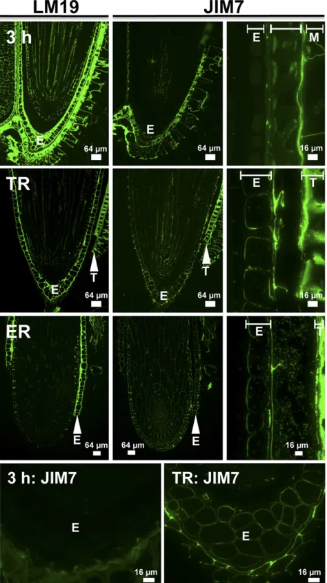
In order to observe if, where, and at which stage during the garden cress seed germination process changes in the degree of pectin methylesterification actually happen in the RAD and CAP, we studied HG epitopes in situ. To distinguish pectic HG in its methylesterified form (Me-HG) from the demethylesterified form (HG), we used a set of well-characterized monoclonal antibodies (www.plantcellwalls.net) in conjunction with fl uores-cence imaging (Fig. 2). Antibody LM19 is specific for demethylesterified HG, and its epitope was detected in seeds 3 h after sowing. The LM19 epitope was ubiqui-tously distributed in the cell walls of the RAD as well as in the CAP (E in Fig. 2), the testa, and the testa-derived mucilage layer. In contrast, the JIM7 (Fig. 2) and LM20 (data not shown) Me-HG epitopes were restricted to the testa and mucilage and were present at reduced levels in the RAD but absent from the CAP. The pattern of the LM19 HG epitope did not change between our sampling times. However, the spatial distribution of Me-HG (JIM7) was altered. The JIM7 signal increased in the RAD and appeared in the inner cell wall of the CAP upon TR (Fig. 2). This suggests that there is new deposition of pectin into the cell wall, as HG is methylesterified in the Golgi and secreted as Me-HG. In testa and mu-cilage, the JIM7 epitope was detectable in all phases during seed germination (Fig. 2).
Molecular Phylogenetic Analyses of Arabidopsis and Garden Cress PMEs and Their Inhibitors
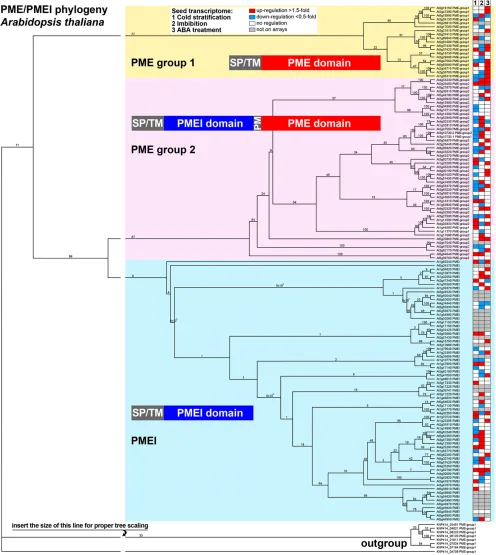
We mined our microarrays for the expression pat-terns of putative PMEs and PMEIs in garden cress seeds (Supplemental Data Sets S1–S3). As the garden cress genome has not been sequenced, we started by identifying 136 Arabidopsis sequences for PMEs and PMEIs by annotation and similarity searches in public databases, with subsequent verification of the exis-tence of specific domains in the predicted proteins. According to our molecular phylogenetic analysis of their full-length predicted protein sequences, these Arabidopsis PMEs and PMEIs cluster into three large groups (Fig. 3): group 1 PME (22 members) and group 2 PME (45 members) contain the PME domain (Pfam01095), which harbors five characteristic sequence motifs impor-tant for PME activity. The group 2 PMEs additionally possess a PMEI domain (Pfam04043) with conserved Cys residues, and both domains are separated by a processing motif that is a putative target for subtilisin-like proteases. The third cluster consists of 69 PMEIs (Fig. 3).
and PMEIs was strongly up-regulated during thefirst few hours of the germination process (2 in Fig. 3). PMEs from both groups as well as PMEIs are up-regulated in the course of the germination process in the presence of abscisic acid (ABA; 3 in Fig. 3).
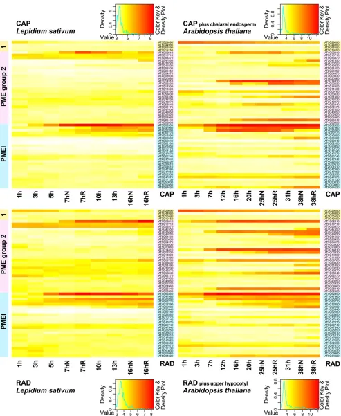
We then compared the expression of PME and PMEI transcripts that we identified in our arrays for garden
cress seeds (four group 1 PMEs, 28 group 2 PMEs, and 25 PMEIs) in CAP and RAD (Fig. 4) with their ho-mologs in Arabidopsis (data extracted from the tran-scriptome of Dekkers et al. [2013]) as defined by the transcripts that bind to the same probe sets. There was an overall stronger differential regulation visible in the Arabidopsis data, which may be due to the heterolo-gous nature of the garden cress arrays or to the fact that the Arabidopsis seed compartments were less confined and contained additional tissues (Fig. 4). However, we could clearly observe that the genes that were strongly differentially regulated in garden cress seed compart-ments were also strongly regulated in Arabidopsis in a similar manner in the same compartments (Fig. 4).
PME Activity in Garden Cress Seed Compartments in Relation to TR and ER
With the large number of PMEs and PMEIs and their diverse expression patterns, it is hard to predict whether there is a net PME activity at any given stage and seed part and how it changes over the course of germination. Therefore, we measured the total PME activity in garden cress RADs and CAPs. The total PME enzyme activity was roughly 10-fold higher in the CAP compared with the RAD during garden cress seed germination (Fig. 5). The PME activities in the CAP were highest during im-bibition and in the very early phase of germination (3–8 h), declined around the time of TR, and then stabilized at a lower activity as the population reached the comple-tion of TR followed by ER (16 h; Fig. 5A). The activity in the RAD also decreased around TR, then increased again as the seeds neared ER, and peaked at a stage where the whole population had reached TR but not yet progressed to ER (16 h).
Endosperm weakening and rupture of garden cress are known to be delayed by exogenous ABA, while the kinetics of TR is unaffected by its presence. In accor-dance with this, when ABA was added to the germi-nation medium, the population only completed ER after about 70 h, but the timing of TR did not change significantly (Fig. 5B). PME activity in the CAP during the early germination phase before TR was similar in seed populations with and without ABA. However, when this seed population neared its delayed ER, PME activity was significantly lower than at the physio-logically equivalent time point without ABA (Fig. 5B). PME activity in the RAD was relatively constant over time and approximately 2-fold lower in the ABA series compared with the control (Fig. 5B).
Mathematical Model of PME Activity in the Garden Cress CAP and RAD
[image:5.585.38.275.73.496.2]As mentioned above, PMEs fall into two groups, one of which (group 2) contains a PMEI domain. In order to explore the significance of the two PME groups, along with the action of the PMEIs, on the total PME activity and, therefore, to the pectin demethylesterification process, we Figure 2. Immunolocalization of the LM19 HG and JIM7 Me-HG pectin
constructed a biologically informed network of reactions (Fig. 6A) and converted it into a system of ordinary dif-ferential equations. Cumulative transcript accumulation for each PME group (Fig. 6B) was used as a proxy for their protein accumulation. The network (Fig. 6) that was used as a basis for our set of ordinary differential equa-tions centers on the demethylesterification of Me-HG, caused by either of the PME groups, with PMEI inhib-iting both groups of PMEs and group 2 PMEs able to inhibit themselves. Several assumptions were made to simplify the system for the purpose of modeling: (1) spatial variations are neglected; (2) negligible deposi-tion of addideposi-tional pectin occurs over the time scale of interest (germination process until approximately 16 h); (3) interactions between PME proteins and their inhib-itors irreversibly remove the proteins from the system; (4) protein production rates are proportional to the levels of the relevant mRNA, the latter being obtained from the transcriptomic data; and (5) a group 2 PME molecule is able to inactivate itself, since it contains both the PME and PMEI domains. These assumptions and the network (Fig. 6A) lead to Equations 1 to 8 (Fig. 6C); for further explanation, see“Materials and Methods.” The model wasfitted to the PME enzyme data shown in Figure 5A, with the resulting param-eters listed in Supplemental Table S3. The resulting PME activity predicted by the model after fitting is shown in Figure 6D in comparison with the measured PME enzyme activity data.
PME and PMEI cumulative transcript accumulation within garden cress seed compartments, our proxy for protein production, appears to be phasic in both CAP and RAD (Fig. 6B):first, the group 1 PMEs are produced, then the PMEI proteins, andfinally the group 2 PMEs. This is especially striking in the RAD, since the group 2 PMEs do not have the same escalation in the CAP, where group 2 PME production peaks around the time of TR before declining. Parameter sensitivity analysis was car-ried out on the model, and it was noted that altering the activating reaction rate,ai, has a greater impact on the model than varying the inhibiting reaction rate, zi. Therefore, our parameter (Supplemental Table S3) sen-sitivity analysis gives insight into which processes are most significant in governing the overall activity and open the way for subsequent application of the model and for its refinement.
Our mathematical model implies that inhibiting processes, in particular by the group 2 PMEs, are less important for the overall activity than their demethy-lesterification function. This suggests that the primary
importance of the group 2 PMEs is in their PME action rather than their PMEI behavior (see “Discussion”). Therefore, analyzing the group 2 PME expression pattern (see below) is relevant to the PME enzyme activity pattern (Fig. 5).
Hormonal and Seed Compartment-Specific Regulation of Garden Cress Group 2 PMEs
With the large number of differentially regulated group 2 PMEs (Fig. 4) in our arrays for both RAD and CAP, as well as the insight from the model (Fig. 6) that the group 2 PMEs are predicted to play a role mostly as PMEs and not as inhibitors, we decided to clone and look in detail at several garden cress group 2 PMEs (Fig. 7). Several group 2 PMEs were dramatically up-regulated in whole Arabidopsis seeds (Fig. 3):At1g11580
increased more than 100-fold, but we did not obtain data corresponding to this transcript from our heterologous ar-ray analysis.At2g26440andAt3g14310were more than 100-fold up-regulated during thefirst 24 h of imbibition in Arabidopsis, but they did not show changes in the
L. sativumarrays in either seed compartment under the conditions we used for our arrays.
We fully cloned and analyzed the complementary DNAs (cDNAs) for the garden cress PME group 2 homolog of ArabidopsisAt1g11580, which we named
LesaPME11580. All other cloned garden cress PME cDNAs were named following the same principle, including PME group 2LesaPME26440,LesaPME14310, and LesaPME51490 (Supplemental Table S4). Taken together, the sequence comparisons (for details, see Supplemental Fig. S2) considering known domains (Markovic and Janecek, 2004; Pelloux et al., 2007) strongly suggest that LesaPME11580 is a functional PME group 2 of garden cress. We also cloned the full-length cDNA of LesaPMEI14890, which shows the typical PMEI domain and other characteristic features of this class of inhibitors (Supplemental Fig. S2), and therefore is most likely a functional PMEI.
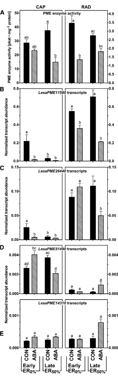
Using quantitative real-time reverse transcription (qRT)-PCR, we analyzed the transcript expression of the garden cress group 2 PMEs (Fig. 7) and a small selection of group 1 PMEs and PMEIs (Supplemental Fig. S3) in the CAP and RAD with and without ex-posure to ABA. In vitro PME activities were measured in the CAP and RAD at two sampling time points, seeds that had just undergone TR (Early ER0%) and seeds just before ER (Late ER50%), for the control and ABA treatments (Fig. 7A). In all conditions tested,
Figure 3. (Continued.)
PME activity in the RAD was significantly lower than in the CAP. In both seed compartments, ABA inhibited PME activity compared with the untreated control (Fig. 7A). Supplemental Figure S3 contains additional data for exposure to 1-aminocyclopropane-1-carboxylic acid (ACC), the precursor of the plant hormone ethyl-ene, which acts antagonistically to ABA in the germi-nation process of garden cress (Linkies et al., 2009). ACC had no effect on total activity in the presence or absence of ABA in the RAD but led to a strong increase in ac-tivity in the CAP at the TR time point (Supplemental Fig. S3).
All transcripts we investigated showed a response to ABA that differed between RAD and CAP, confirming the importance of investigating the seed compartments separately. LesaPME11580 was expressed more abun-dantly in the RAD compared with the CAP (Fig. 7B). The transcript stayed at the same level at the two time points we investigated in the RAD but declined in the CAP between the early and late time points. Treatment with ABA down-regulatedLesaPME11580in all condi-tions tested and in both seed compartments (Fig. 7B), while ACC caused a down-regulation only in the CAP and not in the RAD (Supplemental Fig. S3). In situ mRNA hybridization (Supplemental Fig. S4) confirmed thatLesaPME11580transcripts localized to the RAD and were hardly detectable in the CAP.LesaPME26440was also predominantly expressed in the RAD (Fig. 7C), but contrary to LesaPME11580, it was down-regulated in the presence of ABA specifically at the early time point in the CAP and the late time point in the RAD. In addition, its abundance in the CAP was lower at the later time point than it was at the early time point.
LesaPME51490 showed a predominant expression in the CAP (Fig. 7D), whileLesaPME14310was expressed in both seed compartments at a low level (Fig. 7E).
LesaPME14310expression was sensitive to ABA in the medium only at the late time point in the RAD, whereas ABA had no effect onLesaPME14130at the other time points. Compared with the four group 2 PMEs that we investigated, the group 1LesaPME29090transcript abun-dance was around 10-fold higher, and there was no appreciable down-regulation by ABA (Supplemental Fig. S3).
Exogenous Treatment with PME Enhances Testa Permeability and Promotes TR
Having investigated the endogenous PME transcript abundances, activities, and pectin methylesterification patterns in the garden cress CAP and RAD during germination, we focused on the question of whether
treatment of seeds with exogenous PME affects their germination. Interestingly, addition of 0.2 units of orange (Citrus sinensis) peel PME to the seed incubation medium (0.03 units PME mL21) promoted their TR (Fig. 8A) but did not appreciably affect ER (Supplemental Fig. S5A). In contrast, relatively high PME amounts (ap-proximately 20 units) delayed TR and ER (Supplemental Fig. S5). Earlier work (Linkies et al., 2009) showed that, while treatment of garden cress seeds with ABA or the ethylene precursor ACC affected ER, it did not affect the kinetics of TR. Addition of 0.2 units of PME plus ABA or ACC to the seed incubation medium also promoted TR (Fig. 8A), which suggests that the PME action is a direct effect of the enzyme action and could be associated with an increased testa and/or mucilage permeability.
To assay for testa permeability, we imbibed seeds in tetrazolium assay solution, a method used to analyze Arabidopsistransparent testamutants (Debeaujon et al., 2000) and the effect of myrigalone A on garden cress seeds (Voegele et al., 2012). We imbibed garden cress seeds in tetrazolium salt solution in the absence (CON) or presence of 0.2 units of PME, thus at a concentration of PME that led to earlier TR. After 9 h, the embryos were excised from the seeds, photographed, and cat-egorized (Fig. 8B). For CON, 88% of the embryos were unstained (i.e. the testa was impermeable to the dye) and 12% were yellow, showing a low staining intensity that indicates the low testa permeability. Thus, the testa permeability of CON-imbibed seeds for tetrazo-lium salts was very low. In contrast, for seeds treated with 0.2 units of PME, only 46% of the embryos were pale (impermeable) and 20% stained yellow (low per-meability), while 34% stained partly (either at the COT base or the radicle tip) or almost fully red (highly permeable; Fig. 8B; Supplemental Fig. S6).
PME-mediated demethylesterification of Me-HG in-creases the cell wall HG content. Polygalacturonase (PG) is a pectin-degrading enzyme that cleaves the
a-1,4-D-galacturonosidic linkages of HG chains. Con-certed action of PME and PG, therefore, is known to cause extensive pectin depolymerization (Wakabayashi et al., 2000, 2003). Therefore, we assumed that, if testa and mucilage permeability depend on the state of pectin, PG should further enhance the testa permea-bility when exogenously applied in combination with 0.2 units of PME. This was indeed the case: only 18% of the embryos were pale (impermeable), 20% were stained yellow, and 62% were stained partly or almost fully red upon treatment with PME plus PG (Fig. 8B; Supplemental Fig. S6). We conclude that the promoting effect on TR and ER by low concentrations of PME is at least partially achieved through an enhanced perme-ability of the testa and/or mucilage layer (Fig. 8).
Figure 4. (Continued.)
DISCUSSION
TR Constitutes a Transition between Phases of Gene Expression and Enzyme Activities in the Garden Cress CAP and RAD
[image:10.585.310.549.65.586.2]Our microarray analysis provides a high-resolution picture of seed compartment-specific transcriptome changes during garden cress seed germination. The added density of sampling time points made possible by this technical advance supports the identification of the importance of TR as a transitional event during garden cress seed germination. Many transcripts showed an increase in abundance shortly before or more often just after the TR event. One group of genes strongly up-regulated around TR are cell wall-modifying enzymes, and cell wall-related genes were evident in our GO over-representation analysis. This indicates the importance of
Figure 5.PME enzyme activity during the seed germination of garden cress. Seeds were imbibed in water (control [CON]; A) or 5mMABA (B). The kinetics of garden cress TR and ER at 18˚C in continuous light is shown. PME enzyme activity was measured for protein extracts of CAP and RAD excised from imbibed seeds at the times indicated. Mean values6SEfor four biological replicates with 100 seeds each are
[image:10.585.60.277.277.636.2]presented.
Figure 6. Mathematical model of the contributions of group 1 PMEs, group 2 PMEs, and PMEIs to overall PME activity in RAD and CAP during garden cress seed germination. A, Network diagram, whereaiindicates the rates at
which Me-HG is demethylesterified andzirepresents the rates at which a
cell wall remodeling in the CAP and RAD around and after the time of TR. This increased level of expression is then maintained or further increased for the remaining germination process. While specific groups of genes such as the cell wall-modifying enzymes are up-regulated at TR, the overall number of differentially regulated genes drops drastically once TR is complete. These observa-tionsfit with those made using microarrays with seed compartment-specific RNA of Arabidopsis, where TR also emerged as a central event for transcriptional regulation in seeds (Dekkers et al., 2013). The transi-tional nature of the TR time point was also evident in our principal component analysis (Fig. 1): up to and including TR, the CAP and NME endosperm samples are clearly distinguishable, but after TR from 10 to 16 h with and without ER, the samples cluster together as one group.
As TR is not influenced by the presence of ABA in the germination medium, but ER is strongly delayed by ABA, and the processes that begin at TR are clearly subject to further hormonal regulation once they have been initiated (Linkies et al., 2009). For cell wall-modifying enzymes, this has been observed for PME enzyme activities during Arabidopsis seed germination (Müller et al., 2013). Whole-seed total PME activity in-creased until TR was reached and then declined. When ABA was added to the medium, TR still constituted the highest point of PME activity, but the activity only de-creased with a delay after a plateau phase (Müller et al., 2013).
Cell Wall-Modifying Enzymes Are Differentially Regulated at the Time of TR in Garden Cress in a Seed Compartment-Specific Manner
[image:11.585.57.252.64.692.2]Cell wall modifications are necessary to allow radi-cle elongation and endosperm weakening, the two processes that eventually lead to ER (Schopfer, 2006; Linkies and Leubner-Metzger, 2012). It is also possible that cell wall modifications contribute to TR, as cell wall loosening in the RAD and CAP and a subsequent
Figure 7.Spatial and temporal analysis of transcript abundances of novel garden cress (Lesa) group 2 PMEs in germinating seeds (18˚C and continuous light) by qRT-PCR. A, PME enzyme activities as determined
in Figure 5. Note that the scales for CAP and RAD are different; note further that only intact CAPs (and corresponding RADs) were used at both time points. B to E, Normalized transcript abundances of selected group 2 PMEs. Seeds were imbibed without (control [CON]) or with ABA (5mM). CAP and RAD were excised from seeds; results for CAP (left) and RAD (right) are displayed on identical scales. Early germi-nation indicates seeds after TR but prior to ER (16 h). Late germigermi-nation indicates seeds at ER50%, which was approximately 22 h for control
treatment and approximately 65 h for ABA treatment. Only unruptured CAPs were sampled.Lesa17210,Lesa04320, andLesa20000(Graeber et al., 2011) were used as references genes for the qRT-PCR normali-zation as described in “Materials and Methods.” Mean values6SEfor
volume increase through water uptake could provide the additional force necessary to overcome the break-ing resistance of the testa. The testa is dead tissue, but cell wall-modifying enzymes could be secreted from the underlying endosperm to cause modifications in the inner testa cell walls, which could lead to site-specific weakening. A number of cell wall-modifying genes, and the enzyme activities of their products, have been shown to be differentially regulated before ER in the seeds of various endospermic species, such as b-1,3-glucanase in tobacco (Nicotiana tabacum; Leubner-Metzger et al., 1995; Manz et al., 2005),b -1,4-mannanase in tomato (Solanum lycopersicum; Nonogaki et al., 2000), garden cress (Morris et al., 2011), and Arabidopsis (Iglesias-Fernández et al., 2011), and xyloglucan endotransglycosylases/hydrolases in gar-den cress (Voegele et al., 2011; Graeber et al., 2014) and Arabidopsis (Endo et al., 2012), and species-specific changes in cell wall composition have been observed during the later germination process (Lee et al., 2012), supporting the importance of the cell wall remodeling during seed germination.
We found that PME activity during germination was 1 order of magnitude higher in the CAP than in the RAD. A higher activity in the seed-covering layers than the radicle was also observed in germinating seeds of the conifer yellow cypress (Chamaecyparis nootkatensis[formerly yellow cedar]; Ren and Kermode, 2000). We observed the highest PME activity in the garden cress CAP in the first hours after the start of imbibition (Fig. 5A), which possibly prevents preterm CAP weakening, similar to the observation by Müller et al. (2013) that high PME activity in whole seeds was associated with a delay of ER in Arabidopsis. Contrary to the CAP, the RAD showed an increase of PME activity only after TR.
Our modeling approach (Fig. 6) that used the CAP-and RAD-specific PME activities we measured as well as the compartment-specific transcriptomes predicted that group 2 PMEs, although they possess a PMEI domain, mainly contribute their PME activity, rather than an inhibitory effect on themselves or other PMEs, to the overall PME activity in the distinct seed com-partments. Beyond seed, our mathematical modeling provides support with a completely independent ap-proach for the experimental evidence provided by others (Wolf et al., 2009b) that the PMEI domains of group 2 PMEs are cleaved off during protein maturation and, thus, are not active as inhibitors.
Changes in Pectin Methylesterification around TR Might Account for Transcriptome Changes through Mechanosensing Processes
[image:12.585.61.277.67.516.2]In situ analyses indicated that deesterified HG was present throughout the garden cress seed cell walls during the whole germination process and was partic-ularly abundant in the seed-covering layers, indicating that PMEs are widely active in seed cell walls. Me-HG Figure 8. Treatment of garden cress seeds with low amounts of PME
promotes TR and enhances testa permeability. A, Treatment of imbibed seeds with low amounts (0.2 units; i.e. 0.03 units mL21) of orange peel
PME promoted but did not affect ER. This promotion of TR by low PME amounts was not affected by simultaneous treatment with ABA or ACC. Note that, in contrast to low amounts, relatively high amounts (ap-proximately 20 units) of PME delayed TR and ER (Supplemental Fig. S5). Seeds were imbibed at 18˚C in continuous light; mean values6SEof
epitopes were only detected in the CAP around the time of TR. It is likely that this indicates the addition of new cell wall material in the endosperm, as there is currently no known mechanism by which methylester groups can be added to HG in muro. The fact that there are changes in PME activity as well as in the degree of methylester-ification around the time of TR suggests that this con-tributes to the biomechanical changes that ultimately lead to ER (Linkies et al., 2009; Martínez-Andújar et al., 2012; Dekkers et al., 2013). It has been shown that changes in elasticity caused by changes in pectin methylesterification in the apical meristem are crucial for phyllotaxis (Peaucelle et al., 2011; Braybrook and Peaucelle, 2013). PME action also has been connected with brassinosteroid signaling, as several of the strong phenotypes caused by the over-expression of AtPMEI5 could be suppressed when a brassinosteroid receptor was mutated in the overexpressor (Wolf et al., 2012). Brassinosteroids are also known to promote germination in Arabidopsis (Steber and McCourt, 2001) and tobacco (Leubner-Metzger, 2001); thus, a change in their signaling caused by the regulation of PME activity might contribute to changes in germination behavior.
Exposure to PMEs Changes Seed Coat Permeability and Germination Speed
Low levels of exogenous PME promoted TR and ER, while they was delayed by higher PME concentrations (Fig. 8). The opposing effects of different concentrations of PME might result from differences in the degree and pattern of demethylesterification caused by the different concentrations. It is possible that low PME concentra-tions accelerated water uptake and swelling of the seed due to increased seed coat permeability through cell wall loosening. Indeed, we found an increase in testa and/or mucilage permeability to tetrazolium in the presence of germination-accelerating concentrations of PME (Fig. 8). That this was a direct effect of the PME on the cell wall was demonstrated by a further increase in permeability when PG was also added. Treatment of garden cress seeds with the allelochemical myrigalone A increases the coat permeability (Voegele et al., 2012). In Arabidopsis, mutants with an increased testa per-meability (transparent testa mutants) germinate faster than the wild type (Debeaujon et al., 2000). Moreover, methanol, which is released by PME action, is known to be a hydroxyl radical scavenger. Hydroxyl radicals have been shown to promote cell wall loosening asso-ciated with ER during garden cress seed germination (Müller et al., 2009). Hence, radical-mediated loosening of the CAP may be interfered with by the methanol produced through high PME activity, as would be ex-pected from exposure to high concentrations of PME.
Altogether, on the basis of ourfindings, we conclude that garden cress seed germination entails tightly regu-lated changes in the degree of pectin methylesterification through seed compartment-specific differential expression of PMEs and PMEIs.
MATERIALS AND METHODS
Plant Material and Germination Kinetics
For all experiments, after-ripened seeds of garden cress (Lepidium sativum)
‘FR14’were used (Graeber et al., 2010). Seeds were incubated in petri dishes with two layers offilter paper containing 6 mL of distilled and autoclaved water, sealed with Parafilm, and placed in a climate chamber with continuous light (approximately 100mmol m22s21) at 24°C for the microarrays and at 18°C for the PME experiments. TR and ER were scored at the indicated times. Where indicated, orange (Citrus sinensis) peel PME (P5400; Sigma), (+)cis-trans-ABA (Duchefa), or ACC (Sigma) was added in the indicated concentrations.
RNA Extraction and Microarrays
Garden cress seed compartments (100 RADs, 100 CAPs, 200 NMEs, or 50 COTs) were homogenized in liquid nitrogen with a Precellys homogenizator for two cycles at 6,100 rpm, thawed in 1 mL of CTAB buffer (2% [w/v] hexadecyl-trimethyl-ammonium bromide, 2% [w/v] polyvinylpyrrolidone [molecular weight = 40,000], 100 mMTris-HCl, pH 8, 25 mMEDTA, pH 8, 2M NaCl, and 2% [v/v]b-mercaptoethanol), homogenized again, and then incu-bated at 65°C for 15 min. After two extraction steps with 1 mL of chloroform: isoamyl alcohol (24:1 [v/v]), the volume of the hydrophilic phase was deter-mined, and one-fourth of that volume LiCl (10M) was added. The RNA was precipitated at 4°C overnight. After centrifugation at 13,000 rpm and 4°C, the pellet was resuspended in 600mL of SSTE buffer (1MNaCl, 0.5% [w/v] SDS, 10 mMTris-HCl, pH 8, and 1 mMEDTA), and two more chloroform extractions were performed in PhaseLock vials. The RNA was precipitated with sodium acetate/ethanol at220°C, and the pellet was washed with ethanol, air dried, and resuspended in RNase-free water. An RNeasy column cleanup was per-formed according to the manufacturer’s instructions including the optional DNase. The quality of the resulting RNA was assessed via nanodrop measure-ment, analytical gel electrophoresis, and integrity measurement on a bioanalyzer with a nanochip. RNAs were concentrated to 100 ngmL21and shipped on dry ice to ServiceXS, which performed the antisense RNA synthesis and hybridi-zations to the Arabidopsis (Arabidopsis thaliana) GeneChip ATH1 Genome Array (Affymetrix).
Probe Masking and Normalization
reference gene candidates identified using the garden cress seed compartment-specific Complete Arabidopsis Transcriptome Microarray microarrays (Graeber et al., 2011), one is not on the ATH1 microarrays and nine were kept (Supplemental Fig. S7). These nine reference gene candidates include the three most stable reference genes for which the geometric mean was used for nor-malizing our qRT-PCR analysis; these were validated as being stable in the RNA samples used in this work. The chips were then background corrected and normalized using robust multiarray averaging (Irizarry et al., 2003) with the CDF resulting from the above masking method. The microarray data including the normalized intensity values for each microarray of our garden cress work were deposited in the National Center for Biotechnology Information’s Gene Expression Omnibus with accession number GSE55702. Using the masking method presented here, with a false discovery rate of 0.01, 36.6% of probes and 65.1% of probe sets were retained, leading to 5,793 genes identified as being differentially expressed between 1 and 16 h after sowing in the CAP, and 6,098 genes in the RAD. Conversely, using the method of Hammond et al. (2005), the maximal number of differentially expressed genes was 1,712 at a cutoff of 100. Thus, our method retained a much larger number of differentially expressed genes that could then be used for further analysis.
Immunofluorescence Microscopy
The preparation of plant materials and subsequent immunofluorescence microscopy with antibodies specific for specific cell wall epitopes were conducted as described (Lee et al., 2012).
PME Activity Assay
Activity assays were performed as described (Downie et al., 1998) using esterified pectin from citrus fruit (P9561; Sigma) with more than 85%
esteri-fication with the following modifications: we used 4% (w/v) agarose, the incubation took place at 32°C, and after Ruthenium Red staining, an addi-tional wash step was carried out overnight. For protein extraction, 100 RADs or 150 CAPs were ground in liquid nitrogen, and 200mL of extraction buffer (40 mMsodium acetate, pH 5.2) was added. The samples were incubated on a shaker at 4°C for 10 min and then centrifuged at 10,000 rpm for 5 min at 4°C. For RADs and CAPs, 10 and 20mL, respectively, of the supernatant were loaded directly into the wells of the agarose plate. For each time or treatment, three biological replicates were used. To normalize enzyme activities, protein concentration in the extracts was determined using the Bio-Rad Protein Assay Solution. A standard curve for PME activity was carried out with commercial pectinesterase from orange peel (P5400; Sigma). Stained zones on the agarose plates were analyzed with ImageJ software, and enzyme activity was calculated based on the standard curve (Supplemental Fig. S8).
Mathematical Model of PME Activity in Seed Compartments
A model was developed to describe the action of PME and PMEIs in altering Me-HG into demethylesterified homogalacturonan. The two groups of PMEs (G1 and G2) and the PMEIs may form irreversible complexes (PMEI:G1 and PMEI:G2) as well as the self-inactivation of group 2 PMEs to form an inactive version (iG2). The PME demethylesterification rates of group i PMEs is denotedai, and the binding rates of the PMEI proteins with groupiPMEs are denotedzi. Group 2 PME molecules may inactivate themselves, in a unimo-lecular way, with binding ratez3, whereas the binding between PMEs and PMEIs is obviously bimolecular, as reflected in the nonlinear terms in Equations 1 to 8 (Fig. 6C). Transcript accumulation as measured from the microarrays for the two groups of PMEs and the PMEIs was used as a proxy for protein accu-mulation, withbp(t) being the production of the mRNA corresponding to pro-teinp, andCis a standardizing constant, to describe the relationship between protein and mRNA. To determine the values of the constants, the results from the model werefitted to the garden cress PME enzyme activity data for the RAD and CAP shown in Figure 5A using Matlab’s genetic algorithm. The resulting PME activity predicted by the model afterfitting is shown in Figure 6D. The resultingfitted parameters are shown in Supplemental Table S3.
Testa Permeability Assay Using Tetrazolium
Entire garden cress seeds were incubated for 9 h in continuous light at 18°C with tetrazolium staining solution (Graeber et al., 2010; Voegele et al., 2012)
containing the indicated concentrations of PME (P5400; Sigma) or PG (17389; Sigma). The embryos were subsequently extracted, classified according to their staining intensity and patterns, and photographed.
In Situ mRNA Hybridization
ForLesaPME11580, a forward primer with an additionalBamHI restriction site (59-ATGGATCCAGCAGTGACTGCAGCACCG-39) and a reverse primer with anEcoRI site (59-ATGAATTCCGTGTGAGTGTAGAGCGTGT-39) were used to amplify a probe of 353 bp from reverse-transcribed RNA isolated from garden cress seeds. The probe spanned the pectinesterase domain. After digestion withBamHI andEcoRI, the product was cloned into the pBluescript II KS+ vector. For the sense probe, the plasmid was linearized withXbaI and transcribed with T3 polymerase (Promega), while for the antisense probe,HindIII was used for linearization and T7 polymerase (Promega) was used for transcription with the digoxigenin labeling kit (Roche). Finally, the in situ hybridization was performed as described previously using 400 ng of probe per slide (Mayer et al., 1998).
Sequence Alignments, Molecular Phylogenetic Analysis, and eNorthern Analysis
For all sequence and phylogenetic analyses, the bioinformatic software Geneious 5.0.4. (Biomatters) was used. Multiple sequence alignment on the basis of amino acid sequences of PMEs and PMEIs from different species was performed using the MUSCLE algorithm with default settings. For the phylo-genetic tree, two different algorithms, MAFFT and MUSCLE, were used; two methods also were used for the tree construction, maximum likelihood (PHYML software) and Bayesian inference (MrBayes software). The eNorthern tool based on global transcriptome analysis of Arabidopsis at www.bar.utoronto.ca (Winter et al., 2007) was used for the visualization of transcript expression patterns of Arabidopsis PMEs and PMEI.
Analyses of the Relative Transcript Abundance by qRT-PCR
cDNA synthesis and qRT-PCR were performed as described previously usingLesaG17210,LesaG20000, andLesaG04320as reference genes (Graeber et al., 2011). Analysis of all raw data and calculation of the efficiency (E) and cycle threshold (CT) values were carried out with the PCR Miner software. The relative transcript abundance for every well was calculated as (1 +E)2CTand normalized against the geometric mean of the reference genes. The mean values of four biological replicates6SEare shown. All primers were designed with the Geneious 5.0.4. software. Cloned cDNAs were sequenced and have been de-posited as ESTs or full-length sequences to GenBank. Accession numbers and primer sequences are listed in Supplemental Table S4.
Sequence data from this article can be found in the GenBank/EMBL data libraries under accession numbers provided in Supplemental Table S4.
Supplemental Data
The following supplemental materials are available.
Supplemental Figure S1.Cluster analysis of the garden cress differentially regulated transcriptome over time and seed compartments.
Supplemental Figure S2.Amino acid sequence alignment of garden cress PMEs and PMEIs.
Supplemental Figure S3.Spatial and temporal qRT-PCR analyses of PME and PMEI transcript abundances in germinating garden cress seeds.
Supplemental Figure S4.In situ mRNA hybridization detection of garden cress PME group 2LesaPME11580transcripts.
Supplemental Figure S5.Effect of exogenous treatments of garden cress seeds with PME on TR and ER.
Supplemental Figure S6.Effect of exogenous treatments of garden cress seeds with PME and pectin degradation by PG on testa permeability.
Supplemental Figure S7.Expression of garden cress reference gene candi-dates.
Supplemental Table S1.Overrepresentation analysis of GO terms for CAP genes differentially up-regulated during TR.
Supplemental Table S2.Overrepresentation analysis of GO terms for RAD genes differentially up-regulated during TR.
Supplemental Table S3.Parameter values for the mathematical model.
Supplemental Table S4.GenBank accession numbers and primer sequences.
Supplemental Data Set S1.Normalized log2expression values from individual microarrays for the garden cress seed compartments (13,895 transcripts).
Supplemental Data Set S2.Normalized log2expression mean values for the garden cress seed compartments (13,895 transcripts).
Supplemental Data Set S3.Normalized log2expressionSDvalues for the garden cress seed compartments (13,895 transcripts).
Supplemental Data Set S4.Arabidopsis group 1 and 2 PMEs and PMEIs.
Received July 25, 2014; accepted November 23, 2014; published November 26, 2014.
LITERATURE CITED
Bewley JD(1997) Seed germination and dormancy. Plant Cell9:1055–1066
Braybrook SA, Hofte H, Peaucelle A(2012) Probing the mechanical con-tributions of the pectin matrix: insights for cell growth. Plant Signal Behav7:1037–1041
Braybrook SA, Peaucelle A(2013) Mechano-chemical aspects of organ formation inArabidopsis thaliana: the relationship between auxin and pectin. PLoS ONE8:e57813
Cosgrove DJ, Jarvis MC(2012) Comparative structure and biomechanics of plant primary and secondary cell walls. Front Plant Sci3:204
Dai M, Wang P, Boyd AD, Kostov G, Athey B, Jones EG, Bunney WE, Myers RM, Speed TP, Akil H, et al(2005) Evolving gene/transcript definitions significantly alter the interpretation of GeneChip data. Nucleic Acids Res33:e175
Debeaujon I, Léon-Kloosterziel KM, Koornneef M(2000) Influence of the testa on seed dormancy, germination, and longevity in Arabidopsis. Plant Physiol122:403–414
Dekkers BJ, Pearce S, van Bolderen-Veldkamp RP, Marshall A, Widera P, Gilbert J, Drost HG, Bassel GW, Müller K, King JR, et al(2013) Transcriptional dynamics of two seed compartments with opposing roles in Arabidopsis seed germination. Plant Physiol163:205–215
Dekkers BJ, Willems L, Bassel GW, van Bolderen-Veldkamp RP, Ligterink W, Hilhorst HW, Bentsink L(2012) Identification of reference genes for RT-qPCR expression analysis in Arabidopsis and tomato seeds. Plant Cell Physiol
53:28–37
Downie B, Dirk LM, Hadfield KA, Wilkins TA, Bennett AB, Bradford KJ
(1998) A gel diffusion assay for quantification of pectin methylesterase activity. Anal Biochem264:149–157
Eckardt NA(2005) VANGUARD1: at the forefront of pollen tube growth. Plant Cell17:327–329
Endo A, Tatematsu K, Hanada K, Duermeyer L, Okamoto M, Yonekura-Sakakibara K, Saito K, Toyoda T, Kawakami N, Kamiya Y, et al(2012) Tissue-specific transcriptome analysis reveals cell wall metabolism,flavonol biosynthesis and defense responses are activated in the endosperm of ger-minatingArabidopsis thalianaseeds. Plant Cell Physiol53:16–27
Giovane A, Servillo L, Balestrieri C, Raiola A, D’Avino R, Tamburrini M, Ciardiello MA, Camardella L(2004) Pectin methylesterase inhibitor. Biochim Biophys Acta1696:245–252
González-Carranza ZH, Elliott KA, Roberts JA(2007) Expression of pol-ygalacturonases and evidence to support their role during cell separation processes inArabidopsis thaliana. J Exp Bot58:3719–3730
Graeber K, Linkies A, Müller K, Wunchova A, Rott A, Leubner-Metzger G(2010) Cross-species approaches to seed dormancy and germination: conservation and biodiversity of ABA-regulated mechanisms and the BrassicaceaeDOG1genes. Plant Mol Biol73:67–87
Graeber K, Linkies A, Steinbrecher T, Mummenhoff K, Tarkowská D, Turecková V, Ignatz M, Sperber K, Voegele A, de Jong H, et al(2014) Delay of germination 1 mediates a conserved coat-dormancy mechanism for the temperature- and gibberellin-dependent control of seed germination. Proc Natl Acad Sci USA111:E3571–E3580
Graeber K, Linkies A, Wood ATA, Leubner-Metzger G(2011) A guideline to family-wide comparative state-of-the-art quantitative RT-PCR analysis exemplified with a Brassicaceae cross-species seed germination case study. Plant Cell23:2045–2063
Hammond JP, Bowen HC, White PJ, Mills V, Pyke KA, Baker AJM, Whiting SN, May ST, Broadley MR(2006) A comparison of theThlaspi caerulescensandThlaspi arvenseshoot transcriptomes. New Phytol170:
239–260
Hammond JP, Broadley MR, Craigon DJ, Higgins J, Emmerson ZF, Townsend HJ, White PJ, May ST(2005) Using genomic DNA-based probe-selection to improve the sensitivity of high-density oligonucleotide arrays when applied to heterologous species. Plant Methods1:10
Hyodo H, Terao A, Furukawa J, Sakamoto N, Yurimoto H, Satoh S, Iwai H(2013) Tissue specific localization of pectin-Ca2+cross-linkages and pectin methyl-esterification during fruit ripening in tomato (Solanum lycopersicum). PLoS ONE8:e78949
Iglesias-Fernández R, Rodríguez-Gacio MC, Barrero-Sicilia C, Carbonero P, Matilla A(2011) Three endo-b-mannanase genes expressed in the micropylar endosperm and in the radicle influence germination of Arabidopsis thaliana seeds. Planta233:25–36
Irizarry RA, Hobbs B, Collin F, Beazer-Barclay YD, Antonellis KJ, Scherf U, Speed TP(2003) Exploration, normalization, and summaries of high density oligonucleotide array probe level data. Biostatistics4:249–264
Lamesch P, Berardini TZ, Li D, Swarbreck D, Wilks C, Sasidharan R, Muller R, Dreher K, Alexander DL, Garcia-Hernandez M, et al(2012) The Arabidopsis Information Resource (TAIR): improved gene annota-tion and new tools. Nucleic Acids Res40:D1202–D1210
Lee KJD, Dekkers BJW, Steinbrecher T, Walsh CT, Bacic A, Bentsink L, Leubner-Metzger G, Knox JP(2012) Distinct cell wall architectures in seed endosperms in representatives of the Brassicaceae and Solanaceae. Plant Physiol160:1551–1566
Leubner-Metzger G(2001) Brassinosteroids and gibberellins promote tobacco seed germination by distinct pathways. Planta213:758–763
Leubner-Metzger G, Fründt C, Vögeli-Lange R, Meins F Jr(1995) Class I ß-1,3-glucanases in the endosperm of tobacco during germination. Plant Physiol109:751–759
Linkies A, Leubner-Metzger G(2012) Beyond gibberellins and abscisic acid: how ethylene and jasmonates control seed germination. Plant Cell Rep31:253–270
Linkies A, Müller K, Morris K, Turecková V, Wenk M, Cadman CSC, Corbineau F, Strnad M, Lynn JR, Finch-Savage WE, et al(2009) Eth-ylene interacts with abscisic acid to regulate endosperm rupture during germination: a comparative approach usingLepidium sativumand Ara-bidopsis thaliana. Plant Cell21:3803–3822
Liu PP, Koizuka N, Martin RC, Nonogaki H(2005) The BME3 (Blue Micropylar End 3) GATA zincfinger transcription factor is a positive regulator of Arab-idopsis seed germination. Plant J44:960–971
Manz B, Müller K, Kucera B, Volke F, Leubner-Metzger G(2005) Water uptake and distribution in germinating tobacco seeds investigated in vivo by nuclear magnetic resonance imaging. Plant Physiol138:1538–1551
Markovic O, Janecek S(2004) Pectin methylesterases: sequence-structural features and phylogenetic relationships. Carbohydr Res339:2281–2295
Martínez-Andújar C, Pluskota WE, Bassel GW, Asahina M, Pupel P, Nguyen TT, Takeda-Kamiya N, Toubiana D, Bai B, Górecki RJ, et al
(2012) Mechanisms of hormonal regulation of endosperm cap-specific gene expression in tomato seeds. Plant J71:575–586
Mayer KF, Schoof H, Haecker A, Lenhard M, Jürgens G, Laux T(1998) Role of WUSCHEL in regulating stem cell fate in the Arabidopsis shoot meristem. Cell95:805–815
Mohnen D(2008) Pectin structure and biosynthesis. Curr Opin Plant Biol
11:266–277
Morris K, Linkies A, Müller K, Oracz K, Wang X, Lynn JR, Leubner-Metzger G, Finch-Savage WE(2011) Regulation of seed germination in the close Arabidopsis relativeLepidium sativum: a global tissue-specific transcript analysis. Plant Physiol155:1851–1870
Müller K, Levesque-Tremblay G, Bartels S, Weitbrecht K, Wormit A, Usadel B, Haughn G, Kermode AR(2013) Demethylesterification of cell wall pectins in Arabidopsis plays a role in seed germination. Plant Physiol161:305–316
Müller K, Linkies A, Vreeburg RAM, Fry SC, Krieger-Liszkay A, Leubner-Metzger G(2009) In vivo cell wall loosening by hydroxyl radicals during cress seed germination and elongation growth. Plant Physiol150:1855–
Müller K, Tintelnot S, Leubner-Metzger G (2006) Endosperm-limited Brassicaceae seed germination: abscisic acid inhibits embryo-induced endosperm weakening ofLepidium sativum(cress) and endosperm rupture of cress andArabidopsis thaliana. Plant Cell Physiol47:864–877
Nonogaki H, Gee OH, Bradford KJ(2000) A germination-specific
endo-b-mannanase gene is expressed in the micropylar endosperm cap of tomato seeds. Plant Physiol123:1235–1246
Peaucelle A, Louvet R, Johansen JN, Salsac F, Morin H, Fournet F, Belcram K, Gillet F, Höfte H, Laufs P, et al(2011) The transcription factor BELLRINGER modulates phyllotaxis by regulating the expression of a pectin methylesterase in Arabidopsis. Development138:4733–4741
Pelletier S, Van Orden J, Wolf S, Vissenberg K, Delacourt J, Ndong YA, Pelloux J, Bischoff V, Urbain A, Mouille G, et al(2010) A role for pectin de-methylesterification in a developmentally regulated growth acceleration in dark-grown Arabidopsis hypocotyls. New Phytol188:726–739
Pelloux J, Rustérucci C, Mellerowicz EJ(2007) New insights into pectin methylesterase structure and function. Trends Plant Sci12:267–277
Ren C, Kermode AR(2000) An increase in pectin methyl esterase activity accompanies dormancy breakage and germination of yellow cedar seeds. Plant Physiol124:231–242
Saez-Aguayo S, Ralet MC, Berger A, Botran L, Ropartz D, Marion-Poll A, North HM(2013) PECTIN METHYLESTERASE INHIBITOR6 promotes
Arabidopsismucilage release by limiting methylesterification of homo-galacturonan in seed coat epidermal cells. Plant Cell25:308–323
Schopfer P(2006) Biomechanics of plant growth. Am J Bot93:1415–1425
Slotte T, Holm K, McIntyre LM, Lagercrantz U, Lascoux M(2007) Dif-ferential expression of genes important for adaptation inCapsella bursa-pastoris(Brassicaceae). Plant Physiol145:160–173
Steber CM, McCourt P(2001) A role for brassinosteroids in germination in Arabidopsis. Plant Physiol125:763–769
Tan L, Eberhard S, Pattathil S, Warder C, Glushka J, Yuan C, Hao Z, Zhu X, Avci U, Miller JS, et al(2013) An Arabidopsis cell wall proteoglycan consists of pectin and arabinoxylan covalently linked to an arabinogalactan protein. Plant Cell25:270–287
Thompson DS(2005) How do cell walls regulate plant growth? J Exp Bot
56:2275–2285
Voegele A, Graeber K, Oracz K, Tarkowská D, Jacquemoud D, Turecková V, Urbanová T, Strnad M, Leubner-Metzger G(2012) Embryo growth,
testa permeability, and endosperm weakening are major targets for the environmentally regulated inhibition ofLepidium sativumseed germi-nation by myrigalone A. J Exp Bot63:5337–5350
Voegele A, Linkies A, Müller K, Leubner-Metzger G(2011) Members of the gibberellin receptor gene familyGID1(GIBBERELLIN INSENSITIVE DWARF1) play distinct roles duringLepidium sativumandArabidopsis thalianaseed germination. J Exp Bot62:5131–5147
Wakabayashi K, Chun JP, Huber DJ(2000) Extensive solubilization and depolymerization of cell wall polysaccharides during avocado (Persea americana) ripening involves concerted action of polygalacturonase and pectinmethylesterase. Physiol Plant108:345–352
Wakabayashi K, Hoson T, Huber DJ(2003) Methyl de-esterification as a major factor regulating the extent of pectin depolymerization during fruit ripening: a comparison of the action of avocado (Persea americana) and tomato (Lycopersicon esculentum) polygalacturonases. J Plant Physiol
160:667–673
Wang M, Yuan D, Gao W, Li Y, Tan J, Zhang X(2013) A comparative genome analysis of PME and PMEI families reveals the evolution of pectin metabolism in plant cell walls. PLoS ONE8:e72082
Willats WGT, McCartney L, Mackie W, Knox JP(2001) Pectin: cell biology and prospects for functional analysis. Plant Mol Biol47:9–27
Winter D, Vinegar B, Nahal H, Ammar R, Wilson GV, Provart NJ(2007) An“Electronic Fluorescent Pictograph”browser for exploring and analyzing large-scale biological data sets. PLoS ONE2:e718
Wolf S, Mouille G, Pelloux J(2009a) Homogalacturonan methyl-esterification and plant development. Mol Plant2:851–860
Wolf S, Mravec J, Greiner S, Mouille G, Höfte H(2012) Plant cell wall homeostasis is mediated by brassinosteroid feedback signaling. Curr Biol
22:1732–1737
Wolf S, Rausch T, Greiner S(2009b) The N-terminal pro region mediates retention of unprocessed type-I PME in the Golgi apparatus. Plant J
58:361–375
Yang XY, Zeng ZH, Yan JY, Fan W, Bian HW, Zhu MY, Yang JL, Zheng SJ
(2013) Association of specific pectin methylesterases with Al-induced root elongation inhibition in rice. Physiol Plant148:502–511