Functional and Physical Consequence of Human
Immunodefficiency Virus Transactivator TAT Interaction with
Human Cell Cycle Regulator p53
Janet Duvall and Fatah Kashanchi
*Laboratory of Molecular Virology, National Cancer Institute, National Institute of Health, 9000 Rockville Pike, Bldg. 41, Room B307, Bethesda, Maryland 20894, USA.
ABSTRACT
Human immunodeficiency virus (HIV) transactivator Tat is a potent activator of both viral and cellular genes. Tat has also been implicated in the development of AIDS-related malignancy. Here, we show that Tat physically and functionally is able to sequester the cell cycle check point protein p53. This sequestration results in non-functional promoter activity of cyclin-dependent kinase/cyclin inhibitor, namely p21 (Waf1). Therefore, it is proposed that a Tat/p53 complex in vivo may deregulate p53 responsive genes, hence allowing for deregulation of check point, which may lead to development of malignancies.
Keywords: HIV, TAT, Transcription, Cell cycle, p53
INTRODUCTION
Human immunodeficiency virus (HIV) is the etiological agent for acquired immuno-deficiency syndrome (AIDS)[1]. The HIV retrovirus is ap-proximately 10 kilobases (kb) in size and contains gag, pol, and env genes which are flanked by long terminal repeat (LTR) sequences. In addition, the virus also contains at least five other non-structural genes, of which one, Tat, has been extensively studied. The Tat protein of the human immunode-ficiency virus type 1 (HIV-1) plays a key role in virus replication and transformation. Transgenic mice harboring the HIV Tat gene develop Kaposi sarcoma- (KS) like lesions and Tat has recently been shown to synergize with bFGF in inducing angiogenic KS-like lesions in mice [2, 3]. Tran-scriptionally, Tat has been shown to be a potent transactivator of the HIV promoter. Tat down regulation by specific ribozymes inhibit HIV repli-cation [4]. Tat transactivation of the viral LTR is dependent upon the presence of upstream tran-scription factors and the TAR RNA regulatory element. Tat stimulates both transcription initiation and elongation in vivo [5, 6]. In vitro studies of Tat transactivation also support a role for Tat in initia-tion and elongainitia-tion [7, 8]. Bohan et al. [9] have demonstrated that Tat facilitates the formation of the HIV preinitiation complex. Subsequently,
directly with the TBP subunit of TFIID. In agree-ment with these findings, Veschambre et al. [11] have reported a functional interaction between Tat and human TFIID.
The p53 gene is induced in response to DNA damage or under conditions that are unfavorable for DNA synthesis, e.g., an insufficient deoxynucleotide biosynthesis. The p53 protein in turn induces a number of genes whose products function to arrest cell cycle progression, among them the gene encoding the cyclin/cdk inhibitor p21 and the, growth arrest on DNA damage 45 (GADD45) gene. By halting the cell cycle, p53 allows the cell to re-pair its DNA or to await more favorable conditions (e.g., the availability of deoxynucleotides), thus fulfilling the role of a guardian of the genome [12].
Recently few reports have indicated a functional interaction between Tat and p53, which in turn may allow HIV to escape any cell cycle check point [13-15], as well as to increase the chance of mutations in the host genome leading ultimately to malig-nancy. We therefore, were interested in asking if Tat/p53 interaction leads to down regulation of p53-specific transcription and if the down regulation mechanism could be linked to any endogenous p53 responsive genes, which may in turn regulate cell cycle check point. In this study, we present evidence that Tat/p53 interaction does indeed lead to down regulation of p53-specific transcription
dogenous promoters being affected by this inhibition is the cdk inhibitor, p21 (Waf1).
MATERIALS AND METHODS
Transfection of cells and CAT assay. Cells were electroporated as described previously [16]. CEM CD4+ lymphocytes (12D7) were maintained at a density of 0.5 to 0.8 × 10⁶cells/nil with media added daily. Typically, 5 × 10⁶cells were electroporated with DNA (10 µg each) plasmid. Reporter CAT plasmid (5 µg) and the Ga14-p53 (5 µg) were mixed with cells and electroporated using a cell porater apparatus (Gibco/BRL, Gaithersburg, MD). Cell mixtures were electroporated at 800 µF, 230 volts, in RPMI 1640 media without fetal calf serum. Following electroporation, cells were plated in 10 ml of complete media and samples collected 48 hours later for CAT assay. We have consistently observed a 40 to 45 % efficiency of transfection when using the electroporation method as detected by β-galactosidase assay (data not shown). Extracts were prepared 18 hours later for CAT assay. Cells were harvested, washed once with PBS without Ca++and Mg++, pelleted and resuspended in 150 µl of 0.25 M Tris (pH 7.8). Cells were freeze/thawed 3×, with vortexing after each thawing. Tubes were then incubated for 3 min. at 68°C followed by centrifugation. The supernatants were transferred to 1.5 nil Eppendorf tubes. After one final spin, the supernatant was again transferred to 1.5 ml Eppendorf tubes and the protein concentration was de-termined. CAT assays were performed with 30 µg protein according to the previously established method of Gorman et al. [17].
Hela CD4+cells (a generous gift of Dr. Bruce Chesebro NIH, Rocky Mountain) which were used to transfect an epitope-tagged (influenza epitope at the C-terminus of Tat 1-86) plasmid eTat/pCep4, and selected under 200 µg/µ1 of hygromycin. Sin-gle cell dilution resistant clones (either eTat or control pCep4) were maintained up to six months of continuous passage and used to make extracts for in vitro transcription analysis (Kashanchiet al. submitted).
Extracts and In vitro Transcription Assays. Nuclear extracts of Hela cells were made according to the method of Shapiro [18]. Plasmids G5p53CAT and Ga14-p53 arc described elsewhere [19-20]. The super-consensus sequence (SCS) synthetic oligonucleotide used in this study (Operon,
Inc.) contains three adjacent p53 half sites. The sequence of this oligonucleotide is as follows: 5' TCG AGC CGG GCA TGT CCG GGC ATO TCC GGG CAT GTC 3'. A dimer of this sequence was cloned into the pLov-TATA construct [21] at position Narl and Sac1. The resulting plasmid (p53/G-Free) was sequenced for proper orientation. For the G-free in vitro transcription reacions, incubation was at 30°C for 60 min. Transcription buffer (32.5 µl/reaction) contained 3 µl 20% PEG (6000), 3 µ1 50 mM MgCl2, 3 µl 1
mM DTT, Iµl 0.2 M Creatin phosphat (Boehringer Mannheim), 1.5 µl 50 mM ATP/CTP, 1 µ1 20 MM 3' O-Methylguanosine 5' Triphosphate (Pharmacia), 2 µl α-32p-UTP (Amersham, 400 Ci/mMol), 10 units RNase T1 (100 units/µl, Boehringer Mann-heim) and 18 µl of Buffer D containing a final concentration 20 mM HEPES (pH 7.9), 100 mM KCI, 12.5 mM MgCl2, 0.1 mM EDTA, 17% glycerol, and 1 mM DTT. All transcription reactions were terminated by the addition of 20 mM Tris-HCI (pH 7.8), 150 mM NaCI and 0.2% SDS. The quenched reactions were then extracted with an equal volume of phenol-chloroform (50:50) and precipitated with 2.5 vol of ethanol and 0.1 vol of 3M sodium acetate. Following centrifugation, RNA pellets were resuspended in 15 µ1 of formamide denaturation mix containing xylene cyanol and bromophenol blue, heated at 95°C for 3 min and electrophoresed at 400 V in a 4% polyacrylamide (19:1 acrylamide:bis acrylamide) gel containing 7M urea (prerun at 300 V for 30 min) and IX TBE running buffer. Gels were exposed to both Phosphorlmage or Kodak X-OMAT XAR-5 film at -70°C with intensifying screens for autoradiography[21].
Purification of HeLa TFIIA, TFIID and Pol II. For TFIIA and TIM, HeLa nuclear extracts were fractionated using phosphocellulose column chromatography. TFIIA (0.1 M KCI fraction) was purified further on DEAE-Sephacel, Q-Sepharose and Heparin agarose according to established procedures [22]. The TFIIA fraction also contains the elongation transcription factor, TFIIJ. TFIID was prepared from the 1.0 M KCI fraction off of the phosphocellulose fraction and further purified by two sequential rounds of DEAE-52 column chromatography. The semi-purified TFIID has its TAFs, which are known to be needed for transactivation with various activators, namely VPI6, ER, p53, and Tat.
The Pol II was purified first on a DEAE-52 col-umn, followed by HPLC TSK-phenyl and HPLC
DEAE-5PW chromatography as described [23]. The RNA Pol II preparation contains both phos-phorylated and unphosphos-phorylated forms of po-lymerase. Bacula virus p53 was a generous gift of A. Levine [24] which had been tagged with seven histidine residues for purification on a Ni+column. Tat and various GST-Tat fusions have been de-scribed previously [25].
EMSA. Labeling of the p53 oligonucleotides was performed with the large fragment of DNA polymerase and 32P-dCTP. Reaction mixtures for EMSA experiments (30 µl) were carried out with 0.1 pmoles oligonucleotide, 500 ng dI-dC and 1 µg BSA in 2 mM Spermidine, 0.9 mM DTT, 2 mM MgC12, 0.1 mM EDTA (pH 8.0), 25 mM KC1, 20
mM HEPES (pH 7.5), and 10% glycerol. All samples were incubated at room temperature for 30 min. The protein-DNA complexes were resolved on a 4% acrylamide gel which was pre-run at 100V, at 4°C for 2 hours prior to loading. Samples were then electrophoresed at 200V for 3 hrs. Radio-labeled complexes were observed using the ImageQuant program of Phosphorolmager (Molecular Dynam-ics).
GST-fusion protein binding assays. For the in vitro binding assays, various GST fusion proteins
[25] were absorbed onto glutathione Sepharose 4B beads (Pharmacia) in 400 µl of NETN buffer (20 mM Tris-HC1, 150 mM NaCl, 1 mM EDTA and 0.5% NP40, pH 8.0). Samples of each protein bound to Sepharose were incubated with 10 µl of [35S]methionine and cystein-labelled in vitro trans-lated protein and rotated for 2 hrs at room tempera-ture. The beads were then washed four times in NETN buffer and boiled for 3 min in 2X SDS electrophoresis loading buffer before fractionation on a 4-20% Tris-Glycine gel (Novex). The gels were rinsed in 10% acetic acid, dried, and exposed to X-ray film for autoradiography.
Northern blot analysis. Total RNA was ex-tracted using the Trizol reagent (Gibco/BRL). RNA concentration was measured and equivalent amounts of RNA (5 µ g) were loaded on a formal-dehyde-agarose gel. The RNA was blotted onto nitrocellulose and hybridized with randomly primed, 32P-labeled, 3-Actin [26], p53 [27], and p21 [28] probes. Blots were washed, exposed and quantitated using a PhosphoroImager.
Cell cycle analysis. Single color flow cytometric analysis of DNA content was performed on both eTat and control cell lines. Cells were washed with PBS and approximately 2 x 10⁶ cehs were fixed by addition of 500 µl of 70% ethanol. Cell pellets were washed with PBS (3×, 10 ml each time), incubated in 1 ml PBS with 150 µg/m1 RNase A (Sigma) and 20 µg/ml propidium iodide (Sigma) at 37°C for 30 min. The stained cells were analysed for red (FL2) fluorescence on a FACScan (Becton Dickenson) and the distribution of cells in the Gl, S and G2/M phases of the cell cycle was calculated from resulting DNA histogram using Cell FIT software, based on a rectangular S-phase model.
RESULTS
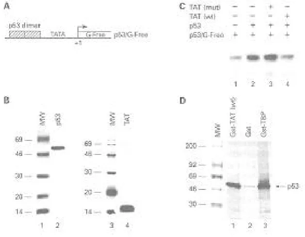
We initially asked if HIV-1 Tat was functionally capable of down-regulating a p53-activated tran-scription in vivo. To do this, we transfected a reporter construct, GP53CAT, either alone or with a p53 activator plasmid, Ga14-p53, which in turn activates transcription (Fig. 1, lanes 1 and 2). The induction by p53 was reproducible and an average of 8- to 10-fold induction was observed on four repeated experiments. However, when wild type Tat plasmid or Tat protein (data not shown) was transfected along with p53 protein, a dramatic drop in transcription was observed (lane 3). The repression was specific, since Tat mutated within the basic domain did not suppress the p53-activated transcription.
Since the in vivo transcription assays in Fig. 1 represents a functional transcriptional and post-transcriptional event, we wanted to determine if the suppression observed by Tat was a direct result of physical interaction between Tat and p53, hence affecting the transcription rate. We first reconsti-tuted a synthetic p53/G-free construct (Figure 2A) and performed in vitro transcription assays using semi-purified pol II factors. Transcription was as-sayed using a G-free cassette, where RNA po-lymerase would normally transcribe random size RNAs off of the template and RNase T1 would trim down all RNAs to only the G-free transcripts. Proteins used for reconstitution were RNA po-lymerase II (pol II, 2 µl), TFIID (1 µl), and TFIIA (3 µ1) from Hela fractions and recombinant TFIIE (100 ng) and TFIIB (10 ng)from E. coli. p53 and Tat were also purified to approximately 80 percent homogeneity as observed by silver stain gels (Fig.
Fig. 1. In vivo effect of p53 and Tat. A) Diagram of G5p53CAT which was synthesized by putting repeated Gal4 binding sites upstream of the p53 binding region. The construct contains a CAT reporter gene downstream of the TATA box, which can be activated by p53. B) CAT assays were performed using the reported plasmid with either Ga14-p53 alone or in combination with wild type Tat or mutant plasmids. Cells were electroporated and CAT assays were performed 18 hr later.
Fig. 2. In vitro transcription analysis of Tat and p53. A) Diagram of p53/G-free plasmid, containing two p53 binding sites inserted upstream of the chicken ovalbumin TATA box. B) Silver stain of 100 ng of either bacula virus wild type p53 (lane 2) or E. coli recombinant Tat (lane 4). Both lanes 1 and 3 represent 1 µg of rainbow 14C molecular weight marker (Amersham). C. In vitro transcription of p53/G-free with semi-purified pol II transcription factors (lane 1), p53 (lane 2), mutant Tat (lane 3), or wild type Tat (lane 4). Transcripts of 32P-labeled RNA were separated on a 8 M urea/4% PAGE and exposed overnight on Phosphorlmage cassette. D) Gst binding assays using35S-labeled p53 protein. Lane 1 represents binding of p53 to Gst-Tat wt protein. Lanes 2 and 3 represent negative (Gst) and positive (Gst-TBP) control binding assays. Products were resolved on a 4-20% SDS/PAGE gradient gel, enhanced in 300 ml of 1 M sodium salicylate, dried and exposed on a cassette.
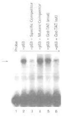
Fig. 3. Band shift assay using 32P-labeled p53 DNA responsive element. Lane 1, probe alone; lane 2, purified p53 binding to probe. Lanes 3 and 4 serve as positive (specific competitor) and negative (mutant competitor) controls for the binding assay. Lanes 5 and 6 indicate binding of p53 in the presence of either Tat mutant, or Gst-Tat wild type protein. Arrow indicates specific DNA/protein complex formation when using conditions described in Materials and Methods.
2B). A simple reconstitution assay using super-coiled p53/G-free construct yields a 362 bp labeled RNA when performing in vitro transcription assays (Fig. 2C, lane 1). A titration of various concentra-tions of p53 and Tat had previously shown that ap-proximately 50 ng of p53 and apap-proximately 100 ng of Tat proteins were sufficient for both activation and suppression respectively (data not shown). When p53 was added to the in vitro transcription system, there was a consistent 3- to 4-fold induction of labeled RNA product (Fig. 2C, lane 2). This ef-fect was abolished when purified Tat was added to the system as observed in lane 4. The observed suppression was specific to the basic domain of Tat, since a Tat mutant deficient in TAR RNA binding (basic domains 49-52) was not capable of down-regulating p53-activated transcription (Fig. 2C, lane 3). Similar results of activated transcription by p53 and suppression by Tat were obtained using Hela nuclear extracts (data not shown).
We next performed Gst binding assays to deter-mine if the down-regulation observed by Tat was the result of direct p53/Tat interaction. A Gst assay was set up using 500 ng of fusion proteins mixed with in
vitro 35S-labeled (Met and Cys) p53 protein. The
results of such an experiment are shown in Fig. 2D, where positive control TBP (TATA binding protein, one of the components of TFIID) bound efficiently (26 percent) to p53 (Fig. 2D, lane 3). The effect was specific since very little labeled p53 bound to the negative control, Gst protein (0.6 percent). However, the binding of p53 to Tat was observed (lane 1), where an average of 18 percent of labeled protein was precipitated. Taken together, these data suggest that Tat may down-regulate p53's function by direct physical interaction, having subsequent secondary effects on the p53 molecule.
Given the suggestion that the p53/Tat complex may down-regulate p53 transcriptional activity, we wished to examine if p53's DNA binding capacity changes once bound to Tat. Band shift experiments were designed where an end-labeled p53 oligonu-cleotide responsive element was used to detect complex formation with purified p53 protein (Fig. 3, lane 2). This complex was specifically competed out with a 100-fold excess amount of the same cold oligonucleotide (lane 3), but not with a mutant cold oligonucleotide (lane 4), indicating the specificity of the complex. Interestingly, when purified un-bound Gst-Tat (20 ng) was pre-incubated with p53 prior to band shift assay, a reduction in p53/DNA complex was observed (lane 6). Tat mutated in the basic domain however, did not compete out the complex to an appreciable degree (lane 5). Similar results were obtained when 20 ng of Gst protein was pre-incubated with p53 prior to complex formation (data not shown). Therefore, an interaction between p53 and Tat may physically hinder p53's DNA binding capacity, inactivating the functional role of p53 as transcriptional activator and guardian of cell cycle check point.
To investigate if the physical and functional in-hibition of p53 by Tat has any physiological rele-vance to endogenous genes in vivo, we investigated the role of p21, a cdc inhibitor, whose expression is activated by p53. The p21/WAF1/Cipl gene is the most well studied p53 response gene and its encoded protein forms part of a quaternary complex found in normal cells along with cyclin/CDKs and the DNA polymerase processivity factor PCNA. At high protein concentrations, p21/WAF1/Cipl inhibits the function of CDKs, particularly those that
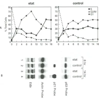
Fig. 4. Cell cycle analysis of Hela/eTat and control cells. A) Cells were blocked with Nocadozole (M phase blocker) for 16 hrs, washed and released using complete media. Samples were collected every 2 hrs and analyzed by cell sorting to determine the population ratios of each phase. The time points show the percent of cells at G1, S and G2/M. Two reproducible peaks of 8 hr and 14 hr were observed only in eTat line. Data points represent cells that had been fixed and stained with propidium iodide prior to cell sorting. B) Northern blot analysis of 8 and 14 hr samples using either p53, p21 or actin as probes. Bottom panel represents ethidium bromide staining of the RNA gel prior to the first hybridization with p21 probe. Two RNA bands correspond to 18 and 28S cellular RNA.
function during the GI phase of the cell cycle. In response to irradiation, p53-dependent G1 arrest is mediated, at least in part, through p53's induction of p21/WAF1/Cipl.
To investigate p21's role in the Tat-containing cell line, we constructed two Hela cell lines which were transfected and selected on hygromycin with either a pCep4 (containing EBNA sequence to maintain high copy number) or pCep4/epitope-tagged Tat plasmids. The first line (pCep4) served as a negative control and the second (pCep4/eTat)
served as the experimental cell line. Both cell lines have been described elsewhere (Kashanchi et al. 1997, submitted). In order to observe any physio-logical changes in these two lines related to cell cycle, we first blocked cells with a mitosis-(M phase) specific blocker, Nocodazole. Nocodazole is a reversible M phase blocker that specifically blocks microtubules, thereby not allowing separa-tion of daughter chromosomes. When cells were blocked with Nocodazole for 18 hours and later released using complete medium, an interesting
Fig. 5. Diagram of p53 binding proteins and proposed model of Tat/p53 inhibition of p21(Waf1). A) Proteins that have been reported to date to interact with p53 (modified from reference Ko and Prives [12]). CK: Casein Kinase; ds DNA PK: double stranded DNA protein kinase; JNK: Member of map kinase family; PKC: protein kinase C; TBP: TATA binding protein; MDM2: Mouse double minute 2; TAF: TBP-associated factor; AdE1B: Adenovirus E1B protein; TFIIH: transcription factor H (belonging to polymerase II category); SV40 Tag: Simian virus 40 T antigen; p53BP: p53 binding protein; XP: Xeroderma Pigmentosium; CSB: Cocayne syndrome complementation group B; TAT: HIV-1 transactivator Tat. B) Proposed model showing how Tat/p53 complex could inhibit p21 transcription, thereby modulating all cyclin-dependent kinases (CdK) and cyclin A, D, and E activities.
pattern of cell cycle progression was observed. The Tat cell line showed an increase of G1 population at 8 hours and increased S phase at 14 hours (Fig. 4A). Subsequent experiments showed that a reproducible G1 peak was present anywhere from 7 1/2 to 8 1/2 hours and an S phase peak was observed from 12 1/2 to 15 hours (data not shown). Therefore, Tat-containing cells have a sharp peak of both G1 and S phase at specific time points following M phase release. The control cells however, showed normal progression through G1 and S as expected.
We then asked if this change in the pattern of cell cycle could be due in part to the sequestration of functional p53. Total RNA was extracted from each of these time points and hybridized against specific probes. Fig. 4B shows the result of such a hybridi-zation experiment, where p53 RNA levels in the eTat line were approximately two-fold less than control cells. When assaying for p21 levels, it was observed that no p21 was expressed in eTat line, but normal expression was seen in control cells (Fig. 2B, p21 probe). Control actin probe showed no difference between any of the samples, indicat-ing that quality and quantity of RNAs loaded were equal. Taken together, these preliminary results
indicated, for the first time, that the p53/Tat com-plex inactivated p21 transcription and may explain the loss of p53 cell cycle check point in the eTat cell line.
DISCUSSION
Tat has been regarded as one of the most interest-ing viral transactivators among human retroviral proteins. Initially, because of Tats small size and not much being known about it (other than activation of viral promoter), Tat was regarded as a regulatory viral protein with limited function. However, data obtained in the past several years from a number of investigators have shed light on the diversity of this protein and its close relatives in other retroviral human, primate and bovine counterparts.
One such diverse finding has been the observation that Tat upregulates, albeit at a much lower rate, non-TAR- containing promoters. Most of these promoters reported to date fall into either viral promoters (CMV, JC and herpes viruses) or cytokine promoters (IL-2, IL-6, TNF-a and TNF-(3). The enhanced activity in cytokine promoters has
prompted investigators to pursue whether Tat could perturb homeostasis of the cell, thus permitting ac-cumulations of chromosomal aberrations and leading to AIDS- related malignancies.
One reported and interesting target of cellular de-regulation by Tat was the p53 protein. In this re-port, we attempted to observe if the Tat/p53 com-plex could indeed down regulate transcription. Re-sults from both in vivo and in vitro studies suggest that a physiological and functional consequence of this interaction is sequestration of the p53 mole-cule. It was of great surprise to us to observe that even though Tat binds to the C-terminal oligomeri-zation domain of p53 [14], it can influence the specific DNA binding domain of p53 located in the middle of this protein. It is not known at this time whether Tat binding to the oligomerization domain of p53 could inhibit tetramer formation of p53 needed for efficient DNA binding. However, pre-liminary results of glycerol gradient sedimentation assays have indicated that Tat can inhibit oli-gomerization of only phosphorylated p53 (F. Ka-shanchi, unpublished results), implying that inhibi-tion of oligomerizainhibi-tion may be one mechanism of Tat/p53 interaction.
The Tat/p53 interaction may have more functional consequence than investigated in this report. We have assayed for p21 activity and observed modulation of this crucial inhibitor of cell cycle kinase activity. With the transcriptional upregulation by p53, the p21 protein is able to inhibit the kinase activities of the G1 cyclin/cdk complexes cdk4/cyclin D and cdk2/cyclin E. The p53 to p21 pathway also inhibits DNA replication by merit of p21's interaction with PCNA. PCNA is involved in the processivity of the DNA polymerase δand also, as a cellular response to DNA damage to ultraviolet radiation, acts in nucleotide excision repair.
Once p53 is sequestered by Tat and down regula-tion of p53 responsive genes such as p21 is carried out, it may then be possible to deregulate the cell cycle pattern as observed in Fig. 4A. Although many possibilities exist for the observed irregular GI and S peak population, one such explanation could be the down-regulation of p21 as observed by northern blots in Fig. 4B. The functional conse-quence of p21 down-regulation is diagrammed in Fig. 5B, where p21 is capable of inhibiting at least three distinct classes of cdk/cyclin complexes. Since p53 upregulates p21 transcription, a Tat/p53 inhibitory complex could theoretically inactivate the
p21 promoter, hence resulting in upregulation of cdk/cyclin complexes. It is not clear at this stage whether p21 inhibition by the Tat/p53 complex is regulating HIV gene expression, or simply aiding in development of AIDS related malignancies. Future experiments on these two issues will undoubtedly shed light on the cell cycle deregulation by the Tat/p53 complex.
ACKNOWLEDGMENTS
The authors wish to acknowledge collaboration with A. Levine and John N. Brady's lab where all of the experiments presented in this manuscript were performed.
REFERENCES
1. Fauci, A.S., Pantaleo, G., Stanley, S., and Weissman, D. (1996) Immunopathogenic mechanisms of HIV infection. Ann. Intern. Med. 24: 654-663.
2. Ensoli, B., Gendelman, R., markham, P., Fiorelli, V., Colombini, S., Raffeld, M., Cafaro, A., Chang, H.K., Brady, J.N., and Gallo, R.C. (1994) Synergy between basic fibroblast growth factor and HIV-1 Tat protein in induction of Kaposi's sarcoma. Nature 371: 674-680. 3. Karp, J.E., Pluda, J.M., and Yarchoan, R. (1996)
AIDS-related Kaposi's sarcoma. A template for the translation of molecular pathogenesis into targeted therapeutic approaches. Hematol. Oncol. Clin. North. Am. 10: 1031-1049.
4. Zhou, C., Bahner, I.C., Larson, G.P., Zaia, J.A., Rossi J.J., and Kohn E.B. (1994) Inhibition of HIV-1 in human T-lymphocytes by retrovirally transduced anti-tat and rev hammerhead ribozymes. Gene 149: 33-39.
5. Keen, N.J., Gait, M.J., and Karn, J. (1996) Human immunodeficiency virus type-1 Tat is an integral component of the activated transcription-elongation complex. Proc. Natl. Acad. Sci. USA 9: 2505-2510.
6. Zhou, Q. and Sharp, P. (1995) Novel mechanism and factor for regulation by HIV-1 Tat. EMBO J. 14: 321-328. 7. Harhaj, E., Blaney, J., Millhouse, S ., and Sun, S . C.
(1996) Differential effects of I kappa B molecules on Tat-mediated transactivation of HIV-1 LTR. Virology 216: 284-287.
8. Cupelli, L.A., and Hsu, M.C. (1995) The human immunodeficiency virus type 1 tat antagonist, Ro 5-3335, predominantly inhibits transcription initia- tion from the viral promoter. J. Virol. 69: 2640-2643. 9. Bohan, C.A., Kashanchi, F., Ensoli, B., Buonaguro, L.,
Boris-Lawrie, K.A., and Brady, J.N. (1992) Analysis
of Tat transactivation of human immunodeficiency virus transcription in vitro. Gene Exp. 2: 391-407. 10. Kashanchi, F., Piras, G., Radonovich, M.F., Duvall,
J.F., Fattaey, A., Chiang, C.M., Roeder, R.G., and Brady, J.N. (1994) Direct interaction of human TFIID with the HIV-1 transactivator tat. Nature 367: 295-299.
11. Veschambre, P., Simard, P., and Jalinot, P. (1995) Evidence for functional interaction between the HIV-1 Tat transactivator and the TATA box binding protein in vivo. J. Mol. Biol. 250: 169-180.
12. Ko, L.J. and Prives, C. (1996) p53: puzzle and paradigm. Genes Dev. 10: 1054-1072.
13. Duan, L., Ozaki, I., Oakes, J.W., Taylor, J.P., Khalili, K., and Pomerantz, R.J. (1994) The tumor suppressor protein p53 strongly alters human immunodeficiency virus type 1 replication. J. Virol. 68: 4302-4313. 14. Longo, F., Marchetti, M.A., Castagnoli, L., Battaglia,
P.A., and Gigliani, F. (1995) A novel approach to protein-protein interaction: complex formation bertween the p53 tumor suppressor and the HIV Tat proteins. Biochem. Biophys. Res. Commun. 206: 326-334.
15. Li, C.J., Wang, C., Friedman, D.J., and Pardee, A.B. (1995) Reciprocal modulations between p53 and Tat of human immunodeficiency virus type 1. Proc. Natl. Acad. Sci. USA 92: 5461-5464.
16. Kashanchi, F., Duvall, J.F., and Brady, J.N. (1992) Electroporation of viral transactivator proteins into lymphocyte suspension cells. Nucleic Acids Res. 20: 4673-4674.
17. Gorman, C.M., Moffat, L.F., and Howard, B.H. (1982) Recombinant genomes which express chlo-ramphenicol acetytransferase in mammalian cells. Mol. Cell. Biol. 2: 1044-1051.
18. Shapiro, D.J., Sharp, P.A., Wahli, W.W., and Keller, M.J. (1988) A high efficiency HeLa cell nuclear transcription extract. DNA 7: 47-55.
19. Newcomb, E.W. (1995) P53 gene mutations in lym-phoid diseases and their possible relevance to drug resistance. Leuk. Lymphoma 17: 211-221.
20. Yamato, K., Oka, T., Hiroi, M., Iwahara, Y., Sugito, S., Tsuchida, N., and Miyoshi, I. (1993) Aberrant
expression of the p53 tumor suppressor gene in adult T-cell leukemia and HTLV-Iinfected cells. Jpn. J. Cancer Res. 84: 4-8.
21. Duvall, J.F., Kashanchi, F., Cvekl, A., Radonovich, M.F., Piras, G., and Brady, J.N. (1995) Transactiva-tion of the human T-cell lymphotropic virus type 1 Taxl-responsive 21-base-pair repeat requires Holo-TFIID and TFIIA. J. Virol. 69: 5077-5086.
22. Flores, 0., Ha, I., and Reinberg, D. (1990) Factors involved in specific transcription by mammalian RNA polymerase II. J. Biol. Chem. 265: 5629-5634.
23. Lu, H., Flores, 0., Weinmann, R., and Reinberg, D. (1991) The non-phosphorylated form of RNA po-lymerase II preferentially associates with the preini-tiation complex. Proc. Natl. Acad. Sci. USA 88: 10004-10008.
24. Lin, J, Chen, J., Elenbaas, B., and Levine, A.J. (1994) Several hydrophobic amino acids in the p53 amino-terminal domain are required for transcrip-tional activation, binding to mdm-2 and the adenovirus 5 E1B 55-1(D protein. Genes Dev. 8: 12351246.
25. Kashanchi, F., Khleif, S.N., Duvall, J.F., Sadaie, M.R., Radonovich, M.F., Cho, M., Martin, M.A., Chen, S.Y., Weinmann, R., and Brady, J.N. (1996) Interaction of human immunodeficiency virus type 1 Tat with a unique site of TFIID inhibits negative co-factor Drl and stabilizes the TFIID-TFIIA complex. J. Virol. 70: 5503-5510.
26. Kashanchi, F., Sadaie, M.R., and Brady, J.N. (1997) Inhibition of HIV-1 transcription and virus replication using soluble Tat peptide analogs. Virology 227: 431-438.
27. Chang, H., Benchimol, S., Minden, M.D., and Mess-ner, H.A. (1994) Alterations of p53 and c-myc in the clonal evolution of malignant lymphoma. Blood 83: 452-459.28.
28. Huppi, K., Siwarski, D., Dosik, J., Michieli, P., Chedid, M., Reed, S., Mock, B., Govol, D., and Mushinski, J.F. (1994) Molecular cloning, sequenc-ing, chromosomal localization and expression of mouse p21 (Waft) . Oncogene 9: 3017-3020.