0022-538X/07/$08.00⫹0 doi:10.1128/JVI.00027-07
Copyright © 2007, American Society for Microbiology. All Rights Reserved.
Defect of Human Immunodeficiency Virus Type 2 Gag Assembly in
Saccharomyces cerevisiae
䌤
Yuko Morikawa,
1* Toshiyuki Goto,
2Daisuke Yasuoka,
1Fumitaka Momose,
1and Tetsuro Matano
3Kitasato Institute for Life Sciences and Graduate School for Infection Control, Kitasato University, Shirokane 5-9-1, Minato-ku, Tokyo 108-8641,1School of Health Science, Faculty of Medicine, Kyoto University, Kawahara-cho 53, Shogoin, Sakyo-ku,
Kyoto 606-8507,2and Graduate School of Medicine, The University of Tokyo,
Hongo 7-3-1, Bunkyo-ku, Tokyo 113-0033,3Japan
Received 5 January 2007/Accepted 25 June 2007
We have previously shown that the expression of human immunodeficiency virus type 1 (HIV-1) Gag protein inSaccharomyces cerevisiaespheroplasts produces Gag virus-like particles (VLPs) at the plasma membrane, indicating that yeast has all the host factors necessary for HIV-1 Gag assembly. Here we expand the study by using diverse primate lentiviral Gags and show that yeast does not support the production of HIV-2 or simian immunodeficiency virus SIVmac Gag VLPs but allows the production of SIVagm and SIVmnd Gag VLPs. Particle budding was observed at the surfaces of cells expressing SIVagm and SIVmnd Gags, but cells expressing HIV-2 and SIVmac Gags showed only membrane-ruffling structures, although they were accompa-nied with electron-dense submembrane layers, suggesting arrest at an early stage of particle budding. Com-parison of HIV-1 and HIV-2 Gag expression revealed broadly equivalent levels of intracellular Gag expression and Gag N-terminal myristoylation in yeast. Both Gags showed the same membrane-binding ability and were incorporated into lipid raft fractions at a physiological concentration of salt. HIV-2 Gag, however, failed to form a high-order multimer and easily dissociated from the membrane, phenomena which were not observed in higher eukaryotic cells. A series of chimeric Gags between HIV-1 and HIV-2 and Gag mutants with amino acid substitutions revealed that a defined region in helix 2 of HIV-2 MA (located on the membrane-binding surface of MA) affects higher-order Gag assembly and particle production in yeast. Together, these data suggest that yeast may lack a host factor(s) for HIV-2 and SIVmac Gag assembly.
The major structural component of retroviruses is encoded by thegaggene, and Gag is the sole protein required for viral particle assembly. Three discrete Gag regions responsible for virus particle assembly have been identified and termed the membrane-binding (M), interacting (I), and late (L) domains. The M domain is located at the N-terminal matrix/membrane (MA) of Gag and contains a membrane-binding signal which directs the association of Gag with the membrane. The signal is largely composed of N-terminal myristoylation of MA in many mammalian retroviruses, including human immunodefi-ciency virus (HIV), and this modification is necessary for Gag targeting and subsequent binding to the plasma membrane (4, 14, 15). The I domain is essential for Gag-Gag interactions and spans from the central capsid (CA) to the nucleocapsid (NC) of Gag (7, 11, 24, 39). The L domain, responsible for pinching off viral particles from the membrane, is located at either the C-terminal domain of Gag or the MA-CA junction (16, 37).
Because Gag is sufficient for retroviral particle budding, many studies on particle assembly have used Gag expression and shown that expression of the Gag protein alone in higher eukaryotic cells produces a Gag virus-like particle (VLP) mor-phologically identical to the immature form of retroviral par-ticles (14, 19, 44). The fact that Gag self-assembles into a viral
particle suggests that Gag assembly is attributable to the in-trinsic properties of Gag. This view is supported by in vitro studies in which purified Gag protein assembled into a spher-ical particle, analogous to a Gag VLP, in a test tube (5, 6, 22, 27). However, a number of recent studies clearly show that the Gag assembly process involves many host factors, some of which are indispensable for particle budding. These include endosomal sorting molecules, such as TSG101, Nedd4, AIP-1/ ALIX, and AP-3 (9, 12, 46, 52, 53). Such host factors and protein sorting pathways appear to be commonly used machin-ery for intracellular trafficking of diverse retroviral Gags (21, 53). ABCE1/HP68 has also been identified as a host factor that supports multimerization of all primate lentiviral Gags (10, 56). In contrast, the host factors identified as host restriction factors, such as cyclophilin A and TRIM-5␣, appear to be Gag type specific, although they are not involved in particle assem-bly but in uncoating and initiation of reverse transcription (2, 3, 20, 47, 50).
Recent studies on reverse genetics use small interfering RNAs, which specifically silence the expression of their corre-sponding genes. This new technology has made it possible to deplete a host factor of interest in mammalian cells. The study of genetics in eukaryotes has long been carried out with Sac-charomyces cerevisiae, because yeast has the ability to replace the wild-type chromosomal copy of a gene with a mutant or deletion derivative, a property which is not available in other eukaryotic cells. Accordingly, many genetic mutants have been isolated in yeast and made available for the study of cellular factors and machinery. We previously developed a Gag VLP budding system with Saccharomyces cerevisiae in which the
* Corresponding author. Mailing address: Kitasato Institute for Life Sciences and Graduate School for Infection Control, Kitasato Univer-sity, Shirokane 5-9-1, Minato-ku, Tokyo 108-8641, Japan. Phone: 81-3-5791-6129. Fax: 81-3-5791-6268. E-mail: [email protected] .ac.jp.
䌤Published ahead of print on 3 July 2007.
9911
on November 8, 2019 by guest
http://jvi.asm.org/
HIV type 1 (HIV-1) Gag protein simultaneously budded Gag VLPs from the plasma membrane, and we have suggested that a combination of this method and yeast genetics may be a powerful tool for the study of the host factors required for particle production (42). Here we expand this study by using diverse primate lentiviral Gags and show that yeast does not support the production of HIV-2 or simian immunodeficiency virus SIVmac Gag VLPs. Our data suggest that yeast may lack a host factor(s) required for tight membrane binding of HIV-2 Gag to facilitate higher-order assembly.
MATERIALS AND METHODS
Construction and expression of diverse primate lentivirusgaggenes.For
expression in yeast, the full-lengthgaggenes of HIV-1 (HXB2 strain), HIV-2
(ROD strain), SIVmac (mac239 strain), SIVagm (TY01 strain), and SIVmnd (GB1 strain) were amplified by PCRs using relevant forward and reverse
prim-ers. For the Gag-Flag fusion protein, thegaggene (truncated just before the
termination codon) was amplified by PCR using a reverse primer containing a Flag epitope tag sequence. DNA construction of chimeric Gags between HIV-1 and HIV-2 and of Gag mutants containing amino acid substitutions was also carried out by PCRs using the relevant forward and reverse primers. The PCR fragments were cloned into the yeast expression vector pKT10 (48), which is a
2m plasmid containing theURA3gene as a selective marker and the
constitu-tive promoter for the yeast glyceraldehyde-3-phosphate dehydrogenase gene. TheS. cerevisiaestrain RAY3A-D (MATa/␣ura3/ura3 his3/his3 leu2/leu2 trp1/
trp1) (40) was transformed with the yeast expression plasmids.
For expression in higher eukaryotic cells, thegaggenes of HIV-1 and HIV-2
were modified C-terminally with a Flag epitope tag and cloned into the higher eukaryotic expression vector pCAGGS (30), which contains the promoter for the
actin gene. The codon usage of the HIV-1gaggene was optimized. HeLa and
293T cells were transfected with the expression plasmids by using Lipofectamine 2000 (Invitrogen).
Preparation of yeast spheroplasts and subcellular fractionation.The proce-dure for yeast spheroplast formation was described previously (39). In brief, yeast transformants were grown at 30°C in synthetic defined medium without uracil (0.67% yeast nitrogen base, 2% glucose, and amino acid mixtures without uracil).
Yeast cells were suspended in wash buffer (50 mM Tris [pH 7.5], 5 mM MgCl2,
and 1 M sorbitol) containing 30 mM dithiothreitol (DTT) and incubated at 30°C for 20 min with gentle shaking. The cells were resuspended in wash buffer containing 3 mM DTT and 0.4 mg/ml Zymolyase and incubated at 30°C for 20 min with gentle shaking for digestion of the cell wall. Following digestion, the cells were washed with 1 M sorbitol.
Subcellular fractionation of yeast cells was performed by a standard procedure (13). Yeast spheroplasts (10 optical density [OD] units) were resuspended in buffer (50 mM Tris [pH 8.0], 1 mM EDTA, 1 mM DTT, 1 mM
phenylmethyl-sulfonyl fluoride, and 1g/ml pepstatin A), with 150 mM NaCl or without salt,
and homogenized with 15 strokes in a homogenizer. Following clarification at
500⫻gfor 5 min at 4°C, the cell lysates (whole-cell lysates) were subjected to
centrifugation at 13,000⫻gfor 10 min at 4°C. The precipitates were stored as
P13 fractions. The supernatants were centrifuged in a TLA100 rotor (Beckman
Coulter) at 100,000⫻gfor 1 h at 4°C, and the precipitates (P100) and
super-natants (S100) were separated.
Sedimentation analysis.Whole-cell lysates and subcellular fractions were ap-plied to 20 to 70% (wt/vol) sucrose gradients in phosphate-buffered saline (PBS)
and sedimented in an SW55 rotor at 120,000⫻gfor 2 h at 4°C, as described
previously (28). Fractions of the gradients were collected and subjected to so-dium dodecyl sulfate-polyacrylamide gel electrophoresis (SDS-PAGE) followed by Western blotting. The 80S ribosome and the immature form of HIV Gag VLPs purified from Gag-expressing HeLa cells were used as molecular weight markers for sedimentation analysis.
Membrane and lipid raft flotation centrifugation.Equilibrium flotation cen-trifugation with membranes was performed as described previously (32, 36), with minor modifications. The formation of yeast spheroplasts was carried out as described above. Yeast spheroplasts (10 OD units) were resuspended in buffer A (50 mM Tris [pH 8.0], 1 mM EDTA, 1 mM DTT, 1 mM phenylmethylsulfonyl
fluoride, and 1g/ml pepstatin A) containing 150 mM NaCl. Following a brief
sonication, the cell lysates were clarified at 500⫻g for 5 min at 4°C. The
supernatants were adjusted to 70% (wt/vol) sucrose in PBS, layered at the bottom of 70%-65%-10% (wt/vol) sucrose step gradients in PBS, and subjected to equilibrium flotation centrifugation. Centrifugation was performed in an
SW55 rotor (Beckman Coulter) at 4°C at 120,000 ⫻g overnight. In some
experiments, cells were resuspended in buffer A with 500 mM NaCl or without salt. For lipid raft flotation, the cell lysates, after sonication, were treated on ice with 0.5% Triton X-100 for 10 min. Following clarification, the supernatants were subjected to equilibrium flotation centrifugation. Fractions of the gradients were collected and subjected to SDS-PAGE followed by Western blotting. Mem-branes of higher eukaryotic cells were analyzed similarly by equilibrium flotation centrifugation.
Purification of Gag VLPs.Purification of yeast-produced Gag VLPs was car-ried out as described previously (42). Briefly, the culture medium of yeast spheroplasts was clarified and then centrifuged through 30% (wt/vol) sucrose
cushions in an SW28 rotor (Beckman Coulter) at 120,000⫻gfor 1.5 h at 4°C.
The VLP pellets were resuspended and centrifuged in 20 to 70% (wt/vol) sucrose
gradients in PBS in an SW55 rotor (Beckman Coulter) at 120,000⫻governight
at 4°C. Purification of Gag VLPs produced by higher eukaryotic cells was carried out by standard procedures.
Protein detection.Following SDS-PAGE, gels were subjected to either Coo-massie brilliant blue (CBB) staining or Western blotting using an anti-HIV-1, anti-HIV-2, or anti-SIVmac CA mouse monoclonal antibody (Advanced Bio-technologies) or anti-SIVagm monkey serum. For the Gag-Flag fusion protein, Western blotting was carried out using an anti-Flag mouse monoclonal antibody (Sigma). In subcellular fractionation experiments, anti-Pep12 (for endosomes), anti-alkaline phosphatase (for vacuoles), and anti-phosphoglycerate kinase (for cytosol) mouse monoclonal antibodies (Molecular Probes) were used as
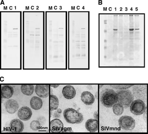
or-FIG. 1. Intracellular expression of diverse lentiviral Gags and pro-duction of Gag VLPs in yeast. Yeast cells were transformed with a pKT10 vector containing the full-lengthgag gene of HIV-1, HIV-2, SIVmac, SIVagm, or SIVmnd. (A) Intracellular Gag expression. Cells (0.5 OD unit) were subjected to SDS-PAGE followed by Western blotting using anti-HIV-1, anti-HIV-2, or anti-SIVmac CA antibody or anti-SIVagm monkey serum. Lanes: M, prestained molecular weight markers; C, cells transformed with the parental vector; 1 to 4, cells transformed with the vector containing thegaggenes of HIV-1, HIV-2, SIVmac, and SIVagm, respectively. (B) Gag VLP production. Follow-ing removal of the cell wall, spheroplasts (200 OD units) were cultured in yeast extract-peptone-dextrose medium containing 1 M sorbitol overnight. Gag VLPs were purified from the culture medium by cen-trifugation on 20 to 70% sucrose gradients and analyzed by SDS-PAGE followed by CBB staining. Lanes: M, prestained molecular weight markers; C, mock fractions prepared from the culture medium of yeast spheroplasts transformed with the parental vector; 1 to 5, Gag VLP fractions purified from culture medium of yeast spheroplasts expressing HIV-1, HIV-2, SIVmac, SIVagm, and SIVmnd Gags, re-spectively. (C) Electron micrographs of Gag VLPs. Purified Gag VLP fractions were subjected to electron microscopic analysis. All micro-graphs are shown at the same magnification. Bar⫽100 nm.
on November 8, 2019 by guest
http://jvi.asm.org/
[image:2.585.301.541.70.287.2]ganelle markers. For the plasma membrane, yeast spheroplasts were incubated with cholera toxin subunit B (CTB), which binds to lipid rafts of the plasma membrane, at 4°C. Following a wash with 1 M sorbitol, subcellular fractionation was carried out as described above. The fractions were subjected to Western blotting using anti-CTB rabbit antibody (Molecular Probes).
For protein myristoylation, yeast cells were metabolically labeled with 500Ci
of [9,10(n)-3H]myristic acid at 30°C for 30 min. Following SDS-PAGE, gels were
subjected to fluorography.
Immunofluorescence staining.Yeast cells were fixed in 3.7% formalin in yeast extract-peptone-dextrose medium at 30°C for 30 min. Following removal of the cell wall, spheroplasts were treated with 0.1% Triton X-100 at room temperature for 5 min for membrane permeabilization. The cells were incubated first with an anti-HIV-1, anti-HIV-2, or anti-SIVmac CA mouse monoclonal antibody (Ad-vanced Biotechnologies) or anti-SIVagm monkey serum and subsequently with mouse immunoglobulin G–Alexa Fluor 488 (Molecular Probes) or anti-monkey immunoglobulin G–fluorescein isothiocyanate. For the Gag-Flag fusion protein, cells were incubated with an anti-Flag mouse monoclonal antibody (Sigma). For the plasma membrane, yeast spheroplasts were first incubated with CTB at 4°C (to label lipid rafts of the plasma membrane but not allow endocy-tosis) and subsequently with anti-CTB rabbit antibody (Vyrant lipid raft labeling kit; Molecular Probes). After fixation with 3.7% formalin, the spheroplasts were permeabilized with 0.1% Triton X-100 and costained with anti-Flag antibody for Gag-Flag.
Electron microscopy.Electron microscopy was carried out by standard proce-dures. Briefly, yeast spheroplasts were fixed in 2% glutaraldehyde in 50 mM cacodylate buffer (pH 7.2) for 2 h and postfixed with 1% osmium tetroxide for 1 h. Cell pellets were embedded in epoxy resin. Ultrathin sections were stained with uranyl acetate and lead citrate and examined with an electron microscope.
RESULTS
Yeast does not support HIV-2 or SIVmac Gag VLP produc-tion.The initial goal of this study was to examine whether Gag proteins of diverse primate lentiviruses produce Gag VLPs
from yeast spheroplasts, as does HIV-1 Gag (42). Primate lentiviruses are classified into the following five equidistant phylogenetic lineages: (i) HIV-1/SIVcpz, (ii) HIV-2/SIVmac/ SIVsm, (iii) SIVagm, (iv) SIVmnd, and (v) SIVsyk (17). We used thegaggenes from four different primate lentivirus lin-eages. Yeast cells were transformed with the yeast expression vector pKT10 containing thegaggene of HIV-1, HIV-2, SIV-mac, SIVagm, or SIVmnd and were grown in synthetic defined medium without uracil. Western blotting using anti-HIV-1, anti-HIV-2, and anti-SIVmac CA antibodies and anti-SIVagm monkey serum revealed individual Gag proteins in the express-ing cells but not in the cells transformed with a parental vector (Fig. 1A). We did not test the cells expressing SIVmnd Gag because an anti-SIVmnd antibody was not available.
[image:3.585.134.451.68.328.2]Following removal of the cell wall, yeast spheroplasts were maintained under isotonic conditions overnight. For purifica-tion of Gag VLPs, the culture medium of the spheroplasts was subjected to centrifugation through a sucrose gradient and subsequent fractionation, as described previously (42). When equivalent volumes of the Gag VLP fractions were subjected to SDS-PAGE followed by CBB staining, Gag VLP production was observed for HIV-1, SIVagm, and SIVmnd Gags, although the yields of produced VLPs varied. In contrast, no Gag VLP production was observed for HIV-2 or SIVmac Gag (Fig. 1B). Western blotting using HIV-2 and SIVmac CA anti-bodies also failed to demonstrate Gag VLP production (data not shown). These findings were not specific to the yeast ex-pression vectors or yeast backgrounds used (data not shown).
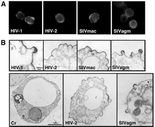
FIG. 2. Immunofluorescence staining and electron microscopy of yeast cells expressing diverse lentiviral Gags. (A) Immunofluorescence detection of Gag antigens. After fixation with 3.7% formalin, the cell wall was removed with Zymolyase and the membrane was permeabilized with 0.1% Triton X-100. Gag antigens were detected using anti-HIV-1, anti-HIV-2, or anti-SIVmac CA antibody or anti-SIVagm monkey serum. (B) Electron micrographs of yeast spheroplasts expressing each of the Gags. Micrographs in upper panels are shown at the same magnification (bar⫽100 nm). Micrographs in lower panels show whole yeast cells taken at the same magnification (bar⫽1m). Cr, yeast cell transformed with the parental vector.
on November 8, 2019 by guest
http://jvi.asm.org/
Electron microscopic analysis confirmed that the produced SIVagm and SIVmnd Gag VLPs were nearly spherical, with electron-dense submembrane layers, similar to HIV-1 Gag VLPs prepared from yeast in parallel (Fig. 1C). The electron-dense submembrane layers, however, were often crescent-shaped or composed of multidomains.
Expression of HIV-2 and SIVmac Gags in yeast causes plasma membrane ruffling but no particle budding.We car-ried out an immunofluorescence study and examined the in-tracellular localization of each Gag protein. Microscopy re-vealed that all Gags tested (HIV-1, HIV-2, SIVmac, and SIVagm Gags) were localized predominantly in proximity to the plasma membrane (Fig. 2A). The data suggest that HIV-2 and SIVmac Gags are capable of targeting the plasma mem-brane in yeast, as are HIV-1 and SIVagm Gags. These findings were later confirmed by experiments in which Gags were costained with lipid rafts of the plasma membrane (Fig. 3C).
Electron microscopic analysis was carried out to examine whether Gag VLPs budded from the cell surfaces of the spheroplasts (Fig. 2B). Consistent with the results of our pre-vious study (42), the spheroplasts expressing HIV-1 Gag showed half-spherical budding structures with electron-dense submembrane layers on the plasma membrane. A similar mor-phology was observed for cells expressing SIVagm Gag. In contrast, the spheroplasts expressing HIV-2 and SIVmac Gags revealed plasma membrane ruffling but no budding particle with a pinch or a thin stalk (Fig. 2B, top panels). The ruffling membrane, especially the area with outward curvature, had an electron-dense submembrane layer, suggesting that Gag pro-teins were gathered and budded but soon arrested at a very early stage of particle budding (Fig. 2B, top and lower middle panels). A yeast spheroplast characteristic of HIV-2 Gag ex-pression is shown in Fig. 2B (lower middle panel). Nearly all the cells observed had plasma membrane ruffling that, in many cases, extended to a broad area of the plasma membrane. This finding was not observed for cells transformed with a parental vector (Fig. 2B, lower left panel) or for cells expressing SIVagm Gag (Fig. 2B, lower right panel).
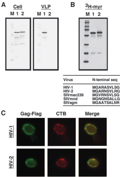
Expression, N-terminal myristoylation, and plasma mem-brane targeting of HIV-2 Gag are comparable to those of HIV-1 Gag in yeast.Since HIV-2 and SIVmac fall into the same primate lentivirus lineage, HIV-2 was chosen and com-pared with HIV-1. We noticed that the sensitivity of the anti-HIV-2 CA antibody used was much lower than that of the anti-HIV-1 CA antibody (data not shown). For normalization, both Gag proteins were modified by adding a Flag epitope tag at the C terminus and were detected by an anti-Flag antibody. Expression of the Gags in yeast and VLP production were carried out as described above. Western blotting using anti-Flag antibody confirmed that no Gag VLP production was observed for HIV-2 Gag, despite the nearly equivalent levels of Gag expression in the cells (Fig. 3A). These data indicate that yeast essentially does not support HIV-2 Gag VLP production. Because the failure was not due to a low level of HIV-2 Gag expression in yeast, we next examined the levels of Gag N-terminal myristoylation. Yeast cells were metabolically labeled with [9,10(n)-3H]myristic acid and subjected to SDS-PAGE
followed by fluorography. In both cases, one major radiola-beled band was detected at a gel position identical to that of the band detected by Western blotting probed with anti-CA
antibody. The data clearly showed that the efficiencies of3H
incorporation were comparable between the two Gags, indi-cating that both Gags were equally myristoylated in yeast (Fig. 3B). A protein myristoylation signal lies on the eight N-termi-nal amino acid residues (51), and the N-termiN-termi-nal amino acid sequences are well conserved between HIV-1 and HIV-2 Gags (Fig. 3B).
[image:4.585.317.523.67.370.2]We observed that both Gags similarly localized in proximity to the plasma membrane (Fig. 2A). To confirm the plasma membrane targeting, lipid rafts of the yeast plasma membrane were probed with CTB and Gag-Flag was costained with anti-Flag antibody, as recent studies have identified the presence of lipid rafts in the yeast membrane (1). Microscopy revealed that HIV-1 Gag-Flag localized on the cell periphery, with partial colocalization with lipid rafts on the plasma membrane. Similar
FIG. 3. N-terminal myristoylation, plasma membrane targeting, and VLP production of HIV-1 and HIV-2 Gags. Yeast cells were transformed with a pKT10 vector containing the HIV-1 or HIV-2gag
gene with a Flag epitope sequence added at the C terminus. (A) In-tracellular Gag expression and Gag VLP production. Preparation of samples was carried out as described in the legend for Fig. 1. SDS-PAGE was followed by Western blotting using anti-Flag antibody. Lanes: M, prestained molecular weight markers; 1 and 2, expression of HIV-1 and HIV-2 Gag-Flag, respectively. (B) N-terminal myristoyl-ation of Gag. HIV-1 and HIV-2 Gag-Flag proteins were metabolically labeled with [3H]myristic acid in yeast and subjected to SDS-PAGE. Lanes: M,14C-labeled molecular weight markers; 1 and 2, cells ex-pressing HIV-1 and HIV-2 Gag-Flag, respectively. (C) Intracellular distribution of Gag. Yeast spheroplasts expressing HIV-1 and HIV-2 Gag-Flag were incubated with CTB and then anti-CTB antibody at 4°C (not to allow endocytosis) (shown in red). After fixation with 3.7% formalin, the spheroplasts were permeabilized with 0.1% Triton X-100 and costained with anti-Flag antibody (shown in green).
on November 8, 2019 by guest
http://jvi.asm.org/
findings were observed for HIV-2 Gag-Flag, indicating that both Gags were capable of being targeted to the plasma mem-brane in yeast (Fig. 3C).
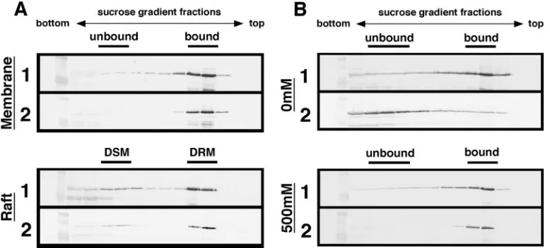
HIV-2 Gag dissociates from yeast membrane in the absence of salt. To test the membrane-binding ability of Gag, mem-brane flotation experiments were carried out using sucrose step gradients. The initial analysis was performed at a physio-logical concentration of salt (150 mM NaCl). When cells ex-pressing HIV-1 Gag-Flag were analyzed, Gag was detected in the interface fractions between the 10% and 65% sucrose layers. A similar flotation profile was observed for HIV-2 Gag-Flag, indicating that both Gags efficiently bound to the yeast membrane (Fig. 4A, top panels). It has been shown that lipid rafts are membrane microdomains that are insoluble by non-ionic detergents and function as a platform for particle assem-bly and budding (29, 33). A similar detergent insensitivity has been reported for lipid rafts of yeast (1). Thus, Gag association with yeast lipid rafts was examined by similar equilibrium flo-tation centrifugation, but after a 0.5% Triton X-100 treatment on ice. When cells expressing HIV-1 Gag-Flag were analyzed, the majority of Gag was distributed in the detergent-resistant membrane fractions, suggesting that a relatively large popula-tion of the membrane-bound Gag was incorporated into the raft fractions. Very similar findings were observed for HIV-2 Gag-Flag (Fig. 4A, bottom panels). These data indicate that the two Gags show equal membrane-binding abilities and in-corporation into lipid raft fractions in yeast.
However, when the sample preparation was carried out in the absence of salt and subjected to membrane flotation anal-ysis, a striking difference was observed between the behaviors of HIV-1 and HIV-2 Gags: HIV-1 Gag displayed membrane binding, but HIV-2 Gag did not (Fig. 4B, top panels). In contrast, no differences were observed when the samples were prepared under high-salt conditions (Fig. 4B, bottom panels).
These results suggest that although both Gags bind efficiently to the yeast membrane, HIV-2 Gag more readily dissociates in the absence of salt than does HIV-1 Gag.
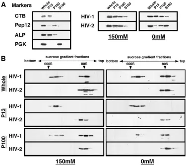
HIV-2 Gag fails to form high-order multimers in yeast.The Gag distribution in yeast was also examined by subcellular fractionation experiments. A differential sedimentation proce-dure (13) yielded P13, P100, and S100 fractions, and the frac-tions were probed for organelle markers by Western blotting. Consistent with previous reports (13), alkaline phosphatase (for vacuoles) was found in the P13 fraction, while Pep12 (for endosomes) was found predominantly in the P100 fraction. Phosphoglycerate kinase is an abundant cytosolic protein and therefore was localized to the S100 fraction. The CTB bound to lipid rafts of the spheroplast surfaces was found in the P13 fraction (Fig. 5A, left panel). When fractions were prepared at a physiological concentration of salt (150 mM NaCl) and sub-jected to Western blotting, for both Gags the majority of Gag was observed in the P13 and P100 fractions, indicating that HIV-2 Gag similarly associated with the yeast membrane. However, when the fractionation was carried out in the ab-sence of salt, the Gag distributions differed. The HIV-2 Gag shifted predominantly to the S100 fraction, while in contrast, HIV-1 Gag was detected in the same fractions as those ob-served in the presence of salt, confirming that HIV-2 Gag easily dissociated from the yeast membrane in the presence of salt (Fig. 5A, right panel).
To further understand these phenomena, the subcellular fractions were subjected to sedimentation analysis on 20 to 70% sucrose gradients and Gag antigens spread within the gradients were detected by Western blotting (Fig. 5B). The initial analysis was performed using the samples prepared in the absence of salt (Fig. 5B, right panels). When the whole-cell lysate from yeast expressing HIV-1 Gag was analyzed, Gag antigens were found in the 25 to 30% and 50% sucrose
frac-FIG. 4. Membrane and lipid raft associations of HIV-1 and HIV-2 Gags in yeast. Protein expression in yeast was carried out as described in the legend for Fig. 3. Panels: 1, cells expressing HIV-1 Gag-Flag; 2, cells expressing HIV-2 Gag-Flag. (A) Membrane and lipid raft associations of Gags at a physiological concentration of salt. Following formation of spheroplasts, cells (10 OD units) were resuspended in buffer with 150 mM NaCl and disrupted by sonication. For analysis of lipid raft association, the cell lysate was treated on ice with 0.5% Triton X-100 for 10 min. The cell lysate was clarified by low-speed centrifugation and subjected to equilibrium flotation centrifugation using a 70%-65%-10% (wt/vol) sucrose step gradient. The gradient fractions were collected from the bottom to the top (left to right) and analyzed by Western blotting using anti-Flag antibody. DSM, detergent-sensitive membrane; DRM, detergent-resistant membrane. (B) Membrane affinity of Gag in the absence of salt or under high-salt conditions. Spheroplasts (10 OD units) were resuspended in buffer, with or without 500 mM NaCl, and disrupted by sonication. After clarification by brief centrifugation, the cell lysate was subjected to equilibrium flotation centrifugation as described above.
on November 8, 2019 by guest
http://jvi.asm.org/
[image:5.585.99.488.69.245.2]tions, likely corresponding to relatively small- and large-mo-lecular-weight complexes, respectively. In contrast, sedimenta-tion analysis using the whole-cell lysate from yeast expressing HIV-2 Gag revealed the presence of a Gag complex in the 25 to 30% sucrose fractions, but no other larger classes of Gag complex were seen. For more physiological conditions, we pre-pared the whole-cell lysate in the presence of 150 mM NaCl and carried out sedimentation analysis (Fig. 5B, left panels). The sedimentation profiles were essentially similar to those of the samples prepared in the absence of salt. These data indi-cate that HIV-2 Gag does not form high-order Gag multimers in yeast, although it cannot be ruled out that sedimentation analysis through 20 to 70% sucrose gradients might lead to dissociation of high-order assembly of HIV-2 Gag. The P13 and P100 fractions were also subjected to sedimentation anal-ysis. In the case of HIV-1 Gag, the P13 fractions included predominantly the higher-order Gag multimers, while the P100 fractions included both high- and low-order Gag multimers, suggesting, though not proving, higher-order Gag assembly at the plasma membrane rather than at the endosome fractions.
As expected, no high-order HIV-2 Gag multimers were de-tected in the P13 or P100 fraction.
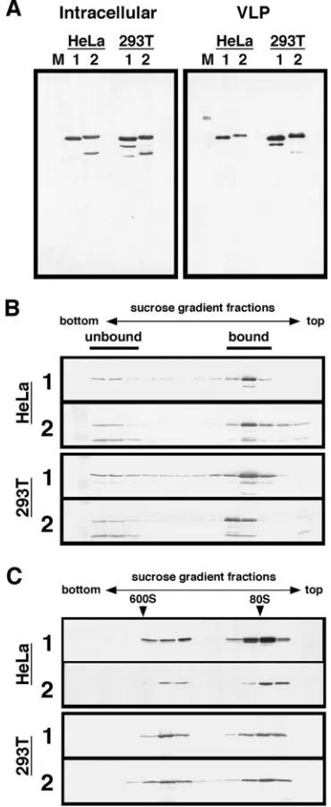
[image:6.585.104.479.68.400.2]Both HIV-1 and HIV-2 Gags efficiently associate with the mammalian cell membrane and form high-order multimers. For comparison, we expressed HIV-1 and HIV-2 Gag-Flag in higher eukaryotic cells, such as HeLa and 293T cells. Intracel-lular expression and particle production were examined by Western blotting using anti-Flag antibody. The expression lev-els in the cells were broadly comparable, and the production of HIV-2 particles was not impaired in either HeLa or 293T cells (Fig. 6A). Consistent with these results, when the membrane-binding affinities of Gags in the absence of salt were analyzed by membrane flotation centrifugation, the majority of Gag was found in membrane-bound fractions for both Gag types (Fig. 6B). Furthermore, when the whole-cell lysate was prepared in the absence of salt and subjected to sedimentation analysis on 20 to 70% sucrose gradients, high-order Gag multimers which sedimented in the 50% sucrose fractions were observed for both Gag types (Fig. 6C). Together, these results indicate that HIV-2 Gag, similar to HIV-1 Gag, bound efficiently to the cell
FIG. 5. Subcellular fractionation of yeast and sedimentation analysis of HIV-1 and HIV-2 Gags. Protein expression in yeast was carried out as described in the legend for Fig. 3. Following the formation of spheroplasts, cells (10 OD units) were resuspended in buffer with or without salt. (A) Subcellular fractionation. Subcellular fractionation was carried out in the presence or absence of salt, and Gag distribution was monitored by Western blotting using anti-Flag antibody (right panels). Subcellular fractions were probed for the following organelle markers: CTB (for lipid rafts on the plasma membrane), Pep12 (for endosomes), alkaline phosphatase (ALP) (for vacuoles), and phosphoglycerate kinase (PGK) (for cytosol) (left panel). (B) Sedimentation analysis of Gag. The whole-cell lysate and subcellular fractions were analyzed on 20 to 70% sucrose gradients by centrifugation at 120,000⫻g for 2 h, and gradient fractions were subjected to Western blotting using anti-Flag antibody. Arrowheads show sedimented positions of the immature form of HIV capsid (600S) and of 80S ribosomes.
on November 8, 2019 by guest
http://jvi.asm.org/
membrane and formed high-order Gag multimers in higher eukaryotic cells and thus that no defect of particle production was observed.
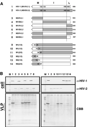
The helix 2 region, but not the trimerization site, in HIV-2 MA abolishes high-order Gag assembly and subsequent par-ticle production in yeast.To map a Gag region responsible for the failure of particle production in yeast, a series of chimeric Gag constructs between HIV-1 and HIV-2 were made (Fig. 7A) and expressed in yeast. Western blotting using anti-HIV-1 and anti-HIV-2 CA antibodies confirmed that each domain of Gag was replaced in the chimeric constructs. Spheroplast for-mation and VLP production were carried out as described above. Equivalent volumes of Gag VLP fractions were ana-lyzed by SDS-PAGE followed by CBB staining. A failure of particle production was observed only when HIV-2 MA was present in the constructs (Fig. 7B, left panels). Further map-ping experiments within MA indicated that the N-terminal one-third of HIV-2 MA (amino acid residues 1 to 44) is es-sentially responsible for the failure of VLP production (Fig. 7B, right panels). However, introduction of the corresponding region of HIV-1 MA into the HIV-2 background [construct M1(1/3)] did not rescue particle production, but a larger region of HIV-1 MA (the N-terminal half; residues 1 to 70) was required for particle production. Conversely, introduction of the N-terminal half of HIV-2 MA into the HIV-1 background [construct M2(1/2)] did not completely abolish particle pro-duction, but the introduction of a larger region of HIV-2 MA [construct M2(2/3)] did. These data suggest but do not prove that other sites affecting particle production may lie in the region between residues 45 and 96 and that the overall struc-tural integrity of MA is not negligible.
Studies by nuclear magnetic resonance (25, 26) and crystal-lography (18, 38) have indicated that primate lentiviral MAs share three-dimensional characteristics, including the follow-ing: (i) the globular domain is composed of five helices (H1 to H5) capped by a-sheet, in which the N-terminal basic resi-dues are clustered; and (ii) in the MA trimer, the N-myristoyl moiety is inserted into a lipid bilayer and the surface-exposed basic residues come into contact with acidic phospholipids, such as phosphatidylinositol 4,5-bisphoshate [PI(4,5)P2], in the
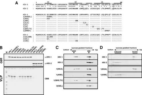
[image:7.585.70.255.73.523.2]membrane (18, 41). Indeed, the gross conformation of HIV-2 MA predicted by homology modeling using the SIVmac MA structure (38) as a template at Geno3D, an automated protein-modeling Web server (http://geno3d-pbil.ibcp.fr), is very simi-lar to that of HIV-1 MA (see the supplemental material at http://www.kitasato-u.ac.jp/lisci/labo/ViralInfection2/1/Fig.S1 .pdf), and the N-terminal half of MA (e.g., the cluster of basic amino acids) is relatively conserved between HIV-1 and HIV-2 compared with the C-terminal half (see the supplemental ma-terial at the website listed above), but our study found that the N-terminal half of MA is a determinant for Gag VLP produc-tion in yeast. To define the residues of MA critical for particle production in yeast, the clusters of nonconserved HIV-1 residues in the N-terminal half of MA were replaced with the correspond-ing HIV-2 residues (Fig. 8A), and the Gag constructs were ex-pressed in yeast. Most of the substitutions had little effect on VLP production, and nearly equivalent levels of VLPs were produced even in the substitution mutant of the N-terminal trimerization site [construct 1(GLA)]. Interestingly, VLP production was se-verely impaired by the introduction of HIV-2 residues 38 to 40,
FIG. 6. Membrane association and multimerization of HIV-1 and HIV-2 Gags in higher eukaryotic cells. HeLa and 293T cells were trans-fected with a pCAGGS vector containing the HIV-1 or HIV-2gaggene with a Flag epitope sequence, and after incubation at 37°C for 48 h, cells and culture medium were harvested for analysis. The culture medium was subjected to centrifugation on 20% sucrose cushions for purification of Gag VLPs. (A) Intracellular Gag expression and Gag VLP production. Transfected cells and Gag VLP fractions were subjected to SDS-PAGE followed by Western blotting using anti-Flag antibody. Lanes: M, molec-ular weight markers; 1 and 2, expression of HIV-1 and HIV-2 Gag-Flag, respectively. (B) Membrane affinity of Gag. Transfected cells were resus-pended in buffer without NaCl and disrupted by sonication. Equilibrium flotation centrifugation and subsequent fractionation were carried out as described in the legend for Fig. 4. The gradient fractions were analyzed by Western blotting using anti-Flag antibody. Panels: 1, cells expressing HIV-1 Gag-Flag; 2, cells expressing HIV-2 Gag-Flag. (C) Sedimentation analysis of Gag. The whole-cell lysate was subjected to sedimentation analysis with a 20 to 70% sucrose gradient by centrifugation at 120,000⫻
gfor 2 h, and gradient fractions were subjected to Western blotting using anti-Flag antibody. Panels: 1, cells expressing HIV-1 Gag-Flag; 2, cells expressing HIV-2 Gag-Flag. Arrowheads show sedimented positions of the immature form of HIV capsid (600S) and of 80S ribosomes.
on November 8, 2019 by guest
http://jvi.asm.org/
located near the end of helix 2 of MA, into HIV-1 Gag [construct 1(ANK)] (Fig. 8B). Introduction of the corresponding sequence of HIV-1 into HIV-2, however, did not confer particle produc-tion, although this was expected from the fact that the M1(1/3) construct did not produce Gag VLP (Fig. 7). We prepared the whole-cell lysates from several of the substitution mutants and examined their ability to form high-order Gag multimers by
[image:8.585.123.463.69.560.2]sed-imentation analysis. The VLP-competent Gag mutants showed both high- and low-order Gag multimers, while in contrast, the VLP-incompetent Gag mutants showed only the low-order form (Fig. 8C). The 1(ANK) Gag construct did not show membrane affinity when samples were prepared in the absence of salt and subjected to membrane flotation analysis (Fig. 8D). These data indicate that a defined region of HIV-2 MA may affect stable
FIG. 7. Mapping of HIV-2 Gag regions inhibitory for Gag VLP production in yeast. (A) Schematic representation of chimeric Gags constructed between HIV-1 and HIV-2. (B) Intracellular Gag expression and Gag VLP production. Yeast cells were transformed with a pKT10 vector containing each Gag construct. Intracellular Gag expression was analyzed by Western blotting using anti-HIV-1 CA and anti-HIV-2 CA antibodies. Production and purification of Gag VLPs were carried out as described in the legend for Fig. 1. Gag VLPs were analyzed by SDS-PAGE followed by CBB staining. Lanes: M, prestained molecular weight markers; 1 and 2, expression of HIV-1 and HIV-2 Gag, respectively; 3 to 14, expression of chimeric Gags M2/I1/L1, M1/I2/L1, M1/I1/L2, M1/I2/L2, M2/I1/L2, M2/I2/L1, M1(1/3), M1(1/2), M1(2/3), M2(1/3), M2(1/2), and M2(2/3), respectively.
on November 8, 2019 by guest
http://jvi.asm.org/
membrane affinity and high-order Gag assembly and, as a result, lead to the failure of VLP production in yeast.
DISCUSSION
The Gag proteins of primate lentiviral lineages have approx-imately 55 to 60% amino acid sequence identity between each of the lineages (8). In this study, we used Gag proteins of four different primate lentiviral lineages and analyzed their ability to produce Gag VLPs in yeast. We found that the Gag protein of the HIV-2/SIVmac lineage failed to release Gag VLPs in yeast, despite the fact that the Gag protein was myristoylated and targeted to the plasma membrane. The failure of HIV-2 Gag VLP production was likely due to the lack of high-order Gag multimerization but was also accompanied by an unstable association of Gag with the membrane. These findings were not observed in higher eukaryotic cells, suggesting that a host factor(s) was involved.
A host factor involved in Gag multimerization, ABCE1/ HP68, has been identified by in vitro translation studies show-ing that this factor associates with relatively high-order Gag multimers but not with a lower-order form and facilitates Gag assembly into a VLP form as a molecular chaperone (56). Immunodepletion of ABCE1/HP68 in the in vitro reactions reduced the higher-order form with the accumulation of lower-order forms (56), a condition which was ostensibly similar to the failure of HIV-2 Gag assembly in yeast. However, this host factor is highly conserved in eukaryotes, including yeast, and has been shown to support all primate lentiviral Gag assembly (10). Later studies have shown that the NC but not MA do-main is critical for Gag-ABCE1/HP68 interaction (23). From these data, it is unlikely that the putative host factor suggested in this study is ABCE1/HP68.
Primate lentiviral MA is composed of five helices, with helices 1 and 2 located on the upper surface of a globular MA structure in which the N-terminal basic residues are clustered.
FIG. 8. Defining the N-terminal HIV-2 MA region inhibitory for Gag assembly and VLP production in yeast. (A) Amino acid alignment of Gag mutants with amino acid substitutions. The trimerization sites are shown in boxes, and helices 1 to 3 are shown as H1 to H3. (B) Intracellular expression and VLP production of Gag mutants. Protein expression and Gag VLP production in yeast were carried out as described in the legend for Fig. 1. Cell samples were subjected to Western blotting using anti-HIV-1 CA and anti-HIV-2 CA antibodies, and Gag VLPs were analyzed by SDS-PAGE followed by CBB staining. Lane M shows prestained molecular weight markers. (C) Sedimentation analysis of Gag mutants. Protein expression in yeast was carried out as described in the legend for Fig. 3. The whole-cell lysate was subjected to sedimentation analysis with a 20 to 70% sucrose gradient by centrifugation at 120,000⫻gfor 2 h, and gradient fractions were subjected to Western blotting using anti-Flag antibody. Arrowheads show sedimented positions of the immature form of HIV capsid (600S) and of 80S ribosomes. (D) Membrane affinities of Gag mutants in the absence of salt. Protein expression in yeast was carried out as described in the legend for Fig. 3, and equilibrium flotation centrifugation was described in the legend for Fig. 4. Gradient fractions were analyzed by Western blotting using anti-Flag antibody.
on November 8, 2019 by guest
http://jvi.asm.org/
[image:9.585.44.541.66.400.2]In the MA trimer model, the myristoyl moiety and N-terminal basic residues act in synergy to stabilize the trimer on an acidic membrane (18). Recent studies have further indicated that PI(4,5)P2, a lipid involved in membrane targeting of Gag (31),
binds to a hydrophobic cleft formed with highly conserved amino acids by helices 2 and 4 and contributes to the mem-brane affinity of MA (41). Our data showed that the dominant region responsible for the failure of HIV-2 Gag VLP produc-tion was mapped near the end of helix 2 of MA, a site distinct from the MA trimerization site or the PI(4,5)P2binding cleft.
The blockage of HIV-2 Gag VLP production in yeast occurred at the process of particle budding rather than Gag transport, and the membrane affinity of Gag was weakened. These facts suggest that such a host factor would be a component(s) in-cluding certain lipids present on or recruited to the plasma membrane, but the lipids would not be PI(4,5)P2, and indeed,
PI(4,5)P2 is widely present in eukaryotes, including yeast.
These data raise the alternative possibility that yeast may have an inhibitory factor(s) for HIV-2 Gag assembly.
Although it has been accepted that Gag oligomerization and membrane targeting are essentially independent events, recent studies have suggested that the membrane affinity of Gag may be promoted by Gag multimerization (43, 55). Another study indicated that the N-terminal myristoyl moiety of MA was exposed when MA formed a trimer (49). However, it is also possible that unstable membrane binding of Gag may not fa-cilitate Gag multimerization, resulting in a lack of high-order assembly. In this study, we observed that the defects in Gag multimerization and membrane affinity occurred concomi-tantly in yeast, but we cannot prove, at present, which directly affected the failure of HIV-2 Gag VLP production in our system. In favor of the former possibility, the lack of a high-order Gag multimer was observed even at a physiological con-centration of salt, a condition under which HIV-2 Gag associ-ated with the yeast membrane. In contrast, the latter possibility would be supported by the data that (i) the region we mapped was located at helix 2 of MA, facing the membrane; (ii) in the MA trimer model (18, 38), this region is located near the center of the trimer, suggesting little possibility of involvement in higher-order Gag assembly; and (iii) the introduction of only three amino acids of HIV-2 Gag (located at the region) into the HIV-1 background, in which Gag multimerization domains are intact, failed to produce Gag VLPs.
The membrane affinity of Gag is regulated by N-myristoyl-ation but also by the basic amino acid clusters in the M domain (4, 14, 15, 35, 54). In our study, the failure of HIV-2 Gag VLP production was accompanied by the dissociation of Gag from the membrane, a phenomenon which was observed only in the absence of salt. In general, relatively high concentrations of salt, such as 500 mM NaCl, disrupt electrostatic protein bonds but do not affect hydrophobic bonds. Conversely, in the ab-sence of salt, hydrophobic but not electrostatic interactions are often disrupted. We observed that neither HIV-1 nor HIV-2 Gag dissociated from the membrane in the presence of 500 mM NaCl, suggesting that the N-terminal myristoyl moiety is a dominant determinant for membrane binding of Gag in yeast and that the membrane affinities of Gag mediated by the myr-istoyl moiety were comparable between the two Gags. In con-trast, HIV-2 Gag dissociated from the yeast membrane in the absence of salt, under which condition HIV-1 Gag remained
associated. These results suggest that the electrostatic interac-tions of HIV-2 Gag with the yeast membrane were weaker than those of HIV-1 Gag, leading to Gag dissociation from the yeast membrane. The amino acid sequences of the MA helix 2 re-gion we mapped for the four primate lentiviral lineages are as follows: HIV-1, SRE; HIV-2, ANK; SIVmac, ANE; SIVagm, GKE; and SIVmnd, KGE.
Mutational studies of the N-terminal region of MA have suggested the involvement of hydrophobicity and electrostatic interactions of MA in Gag relocation and membrane extrusion. Amino acid substitutions in the MA basic domain redirected Gag to the endoplasmic reticulum and endosomes (34, 35, 54). Mutations of hydrophobic residues near the N terminus of MA to less hydrophobic residues severely impaired membrane binding of Gag without inhibiting N-terminal myristoylation, suggesting a failure of membrane insertion of the myristoyl moiety (36). Interestingly, in Mason-Pfizer monkey virus, a prototype for capsid formation prior to membrane relocation, an increase in the hydrophobicity of MA led to arrest at an early stage of particle budding, possibly by inhibiting exposure of the N-terminal myristoyl moiety (45). Thus, it cannot be ruled out that in our study, the absence of salt may have caused a disruption of the gross conformation of Gag (e.g., unfolding of the hydrophobic core of MA) and/or led to sequestration of the N-terminal myristoyl moiety.
ACKNOWLEDGMENTS
We thank S. Morikawa (National Institute of Infectious Diseases, Japan) for supply of thegaggene of SIVagm and the anti-SIVagm monkey serum and T. Miura (Kyoto University, Japan) for supply of the gag gene of SIVmnd. The cDNA of HIV-2 (ROD strain) was provided by the NIH AIDS Research and Reference Reagent Pro-gram. We also thank K. Mizumoto (Kitasato University, Japan) for supply of a yeast expression vector.
This work was supported by an AIDS grant from the Ministry of Health, Labor, and Welfare of Japan and by a grant-in-aid for scientific research from the Japan Society for the Promotion of Science.
REFERENCES
1.Bagnat, M., S. Keranen, A. Shevchenko, and K. Simons.2000. Lipid rafts function in biosynthetic delivery of proteins to the cell surface in yeast. Proc.
Natl. Acad. Sci. USA97:3254–3259.
2.Bieniasz, P. D.2003. Restriction factors: a defense against retroviral
infec-tion. Trends Microbiol.11:286–291.
3.Braaten, D., and J. Luban.2001. Cyclophilin A regulates HIV-1 infectivity,
as demonstrated by gene targeting in human T cells. EMBO J.20:1300–1309.
4.Bryant, M., and L. Ratner.1990. Myristoylation-dependent replication and assembly of human immunodeficiency virus 1. Proc. Natl. Acad. Sci. USA
87:523–527.
5.Campbell, S., and A. Rein.1999. In vitro assembly properties of human immunodeficiency virus type 1 Gag protein lacking the p6 domain. J. Virol.
73:2270–2279.
6.Campbell, S., and V. M. Vogt.1997. In vitro assembly of virus-like particles with Rous sarcoma virus Gag deletion mutants: identification of the p10 domain as a morphological determinant in the formation of spherical
par-ticles. J. Virol.71:4425–4435.
7.Carriere, C., B. Gay, N. Chazal, N. Morin, and P. Boulanger.1995. Sequence requirements for encapsidation of deletion mutants and chimeras of human immunodeficiency virus type 1 Gag precursor into retrovirus-like particles.
J. Virol.69:2366–2377.
8.Desrosiers, R. C.1990. HIV-1 origins. A finger on the missing link. Nature
345:288–289.
9.Dong, X., H. Li, A. Derdowski, L. Ding, A. Burnett, X. Chen, T. R. Peters, T. S. Dermody, E. Woodruff, J. J. Wang, and P. Spearman.2005. AP-3 directs the intracellular trafficking of HIV-1 Gag and plays a key role in
particle assembly. Cell120:663–674.
10.Dooher, J. E., and J. R. Lingappa.2004. Conservation of a stepwise, energy-sensitive pathway involving HP68 for assembly of primate lentivirus capsids
in cells. J. Virol.78:1645–1656.
11.Dorfman, T., A. Bukovsky, A. Ohagen, S. Hoglund, and H. G. Gottlinger.
on November 8, 2019 by guest
http://jvi.asm.org/
1994. Functional domains of the capsid protein of human immunodeficiency
virus type 1. J. Virol.68:8180–8187.
12.Garrus, J. E., U. K. von Schwedler, O. W. Pornillos, S. G. Morham, K. H. Zavitz, H. E. Wang, D. A. Wettstein, K. M. Stray, M. Cote, R. L. Rich, D. G. Myszka, and W. I. Sundquist.2001. Tsg101 and the vacuolar protein sorting
pathway are essential for HIV-1 budding. Cell107:55–65.
13.Gerrard, S. R., B. P. Levi, and T. H. Stevens.2000. Pep12p is a multifunc-tional yeast syntaxin that controls entry of biosynthetic, endocytic and
ret-rograde traffic into the prevacuolar compartment. Traffic1:259–269.
14.Gheysen, D., E. Jacobs, F. de Foresta, C. Thiriart, M. Francotte, D. Thines, and M. De Wilde.1989. Assembly and release of HIV-1 precursor Pr55gag
virus-like particles from recombinant baculovirus-infected insect cells. Cell
59:103–112.
15.Gottlinger, H. G., J. G. Sodroski, and W. A. Haseltine.1989. Role of capsid precursor processing and myristoylation in morphogenesis and infectivity of
human immunodeficiency virus type 1. Proc. Natl. Acad. Sci. USA86:5781–
5785.
16.Gottlinger, H. G., T. Dorfman, J. G. Sodroski, and W. A. Haseltine.1991. Effect of mutations affecting the p6 gag protein on human immunodeficiency
virus particle release. Proc. Natl. Acad. Sci. USA88:3195–3199.
17.Hahn, B. H., G. M. Shaw, K. M. De Cock, and P. M. Sharp.2000. AIDS as
a zoonosis: scientific and public health implications. Science287:607–614.
18.Hill, C. P., D. Worthylake, D. P. Bancroft, A. M. Christensen, and W. I. Sundquist.1996. Crystal structures of the trimeric human immunodeficiency virus type 1 matrix protein: implications for membrane association and
assembly. Proc. Natl. Acad. Sci. USA93:3099–3104.
19.Hoshikawa, N., A. Kojima, A. Yasuda, E. Takayashiki, S. Masuko, J. Chiba, T. Sata, and T. Kurata.1991. Role of thegag andpolgenes of human immunodeficiency virus in the morphogenesis and maturation of retrovirus-like particles expressed by recombinant vaccinia virus: an ultrastructural
study. J. Gen. Virol.72:2509–2517.
20.Keckesova, Z., L. M. Ylinen, and G. J. Towers.2004. The human and African green monkey TRIM5alpha genes encode Ref1 and Lv1 retroviral restriction
factor activities. Proc. Natl. Acad. Sci. USA101:10780–10785.
21.Kikonyogo, A., F. Bouamr, M. L. Vana, Y. Xiang, A. Aiyar, C. Carter, and J. Leis.2001. Proteins related to the Nedd4 family of ubiquitin protein ligases interact with the L domain of Rous sarcoma virus and are required for gag
budding from cells. Proc. Natl. Acad. Sci. USA98:11199–11204.
22.Klikova, M., S. S. Rhee, E. Hunter, and T. Ruml.1995. Efficient in vivo and in vitro assembly of retroviral capsids from Gag precursor proteins expressed
in bacteria. J. Virol.69:1093–1098.
23.Lingappa, J. R., J. E. Dooher, M. A. Newman, P. K. Kiser, and K. C. Klein.
2006. Basic residues in the nucleocapsid domain of Gag are required for interaction of HIV-1 Gag with ABCE1 (HP68), a cellular protein important
for HIV-1 capsid assembly. J. Biol. Chem.281:3773–3784.
24.Mammano, F., A. Ohagen, S. Hoglund, and H. G. Gottlinger.1994. Role of the major homology region of human immunodeficiency virus type 1 in virion
morphogenesis. J. Virol.68:4927–4936.
25.Massiah, M. A., M. R. Starich, C. Paschall, M. F. Summers, A. M. Chris-tensen, and W. I. Sundquist.1994. Three-dimensional structure of the
hu-man immunodeficiency virus type 1 matrix protein. J. Mol. Biol.244:198–
223.
26.Matthews, S., P. Barlow, J. Boyd, G. Barton, R. Russell, H. Mills, M. Cunningham, N. Meyers, N. Burns, N. Clark, S. Kingsman, A. Kingsman, and I. Campbell.1994. Structural similarity between the p17 matrix protein
of HIV-1 and interferon-gamma. Nature370:666–668.
27.Morikawa, Y., T. Goto, and K. Sano.1999.In vitroassembly of human
immunodeficiency virus type 1 Gag protein. J. Biol. Chem.274:27997–28002.
28.Morikawa, Y., T. Goto, and F. Momose.2004. Human immunodeficiency virus type 1 Gag assembly through assembly intermediates. J. Biol. Chem.
279:31964–31972.
29.Nguyen, D. H., and J. E. Hildreth.2000. Evidence for budding of human immunodeficiency virus type 1 selectively from glycolipid-enriched
mem-brane lipid rafts. J. Virol.74:3264–3272.
30.Niwa, H., K. Yamamura, and J. Miyazaki. 1991. Efficient selection for
high-expression transfectants with a novel eukaryotic vector. Gene108:193–
199.
31.Ono, A., S. D. Ablan, S. J. Lockett, K. Nagashima, and E. O. Freed.2004. Phosphatidylinositol (4,5) bisphosphate regulates HIV-1 Gag targeting to
the plasma membrane. Proc. Natl. Acad. Sci. USA101:14889–14894.
32.Ono, A., and E. O. Freed.1999. Binding of human immunodeficiency virus type 1 Gag to membrane: role of the matrix amino terminus. J. Virol.
73:4136–4144.
33.Ono, A., and E. O. Freed.2001. Plasma membrane rafts play a critical role in
HIV-1 assembly and release. Proc. Natl. Acad. Sci. USA98:13925–13930.
34.Ono, A., and E. O. Freed.2004. Cell-type-dependent targeting of human immunodeficiency virus type 1 assembly to the plasma membrane and the
multivesicular body. J. Virol.78:1552–1563.
35.Ono, A., J. M. Orenstein, and E. O. Freed.2000. Role of the Gag matrix domain in targeting human immunodeficiency virus type 1 assembly. J. Virol.
74:2855–2866.
36.Paillart, J. C., and H. G. Gottlinger.1999. Opposing effects of human immunodeficiency virus type 1 matrix mutations support a myristyl switch
model of Gag membrane targeting. J. Virol.73:2604–2612.
37.Parent, L. J., R. P. Bennett, R. C. Craven, T. D. Nelle, N. K. Krishna, J. B. Bowzard, C. B. Wilson, B. A. Puffer, R. C. Montelaro, and J. W. Wills.1995. Positionally independent and exchangeable late budding functions of the Rous sarcoma virus and human immunodeficiency virus Gag proteins. J.
Vi-rol.69:5455–5460.
38.Rao, Z., A. S. Belyaev, E. Fry, P. Roy, I. M. Jones, and D. I. Stuart.1995. Crystal structure of SIV matrix antigen and implications for virus assembly.
Nature378:743–747.
39.Reicin, A. S., S. Paik, R. D. Berkowitz, J. Luban, I. Lowy, and S. P. Goff.
1995. Linker insertion mutations in the human immunodeficiency virus type
1gaggene: effects on virion particle assembly, release, and infectivity. J.
Vi-rol.69:642–650.
40.Ruggieri, R., K. Tanaka, M. Nakafuku, Y. Kaziro, A. Toh-e, and K. Matsu-moto.1989.MSI1, a negative regulator of the RAS-cAMP pathway in Sac-charomyces cerevisiae. Proc. Natl. Acad. Sci. USA86:8778–8782. 41.Saad, J. S., J. Miller, J. Tai, A. Kim, R. H. Ghanam, and M. F. Summers.
2006. Structural basis for targeting HIV-1 Gag proteins to the plasma
mem-brane for virus assembly. Proc. Natl. Acad. Sci. USA103:11364–11369.
42.Sakuragi, S., T. Goto, K. Sano, and Y. Morikawa.2002. HIV type 1 Gag
virus-like particle budding from spheroplasts ofSaccharomyces cerevisiae.
Proc. Natl. Acad. Sci. USA99:7956–7961.
43.Sandefur, S., V. Varthakavi, and P. Spearman.1998. The I domain is re-quired for efficient plasma membrane binding of human immunodeficiency
virus type 1 Pr55Gag. J. Virol.72:2723–2732.
44.Smith, A. J., N. Srinivasakumar, M. L. Hammarskjold, and D. Rekosh.1993.
Requirements for incorporation of Pr160gag-polfrom human
immunodefi-ciency virus type 1 into virus-like particles. J. Virol.67:2266–2275.
45.Stansell, E., E. Tytler, M. R. Walter, and E. Hunter.2004. An early stage of Mason-Pfizer monkey virus budding is regulated by the hydrophobicity of the
Gag matrix domain core. J. Virol.78:5023–5031.
46.Strack, B., A. Calistri, S. Craig, E. Popova, and H. G. Gottlinger.2003. AIP1/ALIX is a binding partner for HIV-1 p6 and EIAV p9 functioning in
virus budding. Cell114:689–699.
47.Stremlau, M., C. M. Owens, M. J. Perron, M. Kiessling, P. Autissier, and J. Sodroski.2004. The cytoplasmic body component TRIM5alpha restricts
HIV-1 infection in Old World monkeys. Nature427:848–853.
48.Tanaka, K., M. Nakafuku, F. Tamanoi, Y. Kaziro, K. Matsumoto, and A. Toh-e.1990.IRA2, a second gene ofSaccharomyces cerevisiaethat encodes a
protein with a domain homologous to mammalianrasGTPase-activating
protein. Mol. Cell. Biol.10:4303–4313.
49.Tang, C., E. Loeliger, P. Luncsford, I. Kinde, D. Beckett, and M. F. Sum-mers.2004. Entropic switch regulates myristate exposure in the HIV-1
ma-trix protein. Proc. Natl. Acad. Sci. USA101:517–522.
50.Towers, G. J., T. Hatziioannou, S. Cowan, S. P. Goff, J. Luban, and P. D. Bieniasz.2003. Cyclophilin A modulates the sensitivity of HIV-1 to host
restriction factors. Nat. Med.9:1138–1143.
51.Towler, D. A., S. P. Adams, S. R. Eubanks, D. S. Towery, E. Jackson-Machelski, L. Glaser, and J. I. Gordon.1988. Myristoyl CoA: protein N-myristoyltransferase activities from rat liver and yeast possess overlapping
yet distinct peptide substrate specificities. J. Biol. Chem.263:1784–1790.
52.VerPlank, L., F. Bouamr, T. J. LaGrassa, B. Agresta, A. Kikonyogo, J. Leis, and C. A. Carter.2001. Tsg101, a homologue of ubiquitin-conjugating (E2) enzymes, binds the L domain in HIV type 1 Pr55(Gag). Proc. Natl. Acad. Sci.
USA98:7724–7729.
53.von Schwedler, U. K., M. Stuchell, B. Muller, D. M. Ward, H. Y. Chung, E. Morita, H. E. Wang, T. Davis, G. P. He, D. M. Cimbora, A. Scott, H. G. Krausslich, J. Kaplan, S. G. Morham, and W. I. Sundquist.2003. The
protein network of HIV budding. Cell114:701–713.
54.Yuan, X., X. Yu, T. H. Lee, and M. Essex.1993. Mutations in the N-terminal region of human immunodeficiency virus type 1 matrix protein block
intra-cellular transport of the Gag precursor. J. Virol.67:6387–6394.
55.Zhou, W., and M. D. Resh.1996. Differential membrane binding of the
human immunodeficiency virus type 1 matrix protein. J. Virol.70:8540–8548.
56.Zimmerman, C., K. C. Klein, P. K. Kiser, A. R. Singh, B. L. Firestein, S. C. Riba, and J. R. Lingappa.2002. Identification of a host protein essential for
assembly of immature HIV-1 capsids. Nature415:88–92.