Int. J. Curr. Res. Biosci. Plant Biol. 2014, 1(3): 17-26
Original Research Article
Pharmacognostical Studies of Various Parts of Couroupita guianensis Aubl.
V. Regina*
Department of Botany, Pachaiyappa's College, Chennai-600 030, Tamil Nadu, India
*Corresponding author.
A b s t r a c t K e y w o r d s
The present study has been aimed to assess the anatomical and histochemical activities of stem, leaf, petiole, lamina, sepal, petal, fruit and seed from an incredible medicinal tree Couroupita guianensis Aubl. Different plant parts of C. guianensis Aubl. tree were collected from the Pachaiappa s college campus, identified and authenticated. All the parts of the plant were sectioned, stained, macerated, mounted and photographed and also various stains were used for histochemical observation. All the parts of the plant possessed many phytoconstituents. From the evaluation it was found that alkaloids, lipids, proteins, saponins and starch grains were detected in the xylem vessels, ground tissues and also in the epidermal layers. The phytoconstituents reported in C.
guianensis Aubl. are needed to be purified for the isolation of active
principles responsible for the medicinal uses.
Couroupita guianensis
Histochemicals
Lecythidaceae Medicinal plant
Phytoconstituents
Introduction
Today no communicable disease represents a leading threat to human health and development. Liver cancer is the world s biggest killer. Strategies for reducing risk factors for cancer aim at providing and encouraging healthy choices for all. Hence the localization, identification and isolation of a compound from the natural healer of any disease are an urgent need. Further medicinal plant parts which play vital roles in healing cancer is an important aspect of this complete biological research processes.
The family Lecythidaceae represents one of the largest families (Aublet, 1775) chiefly tropical and subtropical in distribution (Richter, 1982).
Couroupita guianensis Aubl. (Cannon ball tree) is a
large deciduous, tropical tree growing to 35m high. Leaves occur in clusters at the end of the branches on 0.5 3cm long petioles; lamina simple, 3 10cm, glabrous on upper surface, pubescent on veins on lower surface, base connate and apex acute to acuminate, margin entire with 15 25 pairs of secondary veins (Mitre, 1998). Flowers zygomorphic, 5 6cm across, calyx- 6 broadly ovate lobes with ciliate margins; petal 6 yellow with pink or red bases basally on the lower surface (Thomson, 1921). Fruits- indehiscent, globosely, 12 25cm diameter, woody, dark brown, capsule falling from the tree at maturity. Seeds- up to 300, embedded in pulp, the pulp oxidized to bluish green when exposed to air (Prance and Mori, 1990).
International Journal of Current Research in
Biosciences and Plant Biology
ISSN: 2349-8080 Volume 1 Number 3 (October-2014) pp. 17-26
Int. J. Curr. Res. Biosci. Plant Biol. 2014, 1(3): 17-26 The seeds are embedded in a six-segmented,
(Matthew, 1983) since the whole plant is medicinally useful and a natural healer, available easily. Stem bark, leaf, root, flower and fruit pulp parts were reported earlier with the limited pharmacognostical studies, but leaf lamina, petiole, seed and fruit rind of this medicinal plant is still unexplored hence selected for pharmacognostical studies. The present study is aimed to analyse the anatomical and histochemical activities of the stem, leaf, flower, fruit and seeds of C. guianensis Aubl. and also to localize the special inclusions.
Materials and methods
Plant collection
C. guianensis Aubl. is found to grow as a
waste-land weed in xeric habitats (now, grown as an ornamental tree in most of the gardens). Specimens were collected during several visits from Pachaiyappa s College boy s hostel block in Chennai, India, authenticated (No. PARC/2010/949) (Nair and Henry, 1983). The collected plant material was free from disease and was also free of contamination of other plants. When the fruit was in ripened stage, the fruits were collected from the tree not from down in the floor, and the fruit characters were used for identification of the plant with the help of Floras (Gamble, 1967; Mayuranathan, 1956). Photomicrographs of the plants in its original habitat were taken in the field. To display the surface features of the leaf, stem, petiole, flower bud, fruit and seed were photographed in closer views with the help of macro-lenses.
Anatomical study
For microscopic investigations, small pieces of fruit rind were mixed in FAA (Formalin: acetic acid: alcohol in 1:1:18 ratio). After 24 hrs of fixing, the specimens were dehydrated employing tertiary butyl alcohol, wax infiltrated and embedded in wax blocks (Sass, 1940). With the help of rotary microtome, serial sections ranging in thickness from 8 12 µm were prepared. Sections were stained with toluidine blue as per the method adopted by O Brien et al. (1964) and de-waxing of the sections was by customary procedure (Johansen, 1940), since toluidine blue is a polychromatic stain. For studying
the epidermal layers and ground tissues of fruit rind partial maceration of the bits fruit rind were prepared with chloral hydrate or sodium hydroxide (5%).
The macerated elements were stained with safranin and mounted in glycerin. Dimensional values of the fibres, fibre-tracheids, tracheids, fibriform vessel, elements and vessel elements were computed with micrometers; for each element 15 measurements were taken from which maximum, minimum and mean values were calculated. Diameter of the individual cells and tissue zones were measured in microtome sections. Microscopic descriptions of tissues are supplemented with micrographs wherever necessary. Photographs of different magnifications were taken with Nikon Lab Photo 2 microscopic units. Magnifications of the figures were indicated by the scale bars and the descriptive terms of the anatomical features were given adopting Esau (1964).
Histochemical localization
Apart from the general anatomical studies, simple histochemical observations in order to localize. alkaloids, lipids, protein, saponins and starch grains, the following staining were used.
Coomassie Brilliant Blue Staining-(CBB)
The sections were stained with Coomassie brilliant blue for 15 60 min. at room temperature. Then the sections were rinsed for 5 min. in water. Proteins when present turned into blue colour (Cawood et al, 1978).
Iodine Potassium Iodide Staining-(IKI)
Presence of starch was tested by using 1% Iodine in potassium iodide solution in which the starch stained purple-blue (Bronner, 1975).
Sudan Red Staining-(SUDAN III)
Int. J. Curr. Res. Biosci. Plant Biol. 2014, 1(3): 17-26 Barium Hydroxide Staining-(BH)
Sections were placed directly in one drop of Conc. H2So4 on a slide, which gives a characteristic
sequence of colour reactions, beginning immediately with yellow, changing to red within 30 minutes and finally becoming violet or blue green in a short time. Sections were put in saturation barium hydroxide solution for about 24 hours and were washed with calcium chloride, the placed in potassium dichromate, yellow colour indicated in the presence of saponin (Momin, 2011)
Results
Anatomy of the leaf
The leaf consists of a prominant midrib and thin lamina. The midrib is somewhat elliptical in cross-sectional view, the abaxial being thick and semicircular. The midrib is 1.45mm thick and 2.2mm wide. The epidermal layer of the midrib is thin comprising small, cubical, slightly papillate and fairly thick walled cells. The ground tissue on the adaxial raised past consists of a thick arc of collenchyma cells. Remaining ground tissue is parenchymatous, the cells being small, thin walled and compact.
The vascular system of the midrib is complex. It is a compound system of three segments of vascular strands. At the adaxial part is a small horizontal band of two larger and two smaller, less prominent vascular strands. In the medial part of the midrib is wider and thicker flat vascular band. At the abaxial part is much wider, bowl shaped line of many discrete bundles.
The adaxial and median bundles are collateral with xylem located on the adaxial arc consists central larger bundle, the bundles decreasing gradually in size on either of the arc. The larger and smaller bundles are collateral with adaxial xylem. The xylem elements of the vascular strands are angular, wide, fairly thick walled and diffuse in distributions. The phloem elements are in continuous bands comprising fairly wide angular sieve elements and parenchyma cells. A thin layer of sclerenchyma occurs abutting the phloem zone of the vascular strands.
Lamina
The lamina is dorsiventral and has prominent vascular strands of the lateral veins. The lateral veins do not project beyond the level of the lamina. The lamina is 70-90mm thick. The adaxial epidermis is thick, the cells being squarish or rectangular. The walls are thin and the cuticle is prominent. The mesophyll is differentiated into single adaxial layer of short cyclindrical palisade cells and abaxial zone of three or four layers lobed spongy parenchyma cells. The vascular strand of the lateral veins consists of small cluster of thick walled angular xylem elements and a few phloem elements. These are narrow prominent arcs of sclerenchyma elements on the upper and lower ends of the vascular strands.
Petiole
The petiole is boat shaped in cross-sectional view with flat adaxial side and semicircular abaxial side. The epidermal layer is thin and the epidermal cells are small. The ground tissue consists of outer zone of about five layers of collenchyma cells and remaining portion being thin walled, compact parenchyma cells. The vascular system of the petiole is multistranded and complex. There is a wide bowl shaped abaxial line of discrete vascular bundles. The upper marginal bundles occupy the wing portion forming the wing bundles. There is a flat plate of smaller bundles at the adaxial parts. In the medullary is a single fairly large horizontal segment of vascular bundles. All vascular bundles are collateral with upper xylem and lower phloem. The xylem elements are in vertical parallel lines, the elements are angular and thin walled phloem elements are in isolated clusters located beneath the xylem strand. A thick arc of sclerenchyma cells occur in all vascular bundles, abutting the phloem zone.
Venation system
Int. J. Curr. Res. Biosci. Plant Biol. 2014, 1(3): 17-26
Fig. 1: Anatomy of C. guianensis Aubl.
Int. J. Curr. Res. Biosci. Plant Biol. 2014, 1(3): 17-26 Epidermal cells and stomata
The epidermal cells, when viewed in surface view, are large, with much sinuous and folded, and fairly thick anticlinal walls. The cells appear amoeboid in outline. Large nuclei are seen in most of the epidermal cells. Stomata are amomocytic type-No specific subsidiary cells are evident. The stomata are elliptical, measuring 15×25-30mm in size.
Stem
Stem measuring about 4.5mm thick was studied. It is circular in sectional view; the surface is smooth excepting one or two places where there are prominent lenticels. The epidermal layer and one or two sub-epidermal layers are compressed into dark tannin filled cells due to radical growth of the stem. Inner to the tanniniferous layer showed a narrow zone of about four layers of periderm comprising thin walled tabular cells. The cortex is 800mm in thickness. It is homocellular with compact thin walled parenchyma cells.
Scattered in the cortical zone there are numerous circular vascular strands of different sizes. These cortical bundles are originally leaf-trace bundles which run vertically from one to several internodes before entering into the leaf-base. The cortical leaf trace bundles possess collateral xylem and phloem and are surrounded by sclerenchyma bundles sheaths.
The vascular cylinder is wide and circular enclosing wide parenchymatous pith. The vascular cylinder is 400mm thick. It consists of outermost thin layer of fibres, wide continous cylinder of xylem. One or two rings of phloem fibres are seen in the phloem zone. Xylem consists of thin and short radial rows of xylem elements with parenchymatous gaps in between. The xylem elements are narrow, angular and thin walled.
Floral parts
Sepal: The sepal is thick in the middle and gradually
tapering towards the margins. The central part is 1.25mm thick. The structure of the sepal in simple. The inner epidermis is thin with small darkly stained cells. The outer epidermis consists of thin, radially oblong cells. The ground tissue is homogenous, parechymatous and compact. The cells in the central portion are loosely arranged with
small intercellular spaces. Small vascular traces are scattered in the ground tissue.
Petal: The petal is 600mm thick with middle and it
gradually tapers towards the margin measuring 50mm thick. The structure of the petal is also simple in structure. The outer and inner epidermal layers are thin with squarish cells. The ground tissue is uniformly distributed and consists of compact small parenchyma cells. Small less conspicuous vascular strands are distributed at random in the ground tissue. The marginal part of the petal has thick and darkly stained epidermal cells and four or five layers small compact parenchyma cells. The extreme margin is reduced to upper and lower epidermal cells and a single layer of mesophyll cells.
Fruit-rind
The fruit wall is thick and fleshy. It is 1.3mm thick. The rind consists of distinct outer and inner epidermal layers of fairly wide rectangular cells. The ground tissue is parenchymatous. The outer and inner zones have compact cells; the middle zone consists of lobed loosely arranged parenchyma cells. Small circular vascular strands are diffusely distributed in the median zone. The vascular strands consist of broken nests of small, thick walled elements, each nest being associated with small phloem elements.
Seed
The seed is elliptical is sectional veiw with thick central portion and narrow ends. The seed is 7mm long and 2mm thick. The seed consists of central endosperm and outer seed coat with complex structure. The seed coat comprises a thin sclerotesta which is made up of a layer of radially extended rectangular cells. The cells have extensive wall thickenings and reduced cell lumen. The walls are 30mm thick they have wide central like pits.
Int. J. Curr. Res. Biosci. Plant Biol. 2014, 1(3): 17-26
Table 1. Percentage levels of phytochemical localization in different parts of C. guianensis Aubl.
Maximum Minimum Mean
Phytoconstituents Plant Part
(in m)
Mid rib 160.2 110.2 142.5
Alkaloid
Fruit rind 225.6 180.4 120.3
midrib 150.9 100.9 110.7
lamina 120.4 040.5 060.3
leaf 140.2 105.0 080.9
Lipid
fruit rind 210.4 130.0 0.75.7
Midrib 140.0 130.0 180.6
Petiole 160.8 102.0 170.4
Protein
Fruit rind 180 105.7 180.7
Saponin Fruit rind 290.7 100.9 280.9
Leaf 200.6 100.9 190.6
Petiole 190.8 180.6 170.7
Starch grain
Midrib 150.9 170.6 190.8
Microscopic observation
The leaf-power was examined under the microscope to study the inclusions in the power following elements were recorded in the powder.
Epidermal peelings: Small fragments of epidermal
peeling are common in the powder. They exhibit epidermal cells with highly wavy and folded anticlinal walls, so that the cells assume amoeboid shape. Stomata are seen on the epidermal layer; they are anomocytic type. The stomata are broadly elliptical and thick walled.
Epidermal trichomes: Nonglandular, multicellular,
unbranched epidermal trichomes are abundant in the powder. They have thick smooth walls and lumen. The lumen is filled with dense granular contents. The trichomes are up to 900mm long and 50mm thick at the base. Uniformly distributed and consists of compact small parenchyma cells.
Small less conspicuous vascular strands are distributed at random in the ground tissue. The marginal part of the petal has thick and darkly stained epidermal cells and four or five layers small compact parenchyma cells. The extreme margin is reduced to upper and lower epidermal cells and a single layer of mesophyll cells.
Histochemical tests
The localisation certain compounds in the specific cells were detected by employing stains which
specifically stain the compounds. The results of histochemical studies are given in Table 1.
Lipids are specifically stained by sudan-III, which
gives red colour to the lipids. Lipids are found to be located in the phloem parenchyma and collenchyma cells of the peripheral zone of midrib. In the lamina, both palisade cells and spongy parenchyma cells turn red indicating the presence of lipids.
In the petiole, lipid is located in the ground parenchyma especially in the outer ground cells. Lipid deposition was observed in the vascular bundles and phloem tissue .In the fruit, the outer portion of the rind is deep red, indicating the presence of lipids. The outer portion of the compressed tissue has more accumulation of lipids. These are also isolated cells in the pericarp which contain lipids.
Protein: For localization of protein CBB was used,
which gives blue colouration to the protein. In the leaf clearing the epidermal layers, the mesophyll cells turn blue. In the midrib, phloem cells and xylem parenchyma cells show blue coloured inclusions. Phloem elements in the petiole showed accumulation of proteins. The isolated ground parenchyma cells in the fruit rind possessed amorphous blue colored inclusions.
Alkaloids: Wagner s reagent was used to localise
Int. J. Curr. Res. Biosci. Plant Biol. 2014, 1(3): 17-26
Starch grains: Starch grains turn black or dark
purple with IKI. Dense accumulation of starch grains was noticed in the leaf mesophyll cells, ground parenchyma cells of the petiole, and ground parenchyma of the midrib.
Saponins: The xylem and phloem tissues of fruit
rind, pericarp and the ground parenchyma cells of the fruit rind showed yellow colouration indicating the presence of saponins.
Discussion
Anatomy
C. guianensis Aubl. of Lecythidceae, is unique in
two respects, namely, the flowers are cauliflorous and the stamens are fused into thick hood which bends over the ovary. Albeit its popularity as ornamental avenue tree, its medicinal properties seem to be little studied. Particularly, a pharmacognosy of the fruit rind of this plant has not been studied. Lecythidaceae is related to Myrtaceae. However, Metcalfe and Chalk (1957) have pointed out that the absence of secretory cavities and intra-xylary phloem, the presence of cortical bundles in the axis, and the frequently complex vascular structure of the petiole serve to differentiate the Lecythidaceae from Myrtaceae.
C. guianensis Aubl. is exhibits several microscopic
features that are unique and specific to the plant. The vascular patterns of midrib of leaf and petiole are characteristically multistranded and the distribution is also specific. The vascular structure consists of an abaxial arc of larger bundles, adaxial plate of smaller bundles and a central large plate of bundles both in the midrib and petiole. The petiole is of considerable importance in taxonomy and diagnosis of species, because its structure appears to be but little affected by ecological changes. In
C.guianensis Aubl. the vascular pattern of the
midrib continues in the petiole with little changes. The lamina is simple in structure. It is dorsiventral and mesomorphic with single layer of short palisade cells while dealing with the structures variations of leaves.
Esau (1964) has said that the structure of lamina was modified in different ways by the environmental factors, especially the intensity of light. The leaves on the same individual plant may exhibit drastic differences in their structure due to
the effects of light intensity. So the leaf anatomy seems to be of less helpful for diagnosis of the plants.
Fink (1983) revealed the endogenous adventitious buds develop in situ from differentiated parenchyma cells was the specialized characteristic features of the trunk bark in the tropical cauliflorous tree C.
guianensis Aubl. Venation pattern of the lamina has
been considered one of reliable diagnostic features. Especially the vein-islets and vein terminations are one of the significant values in the study of foliar drugs. In C. guianensis Aubl., the venation is densely reticulate, the lateral veins and veinlets are thick and straight. The vein terminations are long, simple occasionally forked once and straight curved. Along with other structural features, venation type may be of much helpful in diagnostic, process of the species.
The epidermal cells of the lamina, when viewed in paradermal sections, exhibit features that are of some diagnostic values. The cells may be polygonal in outline with straight walls, or they may possess wavery anticlinal walls rendering cells to assume amoeboid outline. In plant of the present study, the epidermal cells have highly wavy anticlinal walls. It is believed that epidermal cells differ considerably in various parameters in different plants. However the shape of the epidermal cells provides useful confirmatory evidence, if the identity of the Leaf is already suspected on other grounds. Straight or wavy anticlinal walls of the epidermal cells provide such confirmatory due for the identity of fragmentary leaf samples.
Type of crystals and their distribution pattern is much useful characters in the pharmacognostic studies of medicinal plants. Calcium oxalate crystals are predominant in most of the plants. They occur in all parts and in all types of cells of the plant. Among the types of calcium oxalate crystal, druses are more predominant in the plant. They may occur in strands, horizontal bands or scattered. In the plant studied, the druses are scattered and diffuse in distribution. This observation is one of the features useful for diagnostics of the species.
Int. J. Curr. Res. Biosci. Plant Biol. 2014, 1(3): 17-26
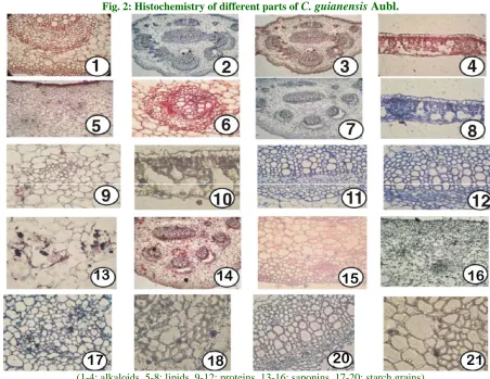
Fig. 2: Histochemistry of different parts of C. guianensis Aubl.
(1-4: alkaloids, 5-8: lipids, 9-12: proteins, 13-16: saponins, 17-20: starch grains)
The sepals and petals of C. guianensis have compact cells and the epidermal cells have no papillae walls; the pericarp of the fruit comprises somewhat arenchymatous ground tissue consist of distinguished inclusions seen and well developed vascular strands.
The fruit morphology is called amphisargam (Greek
amphi double, sarc-fleshy) - a simple, indehiscent
fruit characterized by a pericarp differentiated externally into a dry crust and internally into one or more fleshy layers. Seeds embedded in white, oxidizing bluish green gelatinous pulp Stem, bark, leaves, seed consist of Alpha amyrin, Beta amyrin, Beta sitosterol used in reproductive disorders, infertility, leaf used to cure tumors (Anjaria et al., 1997).
The seeds of C. guianensis are unique with reference to their seed coat. The outer seed coat possesses sclerotic layer. The sclerotesta is uniseriate with radially oblong cells. Their inner tangential walls and the radial (anticlinal) walls are
extremely thick and lignified. These thick walls possess wide canal like pits. The outer tangential wall of each cell produces a long, their walled wide mucilaginous hairs which give Slimy nature of the seeds. The seed structure seems to be an indelible clue for the identity of the seeds of C. guianensis.
Histochemistry
Int. J. Curr. Res. Biosci. Plant Biol. 2014, 1(3): 17-26 might be expected to be particularly valuable as
indicators of taxonomic affinity. In practice there is good evidence in favor of their suggestion.
Metcalfe and Chalk (1957) cites an example to substantiate the above statement in scenario, which in clues a range of habits of herbs, shrubs, succulents and trees, one can find similar secretary structures throughout different species. The presence of a particular compound in the cells of a species will enable one to distinguish it from other species of similar habits.
The histochemical studies also another purpose goes getting a preliminary knowledge of the chemical composition of the species. Based upon the first step of studies we can concentrate for further analysis are the compounds located in the tissues. In the present study, lipids were found to be located in leaf, petiole and pericarp of the fruit (fruit rind). Protein was found to be localized in the phloem and xylem parenchyma and ground parenchyma of the rind of the fruit. Alkaloids occur in the midrib cells and xylem parenchyma and ground cells of the fruit. Saponin is found to be more predominant substance, located in the outer portion of the fruit rind. The above mentioned discussions on pharmacognostic parameters aid in the correct identification of the drug plant, C. guianensis Aubl.
Conclusion
Among the several microscopic features observed from various parts of this plant which were already studied. The most striking character was a special crystals and inclusions in the pericarp of the fruit, a feature restricted to limited number of plants of certain families. Calcium oxalate crystal druses are more predominant, scattered, strands like, diffuse in distribution in the vegetative parts and the reproductive parts varied cells in the parenchyma layers in ground tissue of fruit rind were other useful features of distinction. Histochemically the fruit rind parenchyma cells were yellow amorphous filled cells were useful observation for botanical identity of Couroupita guianensis Aubl.
Acknowledgements
The authors are thankful Dr. P. Jayaraman, Director, PARC-Plant Anatomy Research Centre
(Pharmacognosy Institute), West Tambaram, Chennai for providing medicinal plant and also for his kind assistance in anatomical and histochemical aspects.
References
Anjaria, J., Parabia, M., Bhatt, G., Khamar, R., 1997. Nature Heals-A Glossary of Selected Indigenous Medicinal Plants of India. SRISTI Innovations, Ahmedabad, India. p.15.
Aublet, J.F., 1775. Historic des plantes de la Guiane Francoise. Didot., Paris.
Bronner, R., 1975. Simultaneous demonstration of lipids and starch in plant tissues. Stain. Technol. 50, 1-4.
Cawood, A.H., Potter, U., Dickinson, H.G., 1978. An evaluation of Coomassie blue as a stain for quantitative microdensitometry of protein in section. J. Histochem. Cytochem. 26, 645-650. Chiffelle, T.L., Putt, F.A., 1951. Propylene and
ethylene glycol as solvents for Sudan blue and Sudan black B. Stain. Technol. 26, 51-56. Easu, K., 1964. Anatomy of Seed Plants. John
Wiley and Sons, New York. 550p.
Fink, S., 1983. The occurrence of adventitious and preventitious buds within the bark of some temperate and tropical trees. Am. J. Bot. 70(4), 532 542.
Gamble, J.S., 1967. Flora of Presidency of Madras Vol. II Rep. Ed. Botanical Survey of India, Calcutta. 942p.
Johanson, D.A., 1940. Plant Microtechicque, MC Graw Hill Book Co., New York 523 p.
Matthew, K.M., 1983. The Flora of the Tamilnadu Carnatic, Vol.I, The Rapinat Herbarium, Tiruchirappalli, India. 598 p.
Mayuranathan, P.V., 1956. The flowering plants of Madras City and its immediate neighbourhood. Bull. Madras Govt. Mus., Madras. 116 (FTN 1:58) 563. (Revised by C. Livingston and A.N. Henry).
Metcalfe, C.R., Chalk, L., 1957. Anatomy of the Dicotyledons. Vo1.I, Clarendon Press, Oxford. 276p.
Mitré, M. 1998. Couroupita guianensis. The IUCN Red List of Threatened Species. www.iucnredlist.org.
Momin, R.K., Kadam, V.B., 2011. Histochemical investigation of different organs of genus
Sesbania of Maratwadon region in Maharastra.
Int. J. Curr. Res. Biosci. Plant Biol. 2014, 1(3): 17-26 Nair, N.C., Henry, A.N., Kumari, G.R., Chithra, V.,
1983. Flora of Tamil Nadu, India-Vol.I. 158p. O'Brien, T. P., Feder, N., Mecully, M.E., 1964.
Polychromatic staining of plant cell walls by toluidine blue. O. Protoplasm. 59, 367 373. Prance, G.T., Mori, S.A., 1990. Lecythidaceae
Part II. The zygomorphic flowered new wood genera Couroupita. Fl. Neotrop. 21(2), 1 376.
Richter, H.G., 1982. The wood structure of
Couratari and Couroupita Aubl. (Lecythidaceae). I.A.W.A.Bull., N.S 3: 45 55. Sass, J.E., 1940. Elements of Botanical
Microtechnique, Iowa, USA, 221p.
Thompson, J.M. 1921. Studies in floral morphology II. The staminal zygomorphy of Couroupita