IJBCP
International Journal of Basic & Clinical Pharmacology
Original Research Article
Antidepressants-a possibly risk factor for cataract development:
a cross-sectional study
Rajnish Raj
1*, Anuradha Raj
2, Rohit Garg
1INTRODUCTION Cataract is the leading cause of blindness worldwide.
Oral and inhaled steroids increase risk of cataract.1
DOI: http://dx.doi.org/10.18203/2319-2003.ijbcp20171657
ABSTRACT
Background: Few studies have reported the role of antidepressants as cataractogenic in humans.
Methods: It’s a hospital based descriptive, cross-sectional study. 45 Patients were screened for antidepressant use and diminution of vision, 6 were dropped out. 39 patients with 78 eyes were finally enrolled. They were divided into two goups i.e., Group-I, with cataract (N=53) and Group-II, without cataract (N=25). Three clusters of antidepressants were assessed e.g., SSRI, SNRI and TCA with therapeutic dose (TD) and non-therapeutic dose (NTD) range. Psychiatric illness was diagnosed on DSM-5 and severity of depression on HAM-D. Best corrected visual acuity (BCVA) was converted from Snellen units to logarithm of minimal angle of resolution (log MAR) for statistical analysis. Cataract changes in eyes were seen on slit-lamp and classified on Lens opacities classification system-III (LOCS-III) criteria.
Results: A total 78 eyes of thirty-nine (39) patients were evaluated. Thirty (38.46%) and forty-eight (61.53%) eyes belonged to males and females, respectively. Mean age of males (n=7) was 41.8±2.3 years and females (n=32) 40.2±1.0 years. In Group-I, out of (N=53) eyes that developed cataract 33 were females (62.26%) as compared to males 20 (37.7%). Group -II, out of (N=25) eyes, females without cataract were 15 (60%) as compared to males 10 (40%). Therapeutic dose (TD) of antidepressants (AD) in Group-I had more cataract 37 (69.81%) as compared to non-therapeutic dose (NTD) 16 (30.1%). Most of the eyes with cataract 35 (66.03%) had AD exposure of more than 1 year that was possibly associated with increased risk of cataract development (OR 2.10; 95% CI, 0.79-5.55). Amongst users of antidepressants, SSRI was associated with increased risk of cataract development (OR 2.4; 95% CI, 0.72–7.94) with a female preponderance (OR 1.1; 95% CI, 0.41–2.91). Maximum number of eyes 34 (64.15%) that developed cataract had BCVA of ≥6/12 and minimum of 2 (3.77%) eyes had BCVA of ≤6/36. LOCS-III revealed 38 (71.69%) eyes (71.69%) having peripheral cortical cataract and 15 (28.30%) posterior sub-capsular cataract.
Conclusions: There is a possible risk of association of cataract amongst user of antidepressants. The AD use of more than 1 year or longer had increased risk for development of cataract with a female preponderance. The highest risk was observed in the users of SSRI as compared to SNRI and TCA. Treatment exposure with antidepressant was longer for mild depression having more than two episodes.
Keywords: Diagnostic and statistical manual of mental health-5, Hamilton rating scale for depression, Lens opacities classification system-III, Selective serotonin reuptake inhibitor, Selective nor-epinephrine reuptake inhibitor, Tricyclic antidepressant
1Department of Psychiatry,
Government Medical College, Patiala, Punjab, India
2Department of Ophthalmology,
Himalayan Institute of Medical Sciences, Swami Rama Himalayan University, Dehradun, Uttarakhand, India
Received: 06 March 2017
Accepted: 03 April 2017
*Correspondence to:
Dr. Rajnish Raj, E-mail:
Recently, beta blockers are found to be cataractogenic.2
Beaver Dam Eye Study postulated the association of amitriptyline with increased risk of cortical cataract.3 The
risk of cataract with newer generation antidepressants (AD) is unclear. Selective serotonin reuptake inhibitors (SSRIs) are the third most prescribed class of drug in the world and are associated with cataract gastrointestinal bleeding and fractures.4-8 Etminan and colleagues
analyzed the data of 18,784 Quebec residents age more than ≥65 years old with cataract and compared their health records with 187,840 aged-matched controls without cataract.9 They reported SSRIs having increased
risk of cataract formation by about 15%. Three antidepressants, which were commonly used in United States of America for depression led to possible increased risk of cataract development i.e., paroxetine had 23%, venlafaxine 33% and fluvoxamine 39%. Some of the antidepressants did not appear to be associated with cataract risk, possibly due to a chance factor.
Serotonin receptors (5-HT) are found in the lens of eyes. 5-HT represents a precursor for melatonin production in the pineal gland, crystalline lens and animal studies has shown that excess serotonin can make the lens opaque and lead to cataract formation.10 In the animal studies,
serotonin neurotransmitter has shown to play a vital role in transparency of the lens and can lead in formation of cortical cataracts.5 The discovery of receptors for
serotonin within the eye strongly suggests that 5-HT plays a functional role in the various ocular tissues. Serotonin inhibits the adenosine triphosphatase enzyme (ATPase) at the level of lens epithelium.
The physiological activity of this membrane enzyme represents a crucial point in maintenance of lens transparency.11 Evidence also suggests role of
phosphoinositide cycle and its involvement in cellular signal transduction in the rabbit lens.12 Chidlow and
co-workers have found very low levels in the lens of messenger ribonucleic acid (mRNA) for 5-HT1A and
5-HT7 receptors, indicating that these receptors are
expressed in epithelium.13 Seven distinct families of 5-HT
receptors have been identified (5-HT1 to 5-HT7).14
Similarly, detection of 5-HT receptor subtype mRNAs in numerous human ocular tissues.15
The widespread use of SSRIs by psychiatrists and non-psychiatrists poses a higher risk of cataract development and requires quarterly ophthalmological evaluation of patients for improving health care.
The study was to find out the role of AD as a risk factor and its association in terms of type, dose and duration of its use in development of cataract after controlling for the other associated biases e.g., elderly age, diabetes, hypertension, asthma, hypo or hyperthyroidism, hyperlipidemia, renal pathologies, any other systemic co-morbidity, use of oral steroids and any other ocular ailments.
METHODS
It’s an observational; hospital based; cross-sectional study conducted in tertiary Heath Care Centre of Government Medical College, Patiala. The study lasted for six months i.e., from June 2014 to December 2014. Written informed consent was obtained from all the subjects after they were given explanation of the nature of study. The study was conducted as per the Declaration of Helsinki and Good Clinical Practice Guidelines. The protocol had an approval of college ethical committee and confidentiality of the patient was maintained throughout the study period. The demographic profile of the patients was recorded. Prescription record of patient was retrieved, its strength, quantity, dosage and duration of treatment was extracted and recorded. During the study period, it was ensured that AD dosages are in the therapeutic range. Concomitant treatment with benzodiazepines and other hypnotics were permitted at the discretion of the investigator.
Any ophthalmological examination during the past one year or currently, complaining of blurring of vision or diminution of visual acuity was considered sufficient reason for further evaluation and assessment. Thus, the total of forty-five (45) patients were screened, Six (6) were dropped out as they withdrew consent. Finally, thirty-nine (39) patients with 78 eyes were enrolled. They were further divided into Group -I patients (N=53) with cataract and Group -II patients without cataract (N=25). The goal of the study was to test the null hypothesis (H0) of no association (e.g., OR=1) between AD and development of cataract. However, if disapproved an alternate hypothesis (H1) stands validated for a possible association (OR <1 or OR >1 or OR≠1) between antidepressant as a risk factor and an explanatory cause for the development of cataract i.e., an observed event.
Inclusion criteria
• All patients were below the age of 50 years who took antidepressants. They were further divided into three clusters/subclasses e.g., Selective serotonin reuptake inhibitor (SSRI), Selective nor-epinephrine reuptake inhibitor (SNRI) and Tricyclic antidepressant (TCA) e.g. sertraline, duloxetine and amitriptyline respectively.
• They were divided into two groups i.e., Group-I, with cataract and Group -II, without cataract; then based on the time duration, groups were further divided into AD exposure of <1 year and >1 year and also having psychiatric illness minimum of >2 episodes of major depressive disorder or more per year.
aforementioned therapeutic range was considered as NTD.
Exclusion criteria
• The patients having ≥ 50 years of age.
• The patients with other co-morbidities like diabetes, hypertension, asthma, hyper or hypothyroidism, hyperlipidemia, oral steroid users and smokers.
• The patients with less than 2 episodes of depression.
• Patients with any other existing ocular pathology like glaucoma, uveitis or ocular trauma.
The psychiatric illness was diagnosed by the consultant Psychiatrist on Diagnostic and Statistical Manual of Mental Health-5 (DSM-5) and severity of depression on Hamilton Rating Scale for Depression (HAM-D).16,17 The
degree of severity for mild, moderate and severe depression was in the range 10-13, 14-17 and >17 respectively. The presence or absence of cataract was assessed and diagnosed by the consultant Ophthalmologist. Battery of ophthalmological examinations were conducted e.g., refraction, intraocular pressure and slit-lamp examination of anterior segment and fundus examination. Best corrected visual acuity (BCVA) was converted from Snellen units to logarithm of minimal angle of resolution (logMAR) for statistical analysis. Cataractous changes were assessed on slit-lamp examination after full dilatation and on retro-illumination. To classify cataract, Lens opacities classification system-III (LOCS-III) was used.18
Statistical analysis
Data were initially entered into an excel spreadsheet and then transferred to SPSS software (Statistical Package for
Social Sciences, version 20, SPSS Inc, Chicago, IL). The result was expressed as mean±standard deviation. Independent t-test was used to compare the age of patient between two groups i.e., Group I and Group II, respectively based on presence and absence of cataract. Qualitative data were expressed as frequency and percentage. Fisher's exact test was used to check the association between AD as a risk factor e.g., their type, dosage, duration of drug intake and development of cataract. Univariate and multivariate logistic regression analysis was used to estimate odds ratio (OR) of developing cataract in different categories. To summarize their relationships, estimated OR with 95% of confidence intervals (CI) was used after adjusting for gender and other confounding variables e.g., types, duration and dosage of AD. Depression was diagnosed on DSM-5 and their severity on HAM-D. Mann-Whitney test was used to compare the BCVA (logMAR) values of patient who developed cataract with AD having duration of exposure <1 year and >1 year. The P-value less than (<0.05) was considered statistically significant.
[image:3.595.51.545.524.728.2]RESULTS
Table 1: Showing distribution of patients in different types of antidepressants.
Drug groups No. of eyes involved (N) % (Eyes)
SSRI 37 47.43
SNRI 24 30.76
TCA’s 17 21.79
Total 78 100.00
SSRI Selective serotonin reuptake inhibitor, SNRI Selective nor-epinephrine reuptake inhibitor, TCA's Tricyclic antidepressants
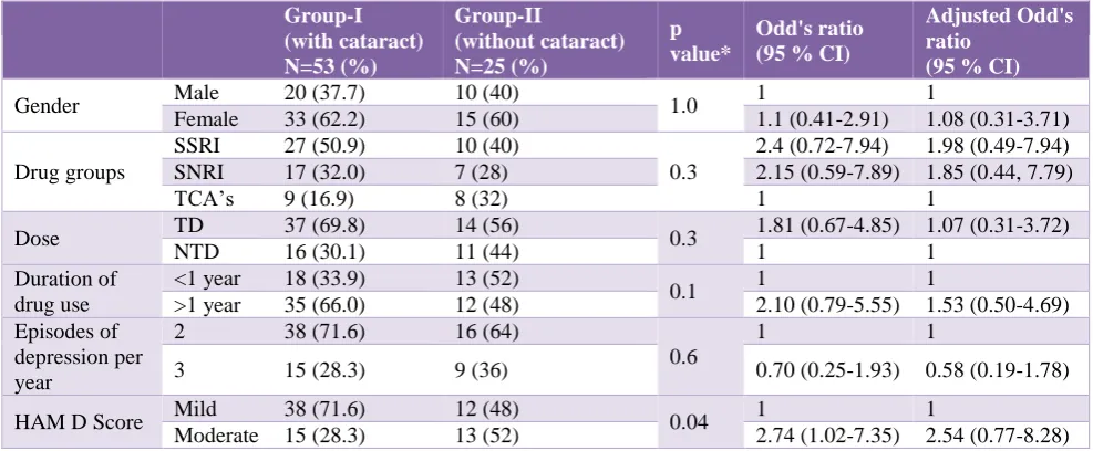
Table 2: Association between development of cataract and various risk factors among patients using various antidepressants.
Group-I (with cataract) N=53 (%)
Group-II
(without cataract) N=25 (%)
p value*
Odd's ratio (95 % CI)
Adjusted Odd's ratio
(95 % CI)
Gender Male 20 (37.7) 10 (40) 1.0 1 1
Female 33 (62.2) 15 (60) 1.1 (0.41-2.91) 1.08 (0.31-3.71)
Drug groups
SSRI 27 (50.9) 10 (40)
0.3
2.4 (0.72-7.94) 1.98 (0.49-7.94) SNRI 17 (32.0) 7 (28) 2.15 (0.59-7.89) 1.85 (0.44, 7.79)
TCA’s 9 (16.9) 8 (32) 1 1
Dose TD 37 (69.8) 14 (56) 0.3 1.81 (0.67-4.85) 1.07 (0.31-3.72)
NTD 16 (30.1) 11 (44) 1 1
Duration of drug use
<1 year 18 (33.9) 13 (52)
0.1 1 1
>1 year 35 (66.0) 12 (48) 2.10 (0.79-5.55) 1.53 (0.50-4.69) Episodes of
depression per year
2 38 (71.6) 16 (64)
0.6
1 1
3 15 (28.3) 9 (36) 0.70 (0.25-1.93) 0.58 (0.19-1.78)
HAM D Score Mild 38 (71.6) 12 (48) 0.04 1 1
Moderate 15 (28.3) 13 (52) 2.74 (1.02-7.35) 2.54 (0.77-8.28)
A total 78 eyes of thirty-nine (39) patients were included for the study. Thirty (38.46%) and forty-eight (61.53%) eyes belonged to males and females, respectively.
Mean age of males (n=7) was 41.8±2.3 years and females (n=32) 40.2±1.0 years. Minimum and maximum age of the patient was 22 and 49 years, respectively. Patients on AD were clustered into three groups e.g. SSRI, SNRI and TCA’s. Their pattern of distribution amongst patients is shown in (Table 1).
In the primary analysis, females had higher risk of cataract development as compared to males. In Group-I, (N=53) eyes with cataract 33 were of females (62.26%)
[image:4.595.54.290.401.537.2]as compared to males 20 (37.7%). Group-II, (N=25) eyes females without cataract were 15 (60%) as compared to males 10 (40%). Group-I with therapeutic dose (TD) of antidepressants (AD) had more cataract 37 (69.81%) as compared to non-therapeutic dose (NTD) 16 (30.1%). Most of the cataractous eyes 35 (66.03%) had AD exposure of more than 1 year that was associated with increased risk of cataract development (OR 2.10; 95% CI, 0.79-5.55). Similarly, amongst users of AD, SSRI was associated with increased risk of cataract development (OR 2.4; 95% CI, 0.72–7.94) with a female preponderance (OR 1.1; 95% CI, 0.41-2.91). Patient with mild degree of depression on HAM-D score showed statistically significant development of cataract (P=0.04) (Table 2).
Table 3: Best corrected visual acuity (BCVA) converted to logarithm of minimal angle of resolution (logMAR) in patients who developed cataract with various antidepressants.
Duration of antidepressant
exposure N Mean± SD
Range
(maximum-minimum) P value
BCVA (logMAR) in patients with cataract
<1 Year 18 0.34±0.11 0.7-0.2
0.07 >1 year 35 0.40±0.14 0.7-0.3
[image:4.595.311.543.560.718.2]P value# calculated by Mann-Whitney test, BCVA-Best corrected visual acuity, logMAR-Logarithm of minimal angle of resolution.
Figure 1: The distribution of BCVA (Snellen acuity) among the eyes which developed cataract.
Figure 2: The distribution of different types of cataract.
In Group-I, the comparison amongst risk of AD users of duration <1 year and >1 year and development of cataract showed mean value for BCVA (logMAR) was 0.34±0.11 and 0.40±0.14 with p-value of 0.07, which was non-significant (Table 3).
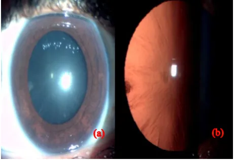
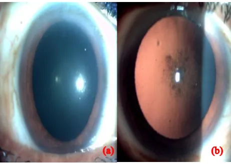
Out of the 53 eyes of the patients, maximum of 34 (64.15%) had cataract with BCVA of ≥6/12 as compare to minimum of 2 (3.77%) having BCVA of ≤6/36 (Figure 1). Lens opacities classification system-III (LOCS-III) revealed 38 (71.69%) peripheral cortical cataract and 15 (28.30%) posterior sub-capsular cataract. (Figure 2, 3 a, b and 4 a, b).
Figure 3 (a, b): Peripheral cortical cataract on diffuse illumination and retro-illumination.
≥ 6/12 6/18 - 6/24 ≤ 6/36
34(64.15%) 2(3.77%)
17(32.07%)
38
71.69
15
28.3
0 20 40 60 80
Number of eyes(N) %(eyes)
[image:4.595.55.294.588.731.2]DISCUSSION
The present study showed that AD was associated with increased risk of cataract at a relatively younger age i.e. <50 years, which cannot be attributed to a bias of senile cataract. Females were at higher risk of cataract as compared to males. SSRIs were strongly associated with cataract as compared to other classes of AD e.g., SNRI and TCA. Drug intake of >1 year was associated with higher rate of cataract formation as compared to <1 year with (OR 2.10, 95% CI 0.79-5.55, P=0.14), which was statistically non-significant. These findings were in agreement with the study by Eries and colleagues that found use of SSRI for 1 or more years associated with an increased risk of cataract (OR 1.36, 95% CI 1.23–1.51; P<0.001). The association between SSRI use and incident of cataract surgery was significant for men (OR 1.34, 95% CI 1.12-1.61) and women (OR 1.37, 95% CI 1.22-1.55). It remained significant after adjusting for risk factors of cataract formation e.g., diabetes mellitus and steroid use (P < 0.001).19
Figure 4 (a, b): Posterior sub-capsular cataract on diffuse illumination and retro-illumination.
Etminan et al., 2010 studied 200,000 residents with age of 65 years and above and reported a possible association between current use to SSRIs i.e., 15% greater relative risk (RR) of cataracts (95% CI 1.08 to 1.23) than non-users after adjusting for confounders e.g., high blood pressure, other medication use and gender etc. The antidepressant drugs e.g., fluvoxamine, venlafaxine and paroxetine had highest risk of cataracts whereas citalopram, fluoxetine and sertraline did not show statistically significant increase.9 In their study, the
average time duration to diagnosis of cataract while patients were on SSRI therapy was 656 days. However, the incidence of community diagnosed cataracts had (adjusted rate ratio 1.06, 95% CI 0.97 to 1.17) with past SSRI use. Amongst SSRIs, the past use of sertraline had 19% elevated risk of cataract (95% of CI 1% to 41%). Any current use of sertraline with cataract diagnosis showed no significant effect on cataract risk (adjusted RR 1.06, 95% CI 0.92 to 1.22).
In the present study, 27 (50.9%) eyes were exposed to SSRI (sertraline) use and developed cataract, which was statistically non-significantly (OR 2.4, 95% CI 0.72-7.94, P=0.3). The findings were consistent as reported earlier by Etminan and colleagues.9 This may suggest
importance of selective serotonin receptors in the formation of cataracts. Catecholamines such as norepinephrine, having catarogenic properties, and pre-marketing clinical trials suggest an association between cataracts and venlafaxine6 whether, it’s a cataractogenic
effect of SSRI or a class effect or limited to specific agents must be investigated in future.
Serotonins have been shown to play a crucial role in lens transparency.12 Animal studies have revealed the role of
serotonin in different areas of eye.5 A wide variety of
serotonin receptors have been identified in the lens of animal models.10,13 Cataractogenic potential of serotonin
was observed in rat model with lens opacification and formation of cortical cataracts after injection of 1% solution of serotonin.5 In the current study, maximum
number of eyes had peripheral cortical cataract, which was visually non-significant but in concordance with the observation by Boerrigter et al.5 One of the postulates for
lens opacification is serotonin interfering with lens metabolism, which compromises ciliary body and anterior chamber. The presence of β receptors in lens and role of cataractogenic properties of catecholamines6
should be further investigated.
In the present study after adjusting for gender and other potential confounding variables e.g., types of AD, duration of drug intake, dosage, episodes of depression and severity of depression, the use of SSRIs for >1 year was associated with increased risk of 35(66%) cataract development (OR 2.10, 95% CI 0.79-5.55, P=0.1) as compared to the duration of <1 year but statistically non-significant. The dosage of AD was a possible risk factor for the development of cataract. The TD showed increase in the development of cataract 37 (69.8%) eyes (OR 1.81, 95% of CI 0.67-4.85) as compared to non-therapeutic dose (NTD) 16 (30.1%). The BCVA for maximum number of eyes 34 (64.15%) was ≥6/12, which required no cataract surgery.
Limitations
• It’s a prescription based study to link the prescription data with psychiatric and ophthalmological diagnoses.
• This study assessed cataract formation rather than cataract surgery and an effort to find an association between the AD and development of cataract.
• The other risk factors that may confound the results were controlled; they were non-significant but statistically or clinically relevant in assessment for development of cataract.
[image:5.595.51.282.324.488.2]Implication
The study highlights the role of SSRI as a risk factor for development of cataract in humans.
CONCLUSION
There is a higher risk of association with cataract formation amongst users of SSRI than SNRI and TCA. SSRI users having exposure more than 1 year or longer for mild degree of depression developed visually non-significant peripheral cortical cataract to non-significant cataract with a female preponderance. Thus, it is recommended that clinicians using AD of longer duration for eligible patients should have periodic follow-up by ophthalmologists to rule out incipient cataract. A larger naturalistic cohort and survival analysis studies are required to establish role of AD having cataractogenic potential before generalizing these results.
ACKNOWLEDGEMENTS
Authors would like to express their sincere thanks to Mr Hem Chandra Sati, Statistical Assistant of All India Institute of Medical Sciences, New Delhi for his immense help in the Biostatistics
Funding: No funding sources Conflict of interest: None declared
Ethical approval: The study was approved by the Institutional Ethics Committee
REFERENCES
1. Ernst P, Baltzan M, Deschenes J, Suissa S. Low-dose inhaled and nasal corticosteroid use and the risk of cataracts. Eur Respir J. 2006;27:1168-74.
2. Kanthan GL, Wang JJ, Rochtchina E, Mitchell P. Use of antihypertensive medications and topical beta-blockers and long- term incidence of cataract and cataract surgery. Br J Ophthalmol. 2009;93:1210-14. 3. Klein BE, Klein R, Lee KE, Danforth LG. Drug use and five-year incidence of age-related cataracts: the beaver dam eye study. Ophthalmol. 2001;108:1670-4.
4. Ludwig J, Marcotte DE, Norberg K. Anti-depressants and suicide. J Health Econ. 2009;28:659-76.
5. Boerrigter RM, Sietsema JV, Kema IP. Serotonin (5-HT) and the rat’s eye: some pilot studies. Doc Ophthalmol. 1992;82(1-2):141-50.
6. Weinstock M, Scott JD. Effect of various agents on drug induced opacities of the lens. Exp Eye Res.1967;6:368-75.
7. Van Walraven C, Mamdani MM, Wells PS, Williams JI. Inhibition of serotonin reuptake by antidepressants
and upper gastrointestinal bleeding in elderly patients: retrospective cohort study. BMJ. 2001;323:655-8.
8. Ginzburg R, Rosero E. Risk of fractures with selective serotonin-reuptake inhibitors or tricyclic antidepressants. Ann Pharmacother 2009; 43: 98-103. 9. Etminan M, Mikelberg FS, Brophy JM. Selective serotonin reuptake inhibitors and the risk of cataracts. Ophthalmology. 2010;117:1251-5.
10. Costagliola C, Parmeggiani F, Sebastiani A. SSRIs and intraocular pressure modifications: evidence, therapeutic implications and possible mechanisms. CNS Drugs. 2004;18(8):475-84.
11. Candia OA, Lanzetta PA, Alvarez LJ, Gaines W. Inhibition of ionic transport and ATPase activities by serotonin analogues in the isolated toad lens. Biochem Biophys Acta. 1980;602:389-400.
12. Vivekanandan S, Lou MF. Evidence for the presence of phosphoinositide cycle and its involvement in cellular signal transduction in the rabbit lens. Curr Eye Res. 1989;8:101-12.
13. Chidlow G, Le Corre S, Osborne NN. Localization of 5-hydroxytryptamine-1A and 5-hydroxytryptamine-7 receptors in rabbit ocular and brain tissues. Neuroscience. 1998;87:675-89.
14. Hoyer D, Hannon JP, Martin GR. Molecular, pharmacological and functional diversity of 5-HT receptors. Pharmacol Biochem Behav. 2002;71:533-54.
15. Sharif NA, Senchyna M. Serotonin receptor subtype mRNA expression in human ocular tissues, determined by RT-PCR. Mol Vision. 2006;12:1040-7.
16. American Psychiatric Association. (DSM-5) Diagnostic and statistical manual of mental disorders (5th ed.) Arlington, VA: American Psychiatric Publishing. 2013:155-88.
17. Hamilton M. Development of a rating scale for primary depressive illness. Br J Soc Clin Psychol. 1967;6(4):278-96.
18. Chylack LT, Wolfe JK, Singer DM et al. Lens Opacities Classification System III. The Longitudinal Study of Cataract Study Group. Arch Ophthalmol. 1993;111:831-6.
19. Erie JC, et al. Selective serotonin reuptake inhibitor use and increased risk of cataract surgery: a population-based, case-control study. Am J Ophthalmology. 2014;158:192-7.