of peer-reviewed research and commentary in the population sciences published by the Max Planck Institute for Demographic Research Konrad-Zuse Str. 1, D-18057 Rostock · GERMANY www.demographic-research.org
DEMOGRAPHIC RESEARCH
VOLUME 9, ARTICLE 8, PAGES 163-196
PUBLISHED 30 OCTOBER 2003
www.demographic-research.org/Volumes/Vol9/8/
DOI: 10.4054/DemRes.2003.9.8
Research Article
Individual Aging and Cancer Risk: How
Are They Related?
Svetlana V. Ukraintseva
Anatoli I. Yashin
1 Introduction 164
2 Cancer incidence rate pattern: typical features and explanations
164
2.1 Age-pattern of overall cancer incidence rate 164 2.2 Mechanisms of the increase in cancer risk with age 167 2.3 Mechanisms of the deceleration and the decline in
cancer risk at old ages
168
3 Aging may not only increase but also reduce cancer risk
172
3.1 How age-associated exposures may increase vulnerability to cancer
172
3.2 Age-related decline in cell proliferation may favor as well as suppress cancer development
173
3.2.1 Age-related decline in cell proliferation may favor survival of malignant tumors in an aging organism
173
3.2.2 Age-related decline in cell proliferation rate may increase risk of malignant transformation
175
3.2.3 How age-related decline in cell proliferation may suppress cancer development
176
3.3 Ontogeny contributes to non-monotonic patterns of cancer morbidity
180
4 Discussion 183
5 Acknowledgements 186
Research Article
Individual Aging and Cancer Risk: How Are They Related?
Svetlana V. Ukraintseva 1
Anatoli I. Yashin 2
Abstract
When individuals get older, the risk of many chronic diseases increases. This increase is in agreement with common theories of aging, such as mutation accumulation, wear and tear, antagonistic pleiotropy, etc. Surprisingly, however, the risk of some chronic conditions (e.g. asthma, arterial hypertension) declines in the old. The cancer incidence rate also declines at old ages after a steep increase during adult life. It contrasts with the continuing increase in total mortality that is often referred to as the aging process. Which forces contribute to a decline in cancer risk in the old? In this paper we review evidence from experimental biology, illustrating the ambivalent role of individual aging in cancer risk, in particular in forming non-monotonic age-patterns of cancer incidence rate. We show that age-associated changes in the organism may contribute not only to the rise, but also to the deceleration and the decline in cancer risk at old ages.
1
Corresponding author: Max Planck Institute for Demographic Research, Rostock, Germany and Research Center for Medical Genetics, Russian Academy of Medical Sciences, 115478 Moscow Tel: +49 (381) 2081-144; Fax: +49 (381) 2081-444. E-mail: [email protected].
2
1. Introduction
Cancer used to be regarded as an aging-related disease. However, in contrast to the increasing pattern of total mortality (often referred to as the aging process), cancer incidence and death rates first increase rapidly and then decelerate or even decline (Source: IARC 1965-1997, Smith 1996, 1999). The risk of many chronic diseases increases with advanced age, and this fact is in agreement with common theories of aging such as mutation accumulation, wear and tear, antagonistic pleiotropy, etc. However, the deceleration or decline in this risk at oldest ages are surprising and unexplained from the point of view of contemporary aging theories. These were shown not only for cancer, but also for a variety of other pathological conditions such as asthma and arterial hypertension (Sankaranarayanan et al.1999, Ukraintseva and Sergeev 2000, Ukraintseva 2000). One explanation of this phenomenon involves effects of a population’s heterogeneity (e.g. related to difference in carcinogenic exposure) (Vaupel and Yashin 1985, 1988). Others require a better understanding of the universal aspects of the aging process, developing at the individual level.
In this paper we show that age-related changes in an organism may not only favor, but also suppress cancer development, and thus they may contribute not only to the rise, but also to the deceleration and the decline in cancer risk at old ages. We analyse findings from human and animal experimental studies in support of this view.
2. Cancer incidence rate pattern: typical features and explanations
2.1. Age-pattern of overall cancer incidence rate
Typical features of the age-pattern of overall cancer incidence rate include (Fig. 1):
i a peak in early childhood ii low rate in youth
iii increase in this rate afterwards
iv the deceleration or decline in the rate at old ages
These features are recurrent over time and place (IARC 1965-1997).
to the increase in cancer risk with age (see e.g. Peto et al. 1975, Rainsford et al. 1985, Volpe and Dix 1986, Dix 1989, Krtolica and Campisi 2002). They ignored other typical features of the cancer rate pattern such as the deceleration and the decline in the rate at old ages. One reason for this might be the use of data on age-specific cancer mortality rather than incidence data. Typical data on cancer mortality are limited by age 75, which does not allow for observation of the decline in the rate at oldest ages (see e.g. EUCAN and GLOBOCAN databases (Ferlay et al. 1999, 2001)). However, the data from special studies on age-specific cancer mortality among the oldest old, combined with available data for earlier ages (Health, US 1997, Smith 1996, 1999), allow us to conclude that the risk of dying from cancer among the oldest old declines with age (Fig. 2).
Figure 2: Cancer incidence rate (1988-92), average annual, and mortality rate (1990) in the USA (IARC 1997, Health US 1996-1997, Smith 1996).
2.2. Mechanisms of the increase in cancer risk with age
Two kinds of mechanisms have been suggested to explain the increase in cancer risk with age.
Exposures. The first explanation refers to simple dose-duration effects of carcinogenic exposures, regardless of any effects of aging. This explanation is supported by the data from rodent experiments with carcinogens and observations on occupational exposure in humans (Doll et al. 1970, Peto et al. 1975, Peto et al. 1985). For example, an experiment involving 950 mice (Peto et al. 1975) showed that regular benzpyrene application to the skin, beginning at 10, 25, 40 or 55 weeks of age, steeply increased the incidence rate of malignant epithelial tumors with time. This increase was associated directly with the duration of exposure but was independent of age at the start of exposure, as were the growth rates of already established tumors. Authors concluded that age equals the duration of exposure to carcinogenic stimuli.
Indeed, universal characteristics of cancer rate patterns suggest an influence of universal factors as well. Carcinogenic exposures might be among such factors only if their effects are similar across human populations and mammalian species. In principle, there are exposures that are universal such as exposure to oxygen. Although these exposures are not directly carcinogenic, they might influence cancer risk through the age-associated change in the internal milieu. However, the mechanism proposed above implies that susceptibility to carcinogens does not increase with age. Thus, the concept suggested by Peto et al (1975) does not involve all kinds of effects of carcinogenic exposures, but only those which do not affect age-associated susceptibility to cancer.
pro-oncogenic tissue microenvironment. DePinho (2000) suggested that cancer-prone phenotype in old age might represent the combined pathogenetic effects of mutation load, telomere dysfunction, and altered stromal milieu. The role of altered stromal milieu was emphasized by most researchers explaining the increase in vulnerability to cancer with age (Cheresov 1997, Rubin 2001, Krtolica and Campisi 2002, Anisimov 2003). These concepts taken together associate the increase in vulnerability to cancer to multiple effects of age. Some of these effects (e.g. changes in tissue microenvironment) actually may arise from age-associated exposures, such as to infectious agents. Although some of the above concepts give acceptable explanations of age-associated increase in cancer risk, they still do not explain the deceleration and the decline in cancer incidence rate at old ages.
2.3. Mechanisms of the deceleration and the decline in cancer risk at old ages
Detection bias. The detection of new cases of cancer often involves complex diagnostic procedures. The use of a number of such procedures (e.g. colonoscopy) may be restricted in the oldest old ages, when individuals are frail, or have multiple chronic conditions. This may create the detection bias since a number of cancers may stay undetected among the oldest old. For this reason the deceleration or decline in the age pattern of cancer incidence rate at oldest old ages, calculated from the available data, may not necessarily reflect the real pattern of changes in cancer risk with age. Several studies have been performed to address this issue.
Stanta et al. (1997) have analysed a group of 507 autopsies of elderly subjects, divided into three age groups, 75-90 years, 95-99, and over 99 (centenarians). The prevalence of cancer was 35% among the younger persons, and 20% and 16% respectively, for two other groups of the oldest old. Accuracy of diagnosis also declined in the oldest old. The authors concluded that both the incidence of cancer and the importance of cancer as a cause of death might decline after age 95. Kuramoto et al. (1993) analysed the prevalence, rate of correct clinical diagnosis, and mortality of cancer in 4,894 consecutive autopsies at the Tokyo Metropolitan Geriatric Hospital from 1972 to 1990. Cancer prevalence decreased with advancing age: 50.0% in the sixties, 47.9% in the seventies, 43.2% in the eighties, and 39.3% in the nineties and over. There is also evidence concerning cancer incidence turnover at old age in laboratory mice (Pompei et al 2001). This significant finding suggests that old age decline in cancer risk is not spurious. Indeed, in the case of experimental animals, such decline can not be related to a diagnostic bias.
rates, many cancer epidemiologists agree on a decelerating and even declining age pattern of these rates at oldest old ages. Some of them have tried to explain such a pattern (Macieira-Coelho 1986, 2001, Vaupel and Yashin 1988, Benson et al. 1996, Ukraintseva and Yashin 2001). The late deceleration in cancer risk could, for instance, reflect diminishing exposure to prevalent carcinogens (e.g. tobacco smoking) at old ages, rather than the effect of somatic aging. Macieira-Coelho (1986) suggested that the fact that cancer risk might level off, or decline at old ages, means that this risk “is related to age rather than to aging”. This point of view implies that physiological aging is not able to decrease cancer risk. Macieira-Coelho (2001) concluded that cancer results from tissue-specific action of genetic, environmental and developmental factors, which are not related to aging.
Selection. Vaupel and Yashin (1988) explained the deceleration and the decline in cancer risk at old ages in terms of differential selection in a heterogeneous population (see also Vaupel and Yashin, 1985 for detail on the concept). Such selection favors survival of individuals who are less prone to cancer (e.g. because they were less exposed to carcinogens during life, or less genetically predisposed to the disease). As a result, a proportion of such individuals increases in an old age population. The change in population structure produces observed effects of the deceleration or the decrease in old age cancer morbidity. This explanation does not involve individual aging as a causal factor.
Individual aging. Few attempts to explain the deceleration and the decline in cancer risk were made in terms of effects of individual aging (Benson et al. 1996, Ukraintseva and Yashin 2001). Benson and co-authors (1996) suggested that the role of aging in cancer incidence be determined by two components. One is responsible for the increase in the incidence rate beginning near age 30 and another for the gradual diminution in this rate of increase. The first component may correspond to the activation of cells with damaged DNA, to the deactivation of DNA repair or to impaired apoptosis. The second one may correspond to the loss of cell division potential with age. The importance of this concept is that it relates aging to both an increase and a deceleration in cancer risk with age. However, the authors have not described mechanisms underlying such a relationship in detail.
We (Ukraintseva and Yashin 2001) suggested dividing age-associated changes in an organism into three groups (time-dependent, basal, and ontogenetic) according to their possibly different influence on risk of age-related diseases, including cancer. Let us briefly describe this idea.
Basal changes represent the effects of universal decline in the rate of physiological processes in the aging organism. The age-related decline in the rate of metabolism, the rate of information processing, and the rate of cell proliferation could be examples (see e.g. Adelman 1971, Buetow 1971, Cameron 1972, Cheron and Desmedt 1980, Grove and Kilgman 1983, Masoro 1985, Katzman 1995, Cerella 1993, Guyton and Hall 1996, Rubin 1997) (Fig. 3).
Figure 3: Human basal metabolism (Guyton and Hall 1996)
Within the framework of basal changes, the main difference between old and young individuals is that the old one lives, thinks and performs basic physiological functions slower than he did when he was young. As a result, some age-related changes, normal as well as pathological, may accumulate at a slower rate with age (Fig. 4). We propose that the basal slowing down may paradoxically contribute to both the increase and the deceleration in the increase in cancer risk with age (see section 3 below).
the organism creates the background for wave-like patterns of chronic morbidity, including that of cancer (Ukraintseva and Yashin 2001).
Figure 4: Examples on decelerated change in physiological parameters with age.
3. Aging may not only increase but also reduce cancer risk
3.1 How age-associated exposures may increase vulnerability to cancer
Available data strongly suggest that changes in tissue microenvironment may favor cancer development (Zimmerman and Carter 1989, McCullough et al. 1994, Cheresov 1997, Bergers and Coussens 2000, Bissell and Radisky 2001, Chang and Werb 2001, Rubin 2001, Krtolica and Campisi, 2002). According to this data, the major roles in such changes are played by: (i) accumulation of harmful substances in extracellular matrix (ECM); (ii) chronic inflammation. Both these conditions can result from age-associated exposures. For example, any organism over the course of its lifetime occasionally deals with substances that can not be utilised by its cells (e.g. talc and heavy metals). Such substances are stored in ECM as non-disposable, and their amount increases with age, (De Duve 1983, Bilych et al. 1999) leading to the disruption of ECM.
3.2 Age-related decline in cell proliferation may favor as well as suppress cancer development
First, one should define what is age-related decline in cell proliferation. It refers to two distinct processes: (i) age-related decline in cell proliferation rate; (ii) an increase in proportion of replicatively senescent cells in an organism with age. The first implies that time between two cellular doublings increases with the age of an organism for cells appertained to the same stage of their replicative life. For instance, stem cells taken from old mice proliferate slower than the stem cells taken from young mice (de Haan et al. 1999). The second implies that cells decline in proliferative potential with each cell doubling. This means that they decrease in the number of divisions left before entering the state of irreversible growth arrest (or, otherwise, terminal non-dividing state). This decrease is often named as “replicative aging”. The terminal non-dividing state is respectively named as “replicative senescence”. Replicatively senescent cells seem to accumulate in the organism with age (Campisi 2000). Each kind of age-related decline in cell proliferation (i.e. decline in cell proliferation rate as well as an increase in proportion of replicatively senescent cells) might have ambivalent influence on cancer risk.
3.2.1. Age-related decline in cell proliferation may favor survival of malignant tumors in an aging organism
disease is twice as frequent in females. Avetisian and Petrova (1996) showed that there was no positive correlation between the high frequency of latent thyroid cancer in the autopsy data and the relatively low annual incidence of clinically evident thyroid cancer in the Ukraine. They have also not found significant differences in the frequency of latent thyroid carcinoma with increased age, while the incidence of clinically evident thyroid cancer increased with age. These results combined suggest that: (i) factors favoring the survival of latent tumors influence cancer risk substantially, (ii) not all-latent tumors survive to contribute to cancer incidence rate, and (iii) the survival of latent tumors depends on age, sex and other factors. Data discussed below suggest that age-related decline in cell proliferation might favor the survival of malignant tumors in an aging organism.
Decline in cell proliferation rate. Farber (1995) stressed that the inhibition of proliferation in normal cells surrounding the tumor is one of the key factors of cancer development. In such an environment, malignant cells may arise and be able to proliferate selectively, relative to the surrounding cells. Tumor cells do not always exhibit a higher proliferation rate than normal host cells (Baserga 1976, Rubin 2001). However, they do not die from apoptosis, and they also do not undergo irreversible growth arrest as normal cells do (Hickman 2002). This gives tumor cells a survival advantage. The age-related decline in proliferation rate of normal cells surrounding the tumor may give tumor cells an additional survival advantage. Kozma (1998) has shown that the normal growth rate of the target tissue needs to be exceeded by the potential tumor. A slowly growing tumor in rapidly growing normal tissue is suppressed. The author concluded that in young organisms (where proliferation rate is high) only fast proliferating tumors might survive, while in old organisms (where proliferation rate of normal cells is low) more cancers, including those with longer doubling time, can establish themselves. This may contribute to the increase in the incidence rate with age.
undergo irreversible growth arrest. Cancer cells are de-differentiated, while the final stage of normal cellular life is terminal differentiation. Cancer cells often exhibit an increased metabolism, while aging cells decline in metabolic activity. Besides, cancer cells may secrete factors promoting angiogenesis, while aging cells do not (Abelev 1997, Hanahan and Weinberg 2000). Many of these cancer features are inherent to most “young” cells in the organism – embryonic cells. Embryonic cells proliferate vigorously, are capable of extensive migration, secrete factors that increase the local supply of blood (i.e. promote angiogenesis), produce enzymes capable of degrading basement membranes (Mintz and Fleischman 1981, Abelev 1997, Bast et al. 2000, Hanahan and Weinberg 2000). One can say that cancer manifests itself as a local, uncontrolled “rejuvenation” in an organism (Ukraintseva 2003, Ukraintseva and Yashin 2003). In this light, the role of organism’s aging in cancer risk seems very peculiar, because it sounds like the aging favors the “rejuvenation”. How does this happen? The fact that cancer cells do not “age”, gives them the proliferative and survival advantage in the surrounding of aging cells. The increase in proportion of senescent cells in an aging organism (Bayreuther et al. 1992, Campisi 2000) might therefore favor survival of already transformed cells and latent tumors, rather than an initial malignant transformation (the latter may actually decline with age, we will discuss this below (see 3.2.3)).
3.2.2 Age-related decline in cell proliferation rate may increase risk of malignant transformation
Age-related decline in the cell proliferation rate may also paradoxically increase the number of loci of chronic proliferation in an organism, which in turn may favor malignant transformation in the old. As mentioned above, the pro-inflammatory status is a typical feature of old age (Franceschi and Ottaviani 1997). It is linked to age-related decline in the immune response. This decline often refers to the age-related decrease in proliferation rate of lymphocytes in response to antigen (Smith and Pereira-Smith 1996). That is, the age-related decrease in the proliferation rate of lymphocytes may favor chronic inflammation accompanied by the increase in local proliferation, and the latter may contribute to an increase in cancer risk in the old.
Collectively, available data imply that age-related decline in cell proliferation might favor cancer development. How is this in accordance with the fact that increased cell proliferation is an established cancer risk factor (Cohen 1991, Butterworth and Goldsworthy 1991, Croy 1993)? First, we have shown above that a decreased cell proliferation rate may paradoxically increase the number of loci of chronic proliferation in the old organism, thus favoring malignant transformation. Next, high proliferation rate, as well as low proportion of senescent (non-proliferating) cells in a young organism, may actually result in higher probability of initial malignant transformation in the young organism, as compared with the old one. At the same time, the probability of survival of earlier transformed cells and latent tumors may be higher in the old organism, due to both a low proliferation rate of normal host cells and a high proportion of senescent (non-proliferating) cells in the tumor’s surrounding. Thus, the dynamic balance between probability of cell transformation and probability of survival of a latent tumor might define the resulting impact of age-related decline in cell proliferation to cancer incidence rate in the old.
3.2.3 How age-related decline in cell proliferation may suppress cancer development
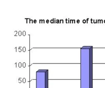
It has been shown that late developing tumors often progress more slowly. They increase in time of tumor doubling, and respectively decrease in the rate of tumor growth with age (Ingram et al. 1992, Peer 1993, Pili et al. 1994, Kozma 1999, Tanaka et al. 2003). This could decelerate the rate of clinical manifestation of respective cancers and contribute to the deceleration of an increase in the cancer risk at old ages (Fig. 5).
Decline in cell proliferation rate. The age-related decrease in the rate of tumor growth might originate from the age-related decline in the rate of physiological processes. As we have stressed above, the general slowing down of physiological processes in an aging organism (e.g. the decline in the rate of metabolism, information processing, and cell proliferation) leads to deceleration in age-related changes of many indices of an organism, normal as well as related to pathology. As a result, pathological effects of such changes may accumulate in the organism at a slower rate with age.
The age-related decline in the rate of physiological processes may directly increase the time between two subsequent doublings of tumor cells in an older organism. For this reason, the time since tumor initiation, till its clinical manifestation, will increase with age.
cancers is worse in a young organism (where the proliferation rate of normal surrounding cells is high) and better in an old organism (where such rate is low). Hence, the proportion of slow growing cancers may increase with the age of the organism. This may create the effect of deceleration of the increase in cancer risk at old ages. The same selection process may be responsible for the increase in the incidence rate with age, because more cancers, including those with longer doubling time, can establish themselves at old ages.
Finally, the age-related decline in the cell proliferation rate may increase time needed to complete all cell divisions required for malignant transformation. This may decelerate the appearance of newly transformed cells in an aged organism, which in turn may contribute to the leveling off in cancer risk at old ages.
Replicative senescence. The increase in the proportion of senescent, non-proliferating cells in an aging organism also may play a role in decreasing cancer risk at old ages. It is well known that non-proliferating cells have low or no probability of malignant transformation. Actually tumors developed from terminally differentiated non-proliferating cells (e.g. mature muscle and nerve cells) are not found (Bast et al, 2000). Therefore, the increase in proportion of senescent (non-proliferating) cells in an old organism would reduce the number of newly transformed cells, “by definition”. Studies of cell cultures support this view, showing that replicative senescence might decrease the risk for neoplastic transformation (Ebbesen 1984, Smith and Pereira-Smith 1996, Campisi1,2 2001).
after exposure to carcinogens), on the grounds of slowed metabolism and rate of information processing in an old organism. An increase in the proportion of damaged proliferating cells in an organism with age may elevate the risk of malignant transformation in the old.
Thus, age-related decline in the cell proliferation rate together with an increase in the proportion of senescent (non-proliferating) cells may reduce the number of newly transformed cells in an old organism, thereby contributing to the deceleration of an increase in the cancer risk at old ages. However, it is not clear if the total of newly transformed cells declines in an old organism. This is because this outcome is also dependent on the proportion of damaged proliferating cells, the proportion of which was shown to increase with age in some tissues of an old organism.
One can conclude that at least three effects of the age-related decline in cell proliferation might contribute to the deceleration of an increase in the cancer risk at old ages:
(i) The age-related decline in the rates of metabolism, information processing and cell proliferation may directly increase the time of tumor doubling in an older organism (Peer 1993, Ingram et al. 1992).
(ii) The survival of slow growing cancers may be worse in a young organism, where proliferation rate of normal surrounding cells is high. Such cancers have higher chances to establish themselves in old individuals (Kozma 1998). (iii) Age-related decline in the rate of cell proliferation, together with the increase
in the proportion of senescent cells, may reduce the number of newly transformed cells in an old organism.
3.3. Ontogeny contributes to non-monotonic patterns of cancer morbidity
After progressive increase during adult life, the overall cancer risk declines in advanced years (IARC 1965-1997). It also exhibits a peak early in life (see Fig. 1 above). The non-monotonic pattern of cancer morbidity might reflect non-monotonic change in vulnerability to the disease with age (i.e. an increase in this vulnerability at early and middle old age, and a decrease at young and oldest old age). Realization of the ontogenetic program could be among factors influencing the vulnerability. This program accounts for rebuilding metabolism at birth, growth, reproductive period and climacteric. It was proposed and supported by a variety of experimental data that the disturbance of neuro-endocrine-immune balance in the organism at critical periods of ontogenesis (e.g. at the climacteric) might increase risk of some cancers (Dilman 1968, 1983, Zimmerman and Carter 1989, Cherezov 1997, Gilbert 1997). However, these studies did not explain the relationship between the ontogenetic changes and the decline in cancer risk at older ages.
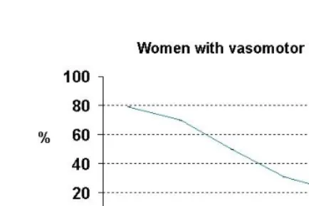
Figure 6: Decline in the proportion of women with vasomotor instability in post-climacteric (Gosden, 1983)
For this reason, morbidity at old ages may sometimes be lower than before. Experimental evidence indicates that in stable periods of ontogenesis, an organism has a mechanism for destruction of the latent tumors, which had appeared in the unstable periods before. The analyses of autopsies taken from people who have died from causes other than cancer showed that latent neuroblastomas of the adrenal gland appeared at a rate of 1:39 in newborn infants (i.e. in the most unstable neonatal period). However, no latent neuroblastomas were found in the glands of infants older than three months (Tulinius 1991).
Figure 7: Age-related change in serum estradiol in women (Dean, 1988).
The low estrogens may protect women from ovarian and endometrial cancers developing later in life, and thus contribute to the decline in risk of these cancers observed at older ages (IARC 1965-1997) (Fig. 8). Thus, new metabolic parameters that become established in the post-reproductive period, may themselves favor the decline in risk of some cancers at old ages.
4. Discussion
We have shown that age-associated changes in an organism may favor as well as suppress cancer development and have suggested several mechanisms explaining such contradictory influence. Among these mechanisms, the ambivalent role of the age-related decline in the rate of physiological processes (in particular, in the rate of cell proliferation) in cancer risk seems to be most intriguing. Still, there are a number of open questions. The first open question concerns the relative contribution of different aging components (i.e. basal, ontogenetic and time-dependent in our terminology) in the forming of non-monotonic patterns of cancer incidence rate. It needs to be defined.
information processing is not clear. A similar problem arises with the analysis of causes of age-related decline in metabolism and cell proliferation rate. Both of these declines may be just consequences of the decline in the rate of information processing, and thus have as causal factors all reasons listed above. There might also be additional causes for these declines. For instance, the age-related decline in the cell proliferation rate might be largely due to the decline in proliferative activity of stem cells in an aging organism. Stem cells are responsible for tissue growth and renewal (NIH 2001). Regenerative ability of stem cells taken from an older organism seems to be worse than that taken from a younger organism. Recipient mice, transplanted with hematopoietic stem cells taken from young donors, showed significantly higher survival rates than those mice with transplants from adult donors (Hirayama et al. 2000). Recent data suggest that stem cells may decline in proliferation rate, relative to the age of the organism. For example, hematopoietic stem cells, isolated from old mice, show lower cycling activity than the stem cells isolated from young mice. The age-related decline in proliferation rate of stem cells is stronger in short-living, as compared with long-living mice strains. The initial proliferation rate as well as the rate of decline in cycling activity during aging seem to be both related to genetically determined inner properties of the stem cells (de Haan et al. 1997, 2000). This very significant finding suggests that the age-related decline in the rate of major physiological processes (i.e. basal component of aging in our terminology) may be a part of the genetic program. Stem cells also show the potential to hunt malignant cells. Neural stem cells (NSCs), when implanted into intracranial gliomas in vivo in adult rodents, distribute themselves quickly and extensively throughout the tumor, and migrate uniquely in juxtaposition to widely expanding and aggressively advancing tumor cells. The NSCs surround the invading tumor border while chasing down infiltrating tumor cells (Aboody et al. 2001). Age-related decrease in proliferative activity of the stem cells may primarily contribute to the better chances of survival of malignant tumors in an older organism, being partly responsible for the age-related increase in cancer risk. Respectively, the higher proliferation rate of stem cells in young organisms may primarily contribute to lower cancer risk in the young as well as to commonly better survival of young individuals from diagnosed cancer (IARC 1995).
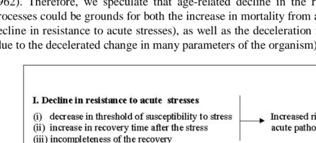
manifestation of some diseases, including cancer, at old ages. Another consequence of the age-related decline in the rate of the physiological processes may be the decline in resistance to acute stresses (such as the flu). This decline in stress resistance is one of the universal features of aging. It is characterized by: (i) an increase in susceptibility to stress, (ii) an increase in recovery time after the stress, and (iii) incompleteness of the recovery (see examples in Cerella 1993, Grove and Kilgman 1983, Ida et al. 1984, O'Neal and Polse 1986, Hall et al. 1989, Lavie et al. 1992, Choi et al. 1995, Hof and Patri 2001). Age-related decreases in rates of metabolism, proliferation and information processing may produce all these features, especially the increase in recovery time after the stress. The age-related decline in stress resistance has a consequence in a higher probability of death following the same stress at older ages. This has been discussed as a major biological reason for the increasing risk of death with age (see e.g. Strehler 1962). Therefore, we speculate that age-related decline in the rate of physiological processes could be grounds for both the increase in mortality from all causes (due to the decline in resistance to acute stresses), as well as the deceleration in chronic morbidity (due to the decelerated change in many parameters of the organism) at old ages (Fig. 9).
The next open question is how to apply these findings to cancer prophylaxis and treatment? One of the above ideas is that factors favoring survival of a malignant tumor might be more contributory to the risk of clinical manifestation of cancer than cell transformation per se. To accept this idea, then prophylactic gauges should include not only avoiding exposure to direct carcinogens, but also methods of suppressing latent tumors in aged organism. For example, one such method could involve a controlled “rejuvenation” of normal tissue in the area near a tumor with stem cell grafting. It would aim to supplant cancer cells rather than to kill them. Of course, this idea is merely speculation. Still, we believe that understanding the ambivalent role of individual aging in cancer risk could be helpful for the development of new therapeutics.
The role of differential selection in a heterogeneous population in forming age-patterns of cancer incidence rate. This is the last, but not least, open question. It was studied in depth by Vaupel and Yashin (1985, 1988). However, additional studies are needed to separate the effects of population heterogeneity from the effects of individual aging on cancer risk in the old.
5. Acknowledgements
References
Abelev, G. I. (1997) [“What a tumor is.”] Sorosovsky Obrazovatelny Journal, 10: 85– 90 (in Russian).
Aboody, K. S., Brown, A., Rainov, N.G., Bower, K.A., Liu, S., Yang, W., Juan, E., Herrlinger, U., Ourednik, V., Black, P., Breakefield, X.O., Snyder, E.Y. (2000) “Neural stem cells display extensive tropism for pathology in adult brain: Evidence from intracranial gliomas.” PNAS, 97, 23: 12846–12851.
Adelman, R.C. (1971) “Age-dependent effects in enzyme induction - a biochemical expression of aging.” Exp Gerontol 6(1): 75-87.
Albanes, D., Winick, M. (1988) “Are cell number and cell proliferation risk factors for cancer?” J Natl Cancer Inst, 80, 10: 772-4.
Anisimov, V. (1987) Carcinogenesis and Aging. Boca Raton, FL: CRC Press.
Anisimov, V. (2003) “The relationship between aging and carcinogenesis: a critical appraisal.” Crit Rev Oncol Hematol., 45(3): 277-304.
Avetisian, I.L., Petrova, G.V. (1996) “Latent thyroid pathology in residents of Kiev, Ukraine.” Environ Pathol Toxicol Oncol, 15, 2-4: 239-43.
Baserga, R. (1976) Multiplication and division in mammalian cells. NY: Marcel Dekker.
Bast, R.C., Kufe, D.W., Pollock, R. E., Weichselbaum, R.R., Holland, J.F., Frei, E., editors. Cancer Medicine. 5th ed. (2000) Canada: BC Decer Inc.
Bayreuther, K., Francz, P.I., Gogol, J., Konterman, K. (1992) “Terminal differentiation, aging, apoptosis and spontaneous transformation in fibroblast stem cell systems in vivo and in vitro.” Ann. New York Acad. Sci, 663: 167–179.
Benson, D., Mitchell, N., Dix, D. (1996) “On the role of aging in carcinogenesis.” Mutation Research/Fundamental and Molecular Mechanisms of Mutagenesis, 356, 2: 209-216.
Bergers, G., Coussens, L.M. (2000) “Extrinsic regulators of epithelial tumor progression: metalloproteinases.“ Current Opinion in Genetics & Development, 10: 120-127.
Bissell, M.J., Radisky, D. (2001) “Putting tumours in context.” Nat Rev Cancer,1, 1: 46-54.
Breslow, N., Chan, C.W., Dhom, G., Drury, R.A., Franks, L. M., Gellei, B., Lee, Y. S., Lundberg, S., Sparke B, Sternby NH, Tulinius H. (1977) “Latent carcinoma of prostate at autopsy in seven areas.” Int J Cancer, 20, 5: 680-8.
Buetow, D.E. (1971) Cellular content and cellular proliferation changes in the tissues and organs of the aging mammal. In: Cameron, I.L., Thrasher, J.D., editors. Cellular and Molecular Renewal in the Mammalian Body. New York: Academic Press: 87–105.
Butterworth, B.E., Goldsworthy, T.L. (1991) “The role of cell proliferation in multistage carcinogenesis.” Proc Soc Exp Biol Med, 198, 2: 683-7.
Cameron, I.L. (1972) “Cell proliferation and renewal in aging mice.” J. Gerontol, 27: 162–172.
1
Campisi, J. (2001) “Cellular senescence as a tumor-suppressor mechanism.” Trends Cell Biol, 11, 11: S27-31.
2
Campisi, J. (2001) “From cells to organisms: Can we learn about aging from cells in culture?” Exp Gerontol, 36, 4-6: 607-18.
Campisi, J. (2000) “Cancer, aging and cellular senescence.” In Vivo,14, 1: 183-8.
Cerella, J. (1993) Adult Information Processing : Limits on Loss. Academic Press.
Chang, C. and Werb, Z. (2001) “The many faces of metalloproteases: cell growth, invasion, angiogenesis and metastasis.” Trends in Cell Biology, 11, 11: S37-S43.
Cheresov, A.E. (1997), Russian, ["Theory of Cancer: The Role of Tissue Homeostasis"]. Moscow: Moscow State University.
Cheron, G., Desmedt, J.E. (1980) “Peripheral and central somatosensory pathways and evoked cerebral potentials during aging.” Rev. Electroencephalogr. Neurophysiol. Clin,10: 146-152.
Choi, S.J., Harii, K., Lee, M.J., Furuya, F., Ueda, K. (1995) Electrophysiological, morphological, and morphometric effects of aging on nerve regeneration in rats. Scand J Plast Reconstr Surg Hand Surg 1995 Jun;29(2):133-40
Cohen, S.M., Purtilo, D.T., Ellwein, L.B. (1991) “Ideas in pathology. Pivotal role of increased cell proliferation in human carcinogenesis.” Mod Pathol, 4, 3: 371-82.
Cohen, S.M., Ellwein, L.B. (1996) “Re: E. Farber, Cell proliferation as a major risk factor for cancer: a concept of doubtful validity.Cancer Res., 55: 3759-3762, 1995.” Cancer Res, 56, 18: 4269-70.
Coussens, L.M., Werb, Z. (2002) “Inflammation and cancer.” Nature, 420(6917): 860-7.
Croy, R.G. (1993) “Role of chemically induced cell proliferation in carcinogenesis and its use in health risk assessment.” Environ Health Perspect,101, Suppl 5: 289-302.
Datta, S.R., Greenberg, M.E.(1998) Molecular mechanisms of neuronal survival and apoptosis. In: O’Malley, B., editor. Hormones and Signaling. San Diego: Academic Press: 257-305.
Dean, W. (1988) Biological Aging Measurement. Los Angeles: Center for Biogerontology.
De Duve, C. (1983) “Microbodies in the living cell.” Sci Am, 248, 5: 74-84.
DePinho, R.A. (2000) “The age of cancer.” Nature, 408, 6809: 248-54.
Dilman, V.M. (1968), [Aging, Climacteric and Cancer]. Leningrad: Medicina. (in Russian).
Dilman, V.M. (1983), [Endocrine Oncology]. Leningrad: Medicina. (in Russian).
Dix, D. (1989) “The role of aging in cancer incidence: an epidemiological study.” J Gerontol, 44, 6: 10-8
Doll R, Morgan LG, Speizer FE. (1970) “Cancers of the lung and nasal sinuses in nickel workers.” Br J Cancer, 24: 624–632.
Dolle, M.E., Giese, H., Hopkins, C.L., Martus, H.J., Hausdorff, J.M., Vijg, J.(1997) “Rapid accumulation of genome rearrangements in liver but not in brain of old mice.” Nat Genet, 17, 4: 431-4.
Ebbesen, E. (1984) “Cancer and normal ageing.” Mech. Ageing Dev, 25: 269-283.
Ferlay, J., Bray, F., Sankila, R., Parkin, D.M. (1999) EUCAN: Cancer Incidence, Mortality and Prevalence in the European Union in 1996, version 3.1. IARC Cancer Base No. 4. Lyon: IARC Press.
Ferlay, J., Bray, F., Pisani, P., Parkin, D.M., Ferlay, I.(2001) GLOBOCAN 2000: Cancer Incidence, Mortality and Prevalence Worldwide, version 1.0. IARC Cancer Base No.5 .Lyon: IARC Press.
Franceschi, C., Ottaviani, E. (1997) “Stress, inflammation and natural immunity in the aging process: a new theory.” Aging Clin. Exp. Res, 9, Suppl to 4: 40-31.
Geiger, H., Van Zant, G. (2002) “The aging of lympho-hematopoietic stem cells.” Nat Immunol, 3(4): 329-33.
Gilbert, C. (1997) “Major human cancers are preventable: physiological stimuli induce a dopamine-thyroid-immune efficient mechanism.” Eur J Cancer Prev, 6, 3: 269-76.
Gosden, R.G. (1985) Biology of Menopause: The Causes and Consequences of Ovarian Ageing. London: Academic Press.
Gosden, R.G., Gosden, R.C. (1997) Biology of Menopause: The Causes and Consequences of Ovarian Aging. London: Academic Press.
de Graaff J., Stolte L.A. (1978) “Age at menarche and menopause of uterine cancer patients. “ Eur J Obstet Gynecol Reprod Biol, 8, 4: 187-93.
Grove, G., Kilgman, A. (1983). “Age-associated changes in human epidermal cell renewal.” J. Geront, 38: 137-142.
Guyton, A.C., Hall J.E. (1996) Textbook of Medical Physiology. Philadelphia: W.B. Saunders Co: 906-909.
de Haan, G., Nijhof, W., Van Zant, G. (1997) “Mouse strain-dependent changes in frequency and proliferation of hematopoietic stem cells during aging: correlation between life span and cycling activity.” Blood, 89, 5: 1543-50.
de Haan, G., Szilvassy, S.J., Meyerrose, T.E., Dontje, B., Grimes, B., Van Zant, G. (2000) “Distinct functional properties of highly purified hematopoietic stem cells from mouse strains differing in stem cell numbers.” Blood, 96, 4: 1374-9.
Hall, D.M., Xu, L., Drake, V.J., Oberley, L.W., Oberley, T.D., Moseley, P.L., Kregel, K.C. (2000) “Aging reduces adaptive capacity and stress protein expression in the liver after heat stress.” Appl Physiol 89, 2: 749-59.
Hasty, P., Vijg, J. (2002) “Genomic priorities in aging.” Science, 296, 5571:1250-1.
Health, United States. (1997) U.S. Department of Health and Human Services Publication No. (PHS) 97-1232, available online at http://www.cdc.gov/nchs/products/pubs/pubd/hus/hus.htm
Hickman, J.A. (2002) “Apoptosis and tumourigenesis.” Current Opinion in Genetics & Development, 12: 67–72.
Hirayama, M., Azuma, E., Jiang, Q., Kobayashi, M., Iwamoto, S., Kumamoto, T., Kisenge, R., Yamamoto, H., Komada, Y. (2000) “The reconstitution of CD45RBhiCD4+ naive T cells is inversely correlated with donor age in murine allogeneic haematopoietic stem cell transplantation.” Br J Haematol, 111, 2: 700-7.
IARC (The International Agency for Research on Cancer [France]). (1965-1997) Cancer Incidence in Five Continents. Volumes I-VII. IARC Sci. Publ. Lyon: IARC.
IARC (1995) Berrino, F., Sant, M., Verdecchia, A., editors. Survival of Cancer Patients in Europe. The EUROCARE Study. IARC Sci. Publ. No.132. Lyon: IARC.
IARC (1997) Parkin, D.M., Whelan, S.L., Ferlay, J., Raymond, L., Young, J., editors. Cancer Incidence in Five Continents. Volume VII. IARC Sci. Publ. No.143. Lyon: IARC.
IARC (1998) Monographs on the Evaluation of Carcinogenic Risks to Humans, Volume 72. Hormonal Contraception and Postmenopausal Hormonal Therapy. Lyon: IARC.
Ida, Y., Tanaka, M., Tsuda, A., Kohno, Y., Hoaki, Y., Nakagawa, R., Iimori, K., Nagasaki, N. (1984) “Recovery of stress-induced increases in noradrenaline turnover is delayed in specific brain regions of old rats.” Life Sci, 34, 24: 2357-63.
Ingram, D.M., Roberts, A., Nottage, E.M. (1992) “Host factors and breast cancer growth characteristics.” Eur J Cancer, 28A, 6-7: 1153-61.
Katzman, R. (1995) Human nervous system. In: Masoro, E., editor. Handbook of Physiology: Section 11, Aging. NY: Oxford Univ. Press: 325-344.
Krtolica, A., Parrinello, S., Lockett, S., Desprez, P.Y., Campisi, J. (2001) “Senescent fibroblasts promote epithelial cell growth and tumorigenesis: a link between cancer and aging.” Proc Natl Acad Sci, 98, 21: 12072-7.
Krtolica A, Campisi J. (2002) “Cancer and aging: a model for the cancer-promoting effects of the aging stroma.” Int J Biochem Cell Biol, 34, 11: 1401.
Kuhn, H. G., Dickinson-Anson, H., Gage, F. H. (1996) “Neurogenesis in the dentate gyrus of the adult rat: age-related decrease of neuronal progenitor proliferation.” J. Neurosci, 16: 2027-2033.
Kuramoto, K., Matsushita, S., Esaki, Y., Shimada, H. (1993), Japanese, [“Prevalence, rate of correct clinical diagnosis and mortality of cancer in 4,894 elderly autopsy cases”] Nippon Ronen Igakkai Zasshi., 30(1):35-40.
Lavie, L., Weinreb, O., Gershon, D. (1992) “Age-related alterations in superoxide anion generation in mouse peritoneal macrophages studied by repeated stimulation and heat shock treatment.” J Cell Physiol, 152, 2: 382-8.
Macieira-Coelho, A. (1986) “Cancer and aging.” Exp Gerontol, 21, 6: 483-95.
Macieira-Coelho, A. (2001) “Neoplastic disease through the human life span.” Biogerontology, 2, 3: 179-92.
Masoro, E. (1985) Metabolism. In: Finch, C., Hayflick, L., editors. Handbook of the biology of aging. NY: Van Nostrand Reinhold: 540-563.
McCullough, K.D., Coleman, W.B., Smith, G.J., Grisham J.W. (1994) “Age-dependent regulation of the tumorigenic potential of neoplastically transformed rat liver epithelial cells by the liver microenvi-ronment.” Cancer Res, 54: 3668-3671.
Miller, D.G. (1980) “On the nature of susceptibility to cancer” Cancer, 46:1307-18.
Mintz, B., Fleischman, R.A. (1981) “Teratocarcinomas and other neoplasms as developmental defects in gene expression.” Adv Cancer Res, 34: 211-78.
NIH (2001) Stem Cells: Scientific Progress and Future Research Directions. Department of Health and Human Services.
O'Neal, M.R., Polse, K.A. (1986) “Decreased endothelial pump function with aging.” Invest Ophthalmol Vis Sci, 27, 4: 457-63.
Peto R, Roe FJ, Lee PN, Levy L, Clack J. (1975) “Cancer and ageing in mice and men.” Br J Cancer, 32, 4: 411-26.
Peto R, Parish SE, Gray RG. (1985) “There is no such thing as ageing, and cancer is not related to it.” IARC Sci Publ, 58: 43-53.
Pili, R., Guo, Y., Chang, J., Nakanishi, H., Martin, G.R., Passaniti, A. (1994) “Altered angiogenesis underlying age-dependent changes in tumor growth.” J Natl Cancer Inst, 86, 17: 1303-14.
Pompei F, Polkanov M, Wilson R. (2001) “Age distribution of cancer in mice: the incidence turnover at old age.” Toxicol Ind Health, 17 (1): 7-16.
Preston-Martin, S., Pike, M.C., Ross, R.K., Henderson, B. E. (1993) “Epidemiological evidence for the increased cell proliferation model of carcinogenesis.” Environ Health Perspect, 101, Suppl 5: 137-8.
Rainsford J, Cohen P, Dix D. (1985) “On the role of aging in cancer incidence: analysis of the lung cancer data.” Anticancer Res, 5, 4: 427-30.
Roitt, I.M. (1997) Roitt’s Essential Immunology. Oxford: Blackwell Science
Rubin, H. (1997) “Cell aging in vivo and in vitro.” Mech. Ageing Dev, 98: 1-35.
Rubin, H. (2001) “The role of selection in progressive neoplastic transformation.” Adv Cancer Res., 83:159-207.
Sankaranarayanan, K., Chakraborty, R., Boerwinkle, E. (1999) “Ionizing radiation and genetic risks. VI. Chronic multifactorial diseases: a review of epidemiological and genetical aspects of coronary heart disease, essential hypertension and diabetes mellitus”, Mutat Res, 436: 21-57.
Smith, D. (1996) “Cancer mortality at very old ages.” Cancer, 77, 7: 1367-72.
Smith, D. (1999) Resistance to causes of death: a study of cancer mortality resistance in the oldest old. In: Robine J-M et al., editors. The Paradoxes of Longevity. Springer-Verlag: 61- 71.
Smith, J. R., Pereira-Smith, O. M. (1996) “Replicative senescence: Implications for in vivo aging and tumor suppression.” Science, 273: 63-66.
Sohal, R., Weindruch, R. (1996) “Oxidative stress, caloric restriction, and aging.” Science, 273: 59-63.
Strehler, B.L. (1962) Time, Cells and Aging. New York: Academic Press.
Suh, Y., Lee, K.A., Kim, W.H., Han, B.G., Vijg, J., Park, S.C. (2002) “Aging alters the apoptotic response to genotoxic stress.” Nat Med, 8, 1: 3-4.
Summerhayes, L.C., Franks, L.M. (1979) “Effects of donor age on neoplastic transformation of adult mouse bladder epithelium in vitro.” J. Natl. Cancer Inst, 62: 1017-1032.
Tanaka, Y., Hongo, K., Tada, T., Sakai, K., Kakizawa, Y., Kobayashi, S. (2003) “Growth pattern and rate in residual non-functioning pituitary adenomas: correlations among tumor volume doubling time, patient age, and MIB-1 index.” J Neurosurg, 98, 2: 359-65.
Tulinius, H. (1991) Latent malignancies at autopsy: a little used source of information on cancer biology. In: Riboli, E., Delendi, M., editors. Autopsy in Epidemiology and Medical Research. IARC Sci Publ. 112: 253-61.
Ukraintseva, S.V. (2000) [“On the role of age in asthma morbidity.”] Clinical Gerontology, 6, 7-8: 29-33 (in Russian).
Ukraintseva, S.V., Sergeev, A.S. (2000) “Analysis of genetic heterogeneity of bronchial asthma as related to the age of onset” Russian Journal of Genetics, 36, 2: 201-205.
Ukraintseva, S.V., Yashin, A. I. (2003) “Opposite phenotypes of cancer and aging arise from alternative regulation of apoptosis and growth signalling pathways.” Annals of the NY Acad. of Science, Forthcoming.
Ukraintseva, S.V. (2003) Cancer and aging: Opposite phenotypes created by common genes. In: Diderich, M., editor. Proceedings of the Apoptosis 2003 meeting. Luxembourg: 462.
Ukraintseva S.V. and Yashin A. I. (2001) “How individual age-associated changes may influence human morbidity and mortality patterns.” Mech. Aging & Development, 122: 1447-1460.
Vaupel, J.W., Yashin, A. I. (1985) “Heterogeneity’s Ruses: some surprising effects of selection on population dynamics.” The American Statistician, 39: 176-185.
Volpe, E.W., Dix, D. (1986) “On the role of aging in cancer incidence: cohort analyses of the lung cancer data.” Anticancer Res, 6, 6: 1417-20.
Whelton, P. (1985) Blood pressure in adults and the elderly. In: Bulpitt, C., editor. Epidemiology of Hypertension. NY: Elsevier: 52-53.
Yancik, R. (1993) “Ovarian cancer.” Cancer, 73: 517-523.
Yancik, R., Ries, L.A. (1994) “Cancer in older persons.” Cancer, 74: 1955-2013.
Yatani, R., Shiraishi, T., Nakakuki, K., Kusano, I., Takanari, H., Hayashi, T., Stemmermann, GN. (1988) “Trends in frequency of latent prostate carcinoma in Japan from 1965-1979 to 1982-1986.” J Natl Cancer Inst, 80, 9: 683-7.
Zimmerman, J.A., Carter, T.H. (1989) “Altered responses to chemical carcinogens in aged animals.” J Gerontol, 6, 44: 19–24.