http://www.arjournals.org/index.php/ijpm/index
Original Research Article
Phytochemical Screening and Toxicity Study of Saraca asoca Bark
Methanolic Extract
Manas Kumar Mukhopadhyay1, Debjani Nath1*
*Corresponding author:
Debjani Nath*
1Department of Zoology, University of Kalyani, Kalyani, Nadia West Bengal,
India, 741235.
A B S T R A C T
The study was conducted to screen for the phytochemicals constituents of bark of Saraca asoca (Roxb.), one of the folk medicinal plants found in West Bengal, Bangladesh and Sri Lanka. The acute and sub- acute toxicity of methanolic extract of S. asoca bark on mice was studied. Phytochemical analysis of S. asoca (bark) methanolic extract revealed the presence of high percentage of flavonoids and reducing sugar in addition with saponins, glycosides, steroids, anthraquinones and volatile oil. In mice, daily single oral doses of S. asoca
methanolic extract (0.3 and 1.2 g/Kg body weight) were well tolerated and no adverse behavioral effect was found as used for 30 days (LD50=6.526 g/Kg for acute administration) and induced no significant changes in body and organs weights. However, hematological parameters showed a significant decrease in platelet count (432±98.3, p<0.05) when the dose is 1.2 g/Kg body weight in relation to control animals, suggesting disturbances in platelet production whereas no effect was found on serum biochemical parameters like serum glutamate oxaloacetate transaminase (SGOT), serum glutamate pyruvate transaminase (SGPT), serum alkaline phosphatase (ALP), and bilirubin, total protein, uric acid, urea, creatinin. This study indicates that Saraca asoca is non-toxic for both acute and sub-acute oral administrations.
Keywords: Saraca asoca, acute and sub acute toxicity, bark extract, toxicity profile.
Introduction
Phytochemicals are secondary plant metabolites that can protect plants from predators, act on animal cells and tissues to affect DNA formation and destroy cell membranes [1]. It is also known that phytochemicals confer pharmacological relevance on plant generally. The growing interest in herbal medicine demands information on various plant preparations used in the treatment of diseases [2, 3]. Scientific evaluation of medicinal plants is important to the discovery of novel drugs and also helps to assess toxicity risks associated with the use of either herbal preparations or conventional drugs of plant origin. Saraca asoca
(Roxb.) De Wilde or S. indica Linn. (Family: Caesalpiniaceae; local names: Ashok, Anganapriya, etc.) is a medicinal plant of West Bengal, Bangladesh and Sri Lanka. It is a medium sized evergreen tree up to 9 m height with numerous branches. Bark extract is bitter in taste and has a stimulatory effect on endometrium and ovarian tissue and used in uterine fibrosis, menorrhagia, bleeding hemorrhoids and hemorrhagic dysentery and also as astringent [4]. Flowers are used in regulation of inflammation, dysentery, and hemorrhoids [5]. A preliminary study showed the presence of catechols, sterols, tannins, glycosides, leukocyanidins and leukopelargonidin in bark extract of Saraca asoca [6]. Saraca asoca is
ISSN: 0975-0185
499 popular in many communities as being used as herbal medicine [7]. But scientific information on its medicinal use and side effects is however lacking. The objective of this study is to determine the phytochemical constituents of Saraca asoca bark and to establish the acute and sub-acute toxicity levels of these chemicals in mice model.
Materials and methods
Animals:Six to eight weeks old Swiss albino mice (Mus musculus) of body weight 15-20 gm were purchased (of the same strain) and were housed in a stainless steel wire cages (Tarsons, India), maintained on a 12 hour light-dark cycle in the animal house of Department of Zoology, University of Kalyani. Pellet diet (West Bengal Diary and Poultry development corp. Ltd., Kalyani Industrial Area, Kalyani) and water was provided ad libitum. Water was supplied ad libitum through the tubing throughout the study. The animals were grouped as 5-6 in number per cage and provided with normal environmental conditions (temperature at 25±1°C and humidity at 55±5%). These mice were kept for a minimum of one week prior to oral administration at the Departmental animal room to allow for their acclimatization to the laboratory conditions.
Bark collection and extraction of phytochemicals:
S. asoca bark was harvested during the month of February-March. The family and species of S. asoca
were confirmed by eminent taxonomists using standard methods. Pieces of bark were air-dried at room temperature until complete drying. They were kept away from direct sun light to avoid destruction of active compounds. The bark pieces were pounded to powder using metallic motor and pestle.
Extraction process
Ten grams of dried crude powder of S. asoca bark was used for extraction by soxhlet extractor. Plant material was defatted by petroleum ether before extraction in methanol. Methanol was evaporated using vacuum evaporator under reduced pressure from the extract. The residue was left to air dry at room temperature for 72 hour. The dry residue was stored at 4°C in air-tight bottle. The extraction yield was 12% (W/W).
Qualitative tests on bark extracts of S. asoca
500 Chemicals:
Chemicals used in this study were purchased from MERCK, India. All the chemicals were of analytical grade.
Acute and sub-acute oral toxicity study:
Toxicity study was performed according to the Organization of Economic Co-operation and Development (OECD) guideline for testing of chemicals (OECD 2001). In order to study acute toxic effect of the plant extract 7 groups of 10 animals (5 male and 5 female) were used. Group I used as control and II to VII used as treatment groups. Last six groups were received a single oral dose of 1, 2, 3, 4, 5 and 6 g/Kg body weight of the methanolic extract respectively for acute toxicity study. All the mice were fasted overnight prior to treatment and body weight of each of them were recorded. All the animals of the last six groups were administered the respective amount of methanolic extract dissolved in 1 ml of distilled water, the control group of animals was administered only distilled water through gastric tubing. On the 15th day, all mice were kept fasted for 15 h, and then sacrificed for necropsy examination. The internal organs were excised and weighed. The gross pathological changes of the tissues were studied and LD50 value was determined. The selection of doses for mice used to study sub-acute toxicity was determined from the equivalence of 15-120 times of normal human dose (10-20 mg/Kg) that also in accordance to the information collected from herbalists. The low dose was 0.3g/kg and the higher dose was selected as 1.2g/kg. The extract was administered daily for 30 days to the mice of both low dose group and higher dose group (each containing 5 male and 5 female animals) through gastric tubing and the control group containing the vehicle. On the 31st day mice were made fasted for 15 h and sacrificed. Blood samples for hematological blood analyses were taken by puncturing heart. The internal organs were weighed to determine relative organs weights, and examined for gross lesions. All tissues were fixed in Buins solution for histological examination.
Behavioral study:
The animals were observed for gross behavioral changes related to CNS depression after dosing (hypo activity, passivity, relaxation and ataxia), CNS stimulation (hyperactivity, irritability, convulsion, straub tail and analgesia) and ANS stimulation (ptosis and exophthalmia) at 1, 2, 3, 4, 12, 48 hours of interval for 14 days after each treatment [11]. The change in body weight and rate of mortality were also recorded for 14 days of observation to test acute toxicity. The deviations in normal behavior, coat conditions, discharge, movement and mortality were also studied for 30 days to test sub-acute toxicity.
Ethical clearance:
The protocol used in this study was approved by the animal ethical committee, Department of Zoology, University of Kalyani (under Committee for the Purpose of Control and Supervision of Experiments on Animals, India).
Hematological parameters:
Collected blood was used to determine hemoglobin concentration (g %) using Sahil’s hemoglobinometer and total WBC, RBC and platelets were counted using hemocytometer chamber [12].
Body weight and organ weights:
The body weight of experimental animals was measured for both acute and sub-acute treatment group. Heart, lung, liver, kidney and pancreas were considered for measurement immediately after sacrifice.
Serum biochemical parameters:
501 Histological studies:
After sacrifice different organs like heart, lung, liver, kidney and pancreas of animals from each group were subjected for histological examinations. After fixing the tissues in Buins solution the tissues were dehydrated by passing through ethanol gradation and made ready for paraffin block preparation. The tissues were sectioned at the thickness of about 5μ. Routine histological studies were performed by using Haemotoxylin stain.
Data analysis:
All values are expressed as mean± SEM. The statistical comparisons were made using students-t test. Values were considered significant at p<0.05 level. From the graph equation of probit against log dose of the methanol extracts, the log dose that responded to probit 5 (50% deaths) was calculated and its anti-log gave the LD50 value [13].
Results
Preliminary phytochemical analysis of Saraca asoca
bark:
The results of qualitative analysis of phytochemicals present in bark of Saraca asoca, is summarized in Table 1 that shows mild, moderate or substantial amount of presence or absence of the phytochemicals.
Table 1: Phytochemicals present in bark of Saraca asoca. Sl. No. Phytochemicals Presence
1 Alkaloids -
2 Anthraquinones +
3 Flavonoids +++
4 Resins -
5 Saponins -
6 Glycosides ++
7 Reducing sugars +++
8 Tannins ++
9 Steroids +
11 Terpinoids -
12 Volatile oil +
(-) = absent (<10mg/Kg),
(+) = mild presence (10mg/Kg to 50mg/Kg),
(++) = moderate presence (50mg/Kg to 100mg/Kg),
(+++) = substantial presence (>100mg/Kg).
Acute oral toxicity:
The effects of S. asoca methanolic extract in mice after oral administration are summarized in Table 2. There was an irregular dose-dependent increase in mortality. The first mouse died at a dose of 3 g/kg, 3 mice died at 4 g/kg, 3 died at 5 g/kg and seven died at 6 g/kg body weight within 2 days of dosing. However, the observed symptoms associated with ingestion of S. asoca
methanolic extract included minor to more noticeable convulsions, disorientation, hypoactivity, hyperventilation and pilo-erection. The body weights were not significantly different during day to day measurement. The non-observed-adverse effect-level was 2 g/kg, the maximum tolerated dose was 5 g/kg and the calculated LD50 = 6.526 g/Kg body weight.
Table 2: Determination of LD50 values by Miller and Tainter method [22] of the methanolic extract of Saraca asoca bark in mice.
Oral dose (mg/kg)
Log dose
Dead/
total % Death
Probit
1000 0 0/10 0 0
2000 3.3 0/10 0 0.078
3000 3.47 1/10 10 0.796
4000 3.6 3/10 30 2.448
5000 3.69 3/10 30 4.463
6000 3.77 7/10 70 6.252
Sub-acute oral toxicity:
Effect of the oral S. asoca methanolic extract on the general behaviour of the mice:
502 with S. asoca methanolic extract was much higher than 1.2 g/kg.
Table 3: Effect of Saraca asoca bark extract on
hematological parameters and body weight after sub-acute
admistration. Data are presented as mean± SEM, *
(p<0.05).
Parameters Control
Lower dose(0.3 g/Kg body weight)
Higher dose (1.2 g/Kg body weight) RBC
(X1012/l) 6.8±0.12 6.5±0.16 6.6±0.13
Hb(g/dl) 12.8±0.22 13.1±0.28 12.9±0.31
WBC(X109/l) 31±0.42 31.2±0.0.38 31.1±0.26
Platelet
(X109/l) 760±41.2 742±46.1 432±98.3* Body weight
(g) 23±1.2 23±1.5 23±1.4
Effect of oral S. asoca methanolic extract on the hematological parameters of the mice:
The hematological parameters (i.e. RBC, haemoglobin, and WBC) showed no significant changes (P > 0.05) in the treated mice compared to the control mice. However, as observed, in the hematological analysis, a significant decrease (P < 0.05) in the platelets count (Table 3) was observed. These statistical differences (i.e. increases and decreases) appeared to be biologically relevant and consistent within the group suggesting they were treatment-related.
Effect of oral S. asoca methanolic extract on organ weights of mice:
The wet weights of mice organs of both treated and control groups are presented in Table 4. The sub-acute oral ingestion of S. asoca methanolic extract over 30 days caused no significant changes in the weights of the organs (i.e. heart, lung, liver, kidney and pancreas) in the treated as compared to the control mice (0 mg/Kg). The slight differences were due to normal biological growth of mice with time.
Table 4: Wet weights of Kidney, Liver, Heart, Lung and Pancreas of sub-acutely treated mice (n=5) with Saraca asoca
bark extract. All data are represented as mean± SEM.
Dose group Kidney (g) Liver (g) Heart(g) Lung(g) Pancreas(g)
Control 1.21±0.63 4.23±1.19 0.12±0.09 0.14±0.08 0.19±0.09
Lower dose(0.3g/Kg) 1.12±0.21 4.21±1.23 0.13±0.05 0.14±0.07 0.18±0.08 Higher dose(1.2g/Kg) 1.19±0.32 4.13±1.08 0.13±0.06 0.15±0.02 0.19±0.04
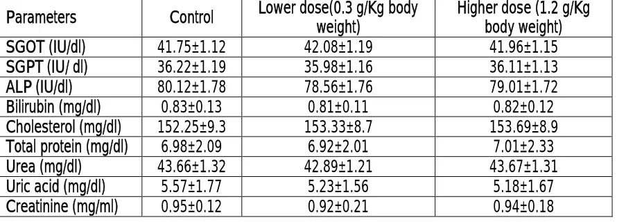
Table 5: Effect of Saraca asoca bark extract on serum biochemical parameters after sub-acute admistration. Data are represented as mean± SEM.
Parameters Control Lower dose(0.3 g/Kg body weight)
Higher dose (1.2 g/Kg body weight)
SGOT (IU/dl) 41.75±1.12 42.08±1.19 41.96±1.15
SGPT (IU/ dl) 36.22±1.19 35.98±1.16 36.11±1.13
ALP (IU/dl) 80.12±1.78 78.56±1.76 79.01±1.72
Bilirubin (mg/dl) 0.83±0.13 0.81±0.11 0.82±0.12
Cholesterol (mg/dl) 152.25±9.3 153.33±8.7 153.69±8.9
Total protein (mg/dl) 6.98±2.09 6.92±2.01 7.01±2.33
Urea (mg/dl) 43.66±1.32 42.89±1.21 43.67±1.31
Uric acid (mg/dl) 5.57±1.77 5.23±1.56 5.18±1.67
503 Effect of oral S. asoca methanolic extract on tissue histopathology of mice:
Histological studies of different mice organ revealed that there were no detectable alterations in treated groups with respect to the control group (data not shown).
Effect of oral S. asoca methanolic extract on serum biochemical parameters of mice:
After 30 days of treatment no significant changes were observed in serum biochemical parameter of mice in S. asoca treated group when compared with control group (Table 5).
Lethal dose (LD50):
The animals having received methanol extract did not exhibit marked behavioral changes but showed weak and less active movement followed by gradual death. The LD50, calculated from equation of probit-log dose of S. asoca methanolic extract was 6.526 g/Kg.
Discussion
There is increasing concern about the safety of use of the medicinal plants. There are general and herb specific concern regarding medicinal plants and their ability to produce toxicity and adverse effects [14]. Toxicity of medicinal plants may be related to the mixture of active compounds that they contain and stability of active ingredients in tissues. Phytotherapy having its pervasive use is substantiated by the affordability, its medicinal value and the belief of their harmlessness [15]. Phytochemical analysis is an important tool to identify and screen plants for their use [6]. The analysis of this plant part (bark), table 1, shows significant presence of flavonoids and reducing sugars in addition to tannins and glycosides. The presence of very small amount of anthraquinones, steroids and volatile oil was also observed.
Generally, the reduction in body weight and internal organ weights are simple and sensitive indices of toxicity after exposure to toxic substances. Body weight changes are indicator of adverse effects of drugs and chemicals and it will be significant if body weight loss occurred is greater than 10% of initial weight [16]. No significant difference in body weight
and organ weight of the experimental animal was found as compared to the between control group of animals (table 3&4). Slight changes in weight may be due to normal physiologic changes of animals.
The haematopoietic system is one of the most responsive targets for toxic compounds and an important manifestation of physiological and pathological status in man and animal [17]. In this study, there was a statistically significant treatment-related decrease in platelet levels of mice in the high dose group when compared to the control group (table 3). This indicates that S. asoca bark extract had an effect on the production of platelets which may be due its modulatory effect of flavonoids on the membrane integrity and function of platelets [18, 19, 20]. All the other hematological parameters did not differ significantly when compared with the control group (table 3), discarding the possibility of anaemia or disturbance in erythrocyte or hemoglobin production.
It is well known that almost all drugs, chemicals, xenobiotics are eliminated through renal excretion hence it is necessary to estimate the effects of methanolic extract on kidney functions. Serum biochemical parameters related to kidney function viz. urea, uric acid creatinine and total protein had no significant changes in experimental groups with respect to control group animals. Other biochemical parameters related to hepatic function like SGOT, SGPT, ALP, billirubin, cholesterol showed no significant difference between control and sub-acutely treated animal group (table 5). Therefore it can be said that the bark extract had no deleterious effect on hepatic and renal function.
The results of the present study indicated that S. asoca
504 suggests that the plant should be considered as practically non-toxic in acute ingestion.
Conclusion
Saraca asoca is highly accepted as universal solution in the ayurvedic medicine. This all-around plant is the source of various types of compounds. Saraca asoca
is age old and consistent source of medicine. It is used in many pharmacological activities like anti cancer, anti menorrhagic, anti oxytoxic, antimicrobial activity and have extend uses in ayurveda, unani and homeopathy [7]. In conclusion, this study provides important data on the acute and sub-acute oral toxicity profile of S. asoca. As the molecular mechanism underlying the links between dietary intake of flavonoids and its structure-function relationship is lacking, it is difficult to conclude about the effect of S. asoca bark extract on platelet count. This result will be beneficial for any future studies. The S. asoca methanolic bark extract is non-toxic in both acute and sub-acute oral administrations for male and female mice. Determination of the effect of this plant extract on pregnant animals is needed to complete the knowledge of safety profile study of this drug.
Acknowledgement
Authors are grateful to Prof. A.K. Banerjee, Vice Chancellor, University of Kalyani for his interest in this research work.
References
1. Cowan MM. Plant Products as antimicrobial agents. Clin. Microbiol Rev. 1999;12:564-82. 2. Atawodi SE. Antioxidant potentials of African
plants. Afr. J. Biotechnol. 2005;4(2):128-133. 3. Sofowora A. Medicinal plants and traditional
medicine. John Wiley & Sons. 1991;pp. 66-79. 4. Ghani A. Medicinal plants of Bangladesh with
chemical constituents and uses. (2nd edn). Asiatic Society of Bangladesh: Dhaka 2003. 5. Nadkarni AK, Nadkarni KM. Indiam Materia
Medica (ed). Popular Prakashan-Bombay; India 2005.
6. Sadhu SK, Khatun A, Phattanawasin P and Masami Ishibashi TO. Lignan glycosides and flavonoids from Saraca asoca with antioxidant activity J Nat Med. 2007;61:480-482.
7. Pradhan P, Joseph L, Gupta V, Chulet R, Arya H, Verma R, Bajpai A. Saraca asoca (Ashoka): A Review. Journal of Chemical and Pharmaceutical Research. 2009;1(1):62-71. 8. Parekh J, Jadeja D, Chanda S. Efficacy of
Aqueous and Methanol Extracts of Some Medicinal Plants for Potential Antibacterial Activity. Turk. J. Biol. 2005;29:203-210. 9. Sofowora A. Medicinal Plants and Traditional
Medicine in Africa(ed). Spectrum Books Limited: Nigeria, 1993;151-153.
10. Trease G, Evans WC. Textbook of Pharmacognosy. (12th edn). Balliere-Tindall: London 1983.
11. Morpugo C. A new design for the screening of CNS active drugs in mice. Arzneim. Forsch. 1971;11:1727-1734.
12. Sharma V, Pandey D. Beneficial Effects of Tinospora cordifolia on Blood Profiles in Male Mice Exposed to Lead. Toxicol Int. 2010;17(1):8–11.
13. Kasolo JN, Bimenya GS, Ojok L and Ogwal-okeng JW. Phytochemicals and acute toxicity of Moringa oleifera roots in mice. Journal of Pharmacognosy and Phytotherapy, 2011;3(3):38-42.
14. Saad B, Azaizeh H, Abu-Hijleh G, Said S. Safety of traditional Arab herbal medicine. Evidence-based Complementary and Alternative Medicine, 2006;3:433–439.
15. Springfield EP, Eagles PKF, Scott G. Quality assessment of South African herbal medicines by means of HPLC fingerprinting. Journal of Ethnopharmacology, 2005, 101, 75–83. 16. Raza M, Al-Shabanah OA, El-Hadiyah TM and
Al-Majed AA. Effect of prolonged vigabatrin treatment on haematological and biochemical parameters in plasma, liver and kidney of Swiss albino mice. Sci. Pharmaceut. 2002;70:135-145.
505 afra in rodents. Journal of Ethnopharmacology, 2007;112:138–144. 18. Guerrero JA, Lozano ML, Castillo J,
Benavente-García O, Vicente V, Rivera J. Flavonoids inhibit platelet function through binding to the thromboxane A2 receptor J Thromb Haemost, 2005;3(2):369-76.
19. Miyuki F, Hironori T, Motohiko N, Toshiyuki T, Ken-ichi N, Munekazu I. Anti-platelet and membrane rigidifying flavonoids in brownish scale of onion Journal of health science. 2003;49(6):475-480.
20. Wright B, Moraes LA, Kemp CF, Mullen W, Crozier A, Lovegrove JA, Gibbins JM. A structural basis for the inhibition of collagen-stimulated platelet function by quercetin and structurally related flavonoids. Br J Pharmacol, 2010;159(6):1312-25.
21. Loomis TA, Hayes AW. Loomis’s Essentials of Toxicology (ed). Academic Press: California, 1996;208–245.
![Table 2: Determination of LD50 values by Miller and Tainter method [22] of the methanolic extract of Saraca asoca bark in mice](https://thumb-us.123doks.com/thumbv2/123dok_us/575103.2056746/4.612.52.283.476.708/table-determination-values-miller-tainter-methanolic-extract-saraca.webp)