O R I G I N A L R E S E A R C H
Open Access
Visualization of ischemic stroke-related
changes on
18
F-THK-5351 positron emission
tomography
Kuo-Lun Huang
1,2†, Jung-Lung Hsu
1,2,3†, Kun-Ju Lin
4,5, Chien-Hung Chang
1,2, Yi-Ming Wu
6, Ting-Yu Chang
1,2,
Yeu-Jhy Chang
1,2, Chi-Hung Liu
1,2, Meng-Yang Ho
7, Shiaw-Pyng Wey
4,5, Tzu-Chen Yen
4,5, Nobuyuki Okamura
8,9,
Ing-Tsung Hsiao
4,5*and Tsong-Hai Lee
1,2*Abstract
Background:The18F-THK-5351 radiotracer has been used to detect the in vivo tau protein distribution in patients with tauopathy, such as Alzheimer’s disease and corticobasal syndrome. In addition,18F-THK-5351 can also monitor neuroinflammatory process due to high affinity to astrogliosis. We aimed to explore18F-THK-5351 distribution patterns and characteristics in patients with recent ischemic stroke.
Results:Fifteen patients received18F-THK-5351 positron emission tomography (PET) and diffusion tensor imaging (DTI) approximately 3 months after ischemic stroke. A region of interest (ROI) was placed in the peri-ischemic area and was mirrored on the contralateral side as the control, and a proportional value was derived from the ratio of the peri-ischemic ROI value over the mirrored ROI value. Increased18F-THK-5351 retention was observed in the areas around and remote from the stroke location. The proportional18F-THK-5351 values were negatively correlated with the proportional fractional anisotropy values (r=−0.39,P= 0.04).
Conclusion:18F-THK-5351 PET imaging provides a potential tool for in vivo visualization of the widespread ischemia-related changes associated with a microstructural disruption in recent ischemic stroke patients.
Keywords:Ischemic stroke,18F-THK-5351, Positron emission tomography, Diffusion tensor imaging, Astrogliosis, Neuroinflammation
Background
Tau protein plays an important role in microtubule assem-bly, and hyperphosphorylated tau (p-tau) protein may ag-gregate into neurofibrillary tangles as the hallmark of Alzheimer’s disease [1]. Total tau (t-tau) protein and p-tau protein accumulation have been interpreted as indicators of ongoing neuronal and axonal degeneration in Alzheimer’s disease [2]. 18F-THK-5351 is a recently developed radio-tracer designed for the in vivo tau protein detection with PET imaging [3].
In addition to the affinity to tau protein,
18
F-THK-5351 has also been reported to bind to monoamine oxidase B (MAO-B), which is attributed to its off-target binding in the striatum, thalamus, and brainstem [4, 5]. Furthermore, MAO-B is largely expressed in astrocytes, and reactive astrocytosis is a neuroinflammatory phenomenon in response to CNS acute injury or chronic neurodegeneration [6]. Over the recent decades, neuroinflammation has been considered as an essential contributor of CNS diseases, and that leads to increasing interest in visualizing neuroinflam-matory changes in a non-invasive manner [7]. The
bind-ing affinity of 18F-THK-5351 to MAO-B has been
applied to visualize neuroinflammation in vivo; in Ishiba-shi’s case report,18F-THK-5351 PET imaging was done 2 years after an ischemic stroke, and it suggests in-creased18F-THK-5351 retention along the pyramidal tract * Correspondence:[email protected];[email protected]
†Kuo-Lun Huang and Jung-Lung Hsu contributed equally to this work.
4Department of Nuclear Medicine and Center for Advanced Molecular
Imaging and Translation, Chang Gung Memorial Hospital, Taoyuan City, Taiwan
1Department of Neurology, Chang Gung Memorial Hospital, Taoyuan City,
Taiwan
Full list of author information is available at the end of the article
may represent Wallerian degeneration [8]. However, it is unknown how early 18F-THK-5351 retention can be ob-served after ischemic stroke. In the present study, we ap-plied 18F-THK-5351 PET imaging and diffusion tensor imaging (DTI) to determine in vivo18F-THK-5351 distri-bution patterns and associated microstructural character-istics in patients with recent ischemic stroke.
Methods Participants
Patients with ischemic stroke were prospectively re-cruited from the Department of Neurology and Stroke Center at Linkou Chang Gung Memorial Hospital,
Taiwan, for 18F-THK-5351 PET imaging from March
2016. Prior to enrollment, all participants were well in-formed of the study objectives and protocol and then provided their written informed consents. The study protocol and procedure for obtaining informed consent comply with the Helsinki Declaration and were approved by the institutional review board of Chang Gung Me-morial Hospital (IRB No. 103-7584A) and the Govern-mental Ministry of Health and Welfare.
The inclusion criteria were as follows: (1) a diagnosis of acute ischemic stroke based on history, neurological exam-ination, and brain MRI performed at the time of hospital ad-mission and (2) no history of a previous disabling stroke. The exclusion criteria were as follows: (1) presence of a de-mentia diagnosis before the index stroke; (2) a mean score of the Chinese version of the Informant Questionnaire on Cognitive Decline in the Elderly≥4; (3) a history of a sub-stantial traumatic brain injury; (4) a history of epilepsy; and (5) cerebellar structural abnormality, such as old stroke and traumatic brain injury. The clinical stroke patterns were summarized by the Oxfordshire Community Stroke Project classification [9].
MRI acquisition
Brain MRI was acquired at admission and 3 months after the stroke. The first MRI scan was acquired at ad-mission mainly for ischemic stroke confirmation, and the scanning protocol included the fluid-attenuated in-version recovery (FLAIR), diffusion-weighted imaging (DWI), apparent diffusion coefficient (ADC), and T1W imaging sequences. Acute ischemic stroke was defined as lesions with hyperintensity on DWI and FLAIR im-ages and hypointensity on ADC and T1W imim-ages.
Follow-up brain MRI scans were acquired around 3 months after the stroke onset. In addition to FLAIR and DWI sequences, the scanning protocol included an axial three-dimensional T1W MP-RAGE sequence and DTI se-quence. The intervals from the index stroke to the MRI scanning were recorded in days. The parameters of each MRI sequence were summarized in Additional file1.
18
F-THK-5351 radiotracer preparation and PET imaging acquisition
The 18F-THK-5351 radiotracer was synthesized with a slight modification of the method by Harada et al. (de-tails in Additional file 1) [3, 10]. Serial 18F-THK-5351 PET scans of the whole brain were acquired on a dedi-cated PET/CT scanner (Siemens Biograph mCT 16; Sie-mens Medical Solutions). Ten 1-min frames of PET images were acquired 50–60 min after an intravenous injection of 378 ± 17 MBq 18F-THK-5351 (18F-THK -5351 images) [11]. PET images were reconstructed using a three-dimensional OSEM (ordered subset expectation-maximization) algorithm (four iterations, 24 subsets; Gaussian filter = 2 mm; zoom = 3) with low-dose CT-based attenuation and scatter and random correc-tion. The intervals from the index stroke to the
18
F-THK-5351 PET study were recorded in days.
Imaging processing and analysis
All the MRI and PET images were co-registered with T1W MP-RAGE images and resliced into 1 × 1 × 1 mm resolution by the SPM8. To obtain the standardized up-take value ratios (SUVRs) from 18F-THK-5351 PET im-ages, the reference region was set at the individual’s cerebellar cortex. The DTI images obtained 3 months after the stroke were reconstructed using the Explor-eDTI toolbox (http://www.exploredti.com) to generate the DTI parametric images including fractional anisot-ropy (FA), mean diffusivity (MD), axial diffusivity (AD), and radial diffusivity (RD).
Region of interest analysis
To delineate the region of interest (ROI),18F-THK-5351 PET SUVR images were overlaid on the co-registered T1W MP-RAGE images, and the DWI images from the acute stroke stage were used as the reference for the acute ischemic regions. An experienced neurologist who was blinded to patients’clinical manifestations created a hand-drawn ROI. The ROIs were drawn on the peri-ischemic areas where substantial18F-THK-5351 up-take was noted; the peri-ischemic area with the maximal diameter of18F-THK-5351 uptake extent was selected as the target slice, and at least three consecutive slices were used for ROI creation. The ROI in the ipsilateral hemi-sphere was mirrored to the contralateral hemihemi-sphere [12]. Because of the heterogeneous nature of infarcts and the topographic distribution of 18F-THK-5351 up-take, the proportional values of the 18F-THK-5351 SUVRs and DTI parameters between bilateral ROIs were calculated (affected side divided by unaffected side) [13]. Statistical analysis
descriptive statistics. The correlations among propor-tional values from 18F-THK-5351 SUVRs and DTI pa-rameters were performed with non-parametric Kendall’s Tau-b correlation analysis. Statistical analyses were per-formed with SAS version 9.2 (SAS Institute Inc., Cary, NC, USA), and aPvalue < .05 was deemed significant.
Results
Fifteen patients were enrolled; no recurrent stroke was noted during the follow-up period, and no adverse event after18F-THK-5351 PET examination was reported. The demographic data, stroke clinical patterns, and vascular risk factors for each patient are summarized in Table1. The median interval from the stroke occurrence to the DTI and18F-THK-5351 scanning was 82 days (IQR, 63– 92 days) and 102 days (IQR, 79–118 days), respectively.
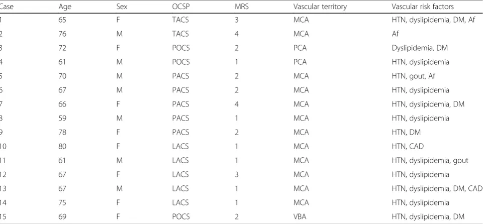
Figure 1 shows representative images of a participant with an acute infarction in the right corona radiata and right temporal lobe (case 6). In addition to the common off-target binding of18F-THK-5351 to the bilateral thal-amus, striatum, and brainstem, more obvious 18 F-THK-5351 retention is noted in peri-ischemic and remote areas over the right central semiovale, basal ganglion, right thalamus, right cerebral peduncle, and right ventral pons while low18F-THK-5351 uptake is noted in the is-chemic core.
Figure 2 shows representative images of another par-ticipant with an acute left frontal cortical infarction (Fig. 2a) (case 5). 18F-THK-5351 retention is decreased in the ischemic core and increased in the peri-ischemic areas (Fig. 2b, c) when overlaid on the DWI image
(Fig. 2d). In the peri-ischemic areas with high
18
F-THK-5351 retention, there is decreased FA on DTI (Fig. 2e) without corresponding white matter change on the follow-up FLAIR image (Fig.2f).
The ROI data from the18F-THK-5351 SUVRs and DTI parameters for each participant are listed in Add-itional file 1: Table S1. The proportional values of the
18
F-THK-5351 SUVRs and DTI parameters are presented in Table 2. The proportional 18F-THK-5351 SUVRs are negatively correlated with the proportional FA values (r=−0.39,P= 0.04) (Fig. 3), but not with other DTI pa-rameters, such as proportional MD, RD, and AD.
Discussion
Ischemic stroke has served as the prototypical models to investigate CNS neuroinflammatory response in both human and animal studies [6, 14]. The recently devel-oped 18F-THK-5351 radiotracer has shown affinity to MAO-B, suggesting 18F-THK-5351 can demonstrate le-sions with astrogliosis [5]. We conducted a case series study to explore the post-ischemic gliosis changes on
18
F-THK-5351 PET imaging and DTI in patients with re-cent ischemic stroke. Firstly, increased 18F-THK-5351 uptake was mainly observed around the DWI-positive region as well as the remote areas. Furthermore, the pro-portional 18F-THK-5351 SUVRs were negatively
corre-lated with the proportional FA values. To our
knowledge, this is the first in vivo human case series study using 18F-THK-5351 PET scanning to determine ischemia-related changes in patients with a recent ische-mic stroke event.
Table 1The demographic data and stroke clinical patterns for each patient
Case Age Sex OCSP MRS Vascular territory Vascular risk factors
1 65 F TACS 3 MCA HTN, dyslipidemia, DM, Af
2 76 M TACS 4 MCA Af
3 72 F POCS 2 PCA Dyslipidemia, DM
4 61 M POCS 1 PCA HTN, dyslipidemia
5 70 M PACS 2 MCA HTN, gout, Af
6 67 M PACS 2 MCA HTN, dyslipidemia
7 66 F PACS 4 MCA HTN, dyslipidemia, DM
8 59 M PACS 1 MCA HTN, dyslipidemia
9 78 F PACS 2 MCA HTN, DM
10 80 F LACS 1 MCA HTN, CAD
11 61 M LACS 1 MCA HTN, dyslipidemia, gout
12 67 F LACS 3 MCA HTN, dyslipidemia
13 67 M LACS 1 MCA HTN, dyslipidemia, DM, CAD
14 75 F LACS 1 MCA HTN, dyslipidemia
15 69 F POCS 2 VBA HTN, dyslipidemia, DM
OCSPOxfordshire Community Stroke Project,MRSModified Rankin Scale,TACStotal anterior circulation syndrome,PACSpartial anterior circulation syndrome,LACS
Despite the high affinity of 18F-THK-5351 for paired helical filaments, off-target binding of 18F-THK-5351 in middle-aged cognitively intact people was noted in the thalamus, striatum, substantia nigra, and periaqueductal gray matter where negligible tau protein was supposed to exist [15]. Similar off-target binding patterns were also reported with another tau PET tracer, 18F-AV-1451, and proposed to be associated with MAO and neurome-lanin [16]. In a recent study by Ng et al., the off-target uptakes of18F-THK-5351 in the thalamus and basal gan-glia were reduced over 50% after administration of the MAO-B inhibitor selegiline [4]. In a recent postmortem Alzheimer’s disease study, regional in vivo 18 F-THK-5351 uptake was significantly correlated with the density of tau aggregates in the neocortex and MAO-B in the whole brain [5]. Therefore, the findings with increased
18
F-THK-5351 retention should be differentiated
between tau protein accumulation and neuroinflamma-tory changes based on the underlying pathophysiology [6,17–20].
A recent report has observed increased18F-THK-5351 retention along the pyramidal tract in a chronic stroke patient [8], and elevated 18F-AV-1451 uptake was also
noted in the peri-infarct areas with white matter changes [21]. Similar 18F-THK-5351 retention patterns in the peri-infarct areas were also noted in our patients with recent ischemic stroke. In fact, reactive astrocytosis is a phenomenon of morphological and functional changes to astrocytes in response to cerebral ischemia, and MAO is abundant in the outer membranes of astrocyte mito-chondria [18, 22]. In a recent report by Thiel et al., the radiotracer, 11C-PK11195, was applied in patients with ischemic stroke, and antegrade increased retention was noted in the peri-lesional and remote areas along the pyramidal tract [13]. Such retention patterns of
11
C-PK11195 may indicate post-ischemic astrogliosis [23], and their imaging presentations are quite similar to the18F-THK-5351 uptake distribution in ischemic stroke patients. In fact, the well-known off-target binding sites of 18F-THK-5351, including the striatum, thalamus, and brainstem, were bilaterally enhanced in our cases, and the 18F-THK-5351 retention of these regions was asym-metrically elevated if they were within the peri-ischemic areas (Fig.1). The superimposed18F-THK-5351 imaging findings in ischemic stroke patients may suggest astro-gliosis caused by different underlying mechanisms.
Fig. 118F-THK-5351 retention patterns in an ischemic stroke patient (case 6).aThe acute ischemic location on diffusion-weighted imaging (DWI). b,cThe FLAIR images and18F-THK-5351 PET images taken 3 months after stroke, respectively.dThe fused DWI and18F-THK-5351 PET images.
Common off-target binding of the18F-THK-5351 is observed in the bilateral striatum, thalamus, and brainstem. Additionally, the18F-THK-5351
Fig. 2Multi-modality imaging characteristics in the patient (case 5) with a left frontal cortical infarction on diffusion-weighted imaging (DWI) (a). Low18F-THK-5351 uptake is noted in the ischemic core, and high uptake is noted in the peri-ischemic area over the corona radiata, the genu of
the corpus callosum, and in the bilateral frontal areas (b,c). The ROI for18F-THK-5351 retention is drawn in the peri-ischemic area, and a mirrored ROI is placed in the contralateral hemisphere as the control (d). There is a decreased fractional anisotropy (FA) on diffusion tensor imaging (e) (filled arrow) in the peri-ischemic area with increased18F-THK-5351 retention as compared to the contralateral side (empty arrow). No
corresponding white matter change is noted on the follow-up FLAIR image (f)
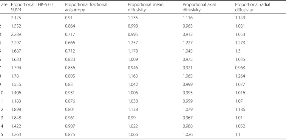
Table 2The proportional18F-THK-5351 SUVR and diffusion tensor imaging parameters
Case Proportional THK-5351 SUVR
Proportional fractional anisotropy
Proportional mean diffusivity
Proportional axial diffusivity
Proportional radial diffusivity
1 2.125 0.91 1.135 1.116 1.149
2 1.552 0.864 0.998 0.963 1.031
3 2.289 0.717 0.995 0.913 1.053
4 2.297 0.666 1.257 1.227 1.273
5 1.687 0.712 1.178 1.045 1.3
6 1.683 0.833 1.009 0.975 1.035
7 1.794 0.836 0.946 0.921 0.963
8 1.78 0.805 1.163 1.065 1.264
9 1.556 0.83 1.042 0.999 1.077
10 1.406 0.931 1.006 0.993 1.016
11 1.183 0.876 1.038 0.999 1.07
12 1.898 0.801 1.138 1.079 1.186
13 1.848 0.961 0.99 0.967 1.01
14 1.422 0.907 1.022 0.988 1.052
15 1.264 0.875 1.066 1.026 1.1
Furthermore, the 18F-THK-5351 retention appeared more widespread as increased uptake was even noted in the subcortical areas and from the genu of the corpus callosum to the contralateral frontal areas (Fig. 2b, c). Such spillover imaging phenomenon may imply that the influence of ischemic injury is not limited to the infarct core and penumbra, and also, other brain regions where no macrostructural changes are observed on the MRI scans. Nevertheless, some possible off-target binding of
18
F-THK-5351 cannot be excluded especially if the up-take is in the subcortical areas.
Although ischemic stroke was reported to induce tau protein accumulation in human postmortem and animal studies [19,20], such tau protein accumulation may be a transient reaction. Tau protein was found to have the highest serum and cerebrospinal fluid concentration within 1 week after stroke onset [24, 25], and its level may gradually return to the baseline level 1 month later [26]. In our cases, the 18F-THK-5351 PET studies were performed about 3 months after stroke onset.
Diffusion tensor imaging was utilized to
characterize 18F-THK-5351 uptake patterns in pa-tients with recent ischemic stroke in our study. DTI has been applied to assess microstructural neuronal damage in ischemic stroke. In patients with a lacu-nar infarct, diffusion abnormalities are present in the affected tract up to 2 cm away from the lacune with decreased FA and increased diffusivity [27]. These findings are consistent with our study results show-ing that peri-ischemic lesions with increased propor-tional 18F-THK-5351 uptake had lower proportional
FA, suggesting axonal degeneration or a demyelinat-ing process in these areas.
There are several limitations to the current study. Firstly, the 18F-THK-5351 PET imaging and DTI were performed 3 months after the stroke event, and there may be dynamic changes of18F-THK-5351 PET and DTI signals after the index stroke. Therefore, a longitudinal study investigating how 18F-THK-5351 retention evolves from the acute to the chronic ischemic stroke stage is warranted. Furthermore, there was heterogeneity in the stroke locations in our series. In order to determine the influence of 18F-THK-5351 retention patterns on stroke outcome, it is necessary to collect different groups of patients based on a pre-defined stroke location or syndrome in a larger sample size. Secondly, there has been a discussion about the tau radiotracer off-target binding characteristics, and the imaging findings of
18
F-THK-5351 PET in stroke patients could be better elucidated if corresponding fluid biomarkers, tissue pathology, or other complement imaging results are available. Finally, further studies are required to evaluate the influence of increased 18F-THK-5351 uptake on long-term stroke prognosis and cognitive outcome.
Conclusions
We have presented the first in vivo 18F-THK-5351 PET report in patients with recent ischemic stroke, in whom increased 18F-THK-5351 retention was noted in the areas around and remote from the stroke location. In le-sions with increased18F-THK-5351 retention, axonal de-generation or demyelination was found on DTI. Further
Fig. 3Correlation of the proportional18F-THK-5351 standardized uptake value ratios (SUVRs) with the proportional fractional anisotropy (FA)
values. The proportional18F-THK-5351 SUVRs are negatively correlated with the proportional FA values, suggesting that peri-ischemic areas with
studies are required to determine the influence of in vivo
ischemia-related changes on 18F-THK-5351 PET on
long-term stroke prognosis and cognitive outcome.
Additional file
Additional file 1:Table S1.The standardized uptake value ratios (SUVRs) of18F-THK-5351 PET and diffusion tensor imaging parameters in
the bilateral cerebral hemispheres. (DOCX 45 kb)
Abbreviations
AD:Axial diffusivity; ADC: Apparent diffusion coefficient; Af: Atrial fibrillation; CAD: Coronary artery disease; DM: Diabetes mellitus; DTI: Diffusion tensor imaging; DWI: Diffusion-weighted imaging; FA: Fractional anisotropy; HTN: Hypertension; IQR: Interquartile range; LACS: Lacunar syndrome; MAO-B: Monoamine oxidase B; MCA: Middle cerebral artery; MD: Mean diffusivity; MRS: Modified Rankin Scale; OCSP: Oxfordshire Community Stroke Project; OSEM: Ordered subset expectation-maximization; PACS: Partial anterior circulation syndrome; PCA: Posterior cerebral artery; PET: Positron emission tomography; POCS: Posterior circulation syndrome;
p-tau: Hyperphosphorylated tau; RD: Radial diffusivity; ROI: Region of interest; SUVR: Standardized uptake value ratio; TACS: Total anterior circulation syndrome; t-tau: Total tau; VBA: Vertebrobasilar artery
Acknowledgements
We would like to thank the Center for Advanced Molecular Imaging and Translation and the Clinical Trial Center of Linkou Chang Gung Memorial Hospital for the technical support and assistance in the administrative affairs under the support from the Ministry of Health and Welfare, Taiwan (MOHW104-TDU-B-212-113003; MOHW105-TDU-B-212-133020; MOHW106-TDU-B-212-113005).
Funding
This study was supported by grants from the Ministry of Science and Technology, Taiwan (grants MOST 105-2314-B-182A-009 and 106-2314-B-182A-074, 106-2314-B-182-017-MY3) and the Research Fund of Chang Gung Memorial Hospital (CMRPG3F2181, BMRPD69, BMRP488).
Availability of data and materials
Please contact the author for data requests.
Authors’contributions
KLH, JLH, KJL, SPW, TCY, NO, ITH, and THL designed the study. KLH, CHC, TYC, YJC, CHL, YMW, and THL contributed to the patient selection and data acquisition. KLH, JLH, KJL, MYH, YMW, NO, ITH, and THL contributed to the data analysis and interpretation. KLH, JLH, KJL, and MYH contributed to the drafting of the manuscript together with the corresponding authors. CHC, YMW, TYC, YJC, CHL, SPW, TCY, and NO contributed to the critical revision of the manuscript. Finally, THL and ITH supervised the whole part of the present study. All authors read and approved the final manuscript.
Ethics approval and consent to participate
All procedures performed in studies involving human participants were in accordance with the ethical standards of the institutional and/or national research committee and with the principles of the 1964 Declaration of Helsinki and its later amendments or comparable ethical standards. Informed consent was obtained from all individual participants included in the study. In addition, the next of kin or guardians of stroke patients also gave their written informed consent if the patients could not comprehend the study protocol or they could not sign their name clearly. The study protocol and procedure for obtaining informed consent were approved by the institutional review board of Chang Gung Memorial Hospital (IRB No. 103-7584A) and the Governmental Ministry of Health and Welfare.
Consent for publication
Not applicable.
Competing interests
The authors declare that they have no competing interests.
Publisher’s Note
Springer Nature remains neutral with regard to jurisdictional claims in published maps and institutional affiliations.
Author details
1Department of Neurology, Chang Gung Memorial Hospital, Taoyuan City,
Taiwan.2College of Medicine, Chang Gung University, Taoyuan City, Taiwan. 3
Graduate Institute of Humanities in Medicine and Research Center for Brain and Consciousness, Taipei Medical University, Taipei, Taiwan.4Department of
Nuclear Medicine and Center for Advanced Molecular Imaging and Translation, Chang Gung Memorial Hospital, Taoyuan City, Taiwan.5Healthy
Aging Research Center and Department of Medical Imaging and Radiological Sciences, Chang Gung University, Taoyuan City, Taiwan.6Department of
Radiology, Chang Gung Memorial Hospital, Taoyuan City, Taiwan.7Graduate
Institute of Behavioral Sciences, Chang Gung University, Taoyuan City, Taiwan.8Division of Neuro-imaging, Institute of Development, Aging and Cancer, Tohoku University, Sendai, Japan.9Department of Pharmacology,
Faculty of Medicine, Tohoku Medical and Pharmaceutical University, Sendai, Japan.
Received: 24 April 2018 Accepted: 27 June 2018
References
1. Arendt T, Stieler JT, Holzer M. Tau and tauopathies. Brain Res Bull. 2016;126: 238–92.
2. Hansson O, Zetterberg H, Buchhave P, Londos E, Blennow K, Minthon L. Association between CSF biomarkers and incipient Alzheimer’s disease in patients with mild cognitive impairment: a follow-up study. Lancet Neurol. 2006;5:228–34.
3. Harada R, Okamura N, Furumoto S, Furukawa K, Ishiki A, Tomita N, et al. 18F-THK5351: a novel PET radiotracer for imaging neurofibrillary pathology in Alzheimer disease. J Nucl Med. 2016;57:208–14.
4. Ng KP, Pascoal TA, Mathotaarachchi S, Therriault J, Kang MS, Shin M, et al. Monoamine oxidase B inhibitor, selegiline, reduces 18F-THK5351 uptake in the human brain. Alzheimers Res Ther. 2017;9:25.
5. Harada R, Ishiki A. Correlations of 18F-THK5351 PET with post-mortem burden of tau and astrogliosis in Alzheimer’s disease. J Nucl Med. 2018;59: 671–4.
6. Burda JE, Sofroniew MV. Reactive gliosis and the multicellular response to CNS damage and disease. Neuron. 2014;81:229–48.
7. Boutin H, Murray K, Pradillo J, Maroy R, Smigova A, Gerhard A, et al. 18F-GE-180: a novel TSPO radiotracer compared to 11C-R-PK11195 in a preclinical model of stroke. Eur J Nucl Med Mol Imaging. 2015;42:503–11. 8. Ishibashi K, Kameyama M, Tago T, Toyohara J, Ishii K. Potential use of
18F-THK5351 PET to identify Wallerian degeneration of the pyramidal tract caused by cerebral infarction. Clin Nucl Med. 2017;42:e523–e4. 9. Bamford J, Sandercock P, Dennis M, Warlow C, Burn J. Classification and
natural history of clinically identifiable subtypes of cerebral infarction. Lancet. 1991;337:1521–6.
10. Hsiao I-T, Lin K-J, Huang K-L, Huang C-C, Chen H-S, Wey S-P, et al. Biodistribution and radiation dosimetry for the tau tracer 18F-THK-5351 in healthy human subjects. J Nucl Med. 2017;58:1498–503.
11. Lin K-J, Hsiao I-T, Hsu J-L, Huang C-C, Huang K-L, Hsieh C-J, et al. Imaging characteristic of dual-phase 18F-florbetapir (AV-45/Amyvid) PET for the concomitant detection of perfusion deficits and beta-amyloid deposition in Alzheimer’s disease and mild cognitive impairment. Eur J Nucl Med Mol Imaging. 2016;43:1304–14.
12. Sahathevan R, Linden T, Villemagne VL, Churilov L, Ly JV, Rowe C, et al. Positron emission tomographic imaging in stroke: cross-sectional and follow-up assessment of amyloid in ischemic stroke. Stroke. 2016;47:113–9. 13. Thiel A, Radlinska BA, Paquette C, Sidel M, Soucy J-P, Schirrmacher R, et al. The temporal dynamics of poststroke neuroinflammation: a longitudinal diffusion tensor imaging–guided PET study with 11C-PK11195 in acute subcortical stroke. J Nucl Med. 2010;51:1404–12.
14. Simats A, García-Berrocoso T, Montaner J. Neuroinflammatory biomarkers: from stroke diagnosis and prognosis to therapy. Biochim Biophys Acta. 2016;1862:411–24.
16. Saint-Aubert L, Lemoine L, Chiotis K, Leuzy A, Rodriguez-Vieitez E, Nordberg A. Tau PET imaging: present and future directions. Mol Neurodegener. 2017; 12:19.
17. Becerra-Calixto A, Cardona-Gómez GP. The role of astrocytes in neuroprotection after brain stroke: potential in cell therapy. Front Mol Neurosci. 2017;10:88.
18. Pekny M, Pekna M. Astrocyte reactivity and reactive astrogliosis: costs and benefits. Physiol Rev. 2014;94:1077–98.
19. Ichihara K, Uchihara T, Nakamura A, Suzuki Y, Mizutani T. Selective deposition of 4-repeat tau in cerebral infarcts. J Neuropathol Exp Neurol. 2009;68:1029–36.
20. Koike MA, Green KN, Blurton-Jones M, LaFerla FM. Oligemic hypoperfusion differentially affects tau and amyloid-β. Am J Pathol. 2010;177:300–10. 21. Lockhart SN, Ayakta N, Winer JR, La Joie R, Rabinovici GD, Jagust WJ.
Elevated 18F-AV-1451 PET tracer uptake detected in incidental imaging findings. Neurology. 2017;88:1095–7.
22. Ding S. Dynamic reactive astrocytes after focal ischemia. Neural Regen Res. 2014;9:2048–52.
23. Lavisse S, Guillermier M, Hérard A-S, Petit F, Delahaye M, Van Camp N, et al. Reactive astrocytes overexpress TSPO and are detected by TSPO positron emission tomography imaging. J Neurosci. 2012;32:10809–18.
24. Bielewicz J, Kurzepa J, Czekajska-Chehab E, Stelmasiak Z, Bartosik-Psujek H. Does serum tau protein predict the outcome of patients with ischemic stroke? J Mol Neurosci. 2011;43:241–5.
25. Hesse C, Rosengren L, Vanmechelen E, Vanderstichele H, Jensen C, Davidsson P, et al. Cerebrospinal fluid markers for Alzheimer’s disease evaluated after acute ischemic stroke. J Alzheimers Dis. 2000;2:199–206. 26. Kaerst L, Kuhlmann A, Wedekind D, Stoeck K, Lange P, Zerr I. Cerebrospinal
fluid biomarkers in Alzheimer’s disease, vascular dementia and ischemic stroke patients: a critical analysis. J Neurol. 2013;260:2722–7.