J Nanostruct 7(2): 155-164, Spring 2017
RESEARCH PAPER
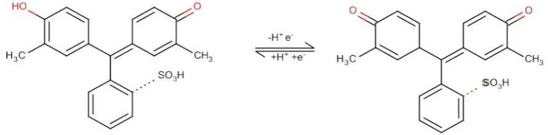
Fabrication of Nano Poly Cresol Red over Glassy Carbon Electrode
and its Application in Selective Determination of Uric acid in the
Presence of Ascorbic Acid
Chinnapan Jayakumar 1, Chinnapan Maria Magdalane 1,2, Kasinathan Kanimozhi 3, Kasinathan Kaviyarasu 4,5, Boniface jeyaraj 1*
1 LIFE, Department of Chemistry, Loyola College (Autonomous), Chennai 600034, India.
2 Department of Chemistry, St. Xavier’s College (Autonomous), Tirunelveli, India.
3 PG Research and Department of Chemistry, Auxilium College (Autonomous), Vellore, India.
4 UNESCO-UNISA Africa Chair in Nanoscience’s/Nanotechnology Laboratories, College of Graduate Studies, University of South Africa (UNISA), Muckleneuk Ridge, Pretoria, South Africa.
5 Nanosciences African network (NANOAFNET), Materials Research Group (MRG), iThemba LABS-National Research Foundation (NRF), 1 Old Faure Road, 7129, Somerset West, Western Cape Province, South Africa.
* Corresponding Author Email: [email protected]
ARTICLE INFO
Article History: Received 10 February 2017 Accepted 21 March 2017 Published 01 April 2017
Keywords: Ascorbic acid Electrocatalytic activity Electron microscopy Nano poly cresol red Uric acid
ABSTRACT
How to cite this article
Jayakumar C, Magdalane C. M, Kanimozhi K, Kaviyarasu K, jeyaraj B. Fabrication of Nano Poly Cresol Red over Glassy Carbon Electrode and its Application in Selective Determination of Uric acid in the Presence of Ascorbic Acid. J Nanostruct, 2017; 7(2):155-164. DOI: 10.22052/jns.2017.02.010
A selective electrochemical method for the determination of uric acid was developed by using nano poly cresol red modified glassy carbon electrode. This new material has been characterized by Scanning Electron Microscopy, cyclic voltammetry and Differential pulse voltammetry. This modified electrode shows excellent electrocatalytic activity towards the oxidation of uric acid in the presence of ascorbic acid in (pH 7.0). Compare to bare electrode the modified electrode able to resolve the both ascorbic acid and uric acid in the solution mixture. The oxidation current of uric acid with the modified electrode is two folds higher that the bare glassy carbon electrode. Using differential pulse voltammetry method, the oxidation current is linear with the uric acid concentration in the range 30 µM - 575 µM with the detection limit of 5 µM (S/N = 3). Moreover uric acid in real samples can be determined using nano poly cresol red without any pretreatment of samples giving satisfactory results.
INTRODUCTION
Uric acid (UA), is a key biomarker for the
diagnosis of several diseases, which is the primary
end product of purine metabolism. The decreasing
and increasing concentration of UA lead to various
diseases such as gout, Lesch–Nyhan syndrome,
hyper uricemia, leukemia and so on [1-3]
Therefore, the monitoring of UA in human blood
and urine is very important for the prevention of
156
C. Jayakumar et al. / Nano Poly Cresol Red over Glassy Carbon Electrode for Selective Determination of Uric acid
J Nanostruct 7(2): 155-164, Spring 2017
Moreover AA at the electrode surface often gives
rise to a serious overlapping peak with uric acid
[4-6]. The different analytical methods were
used for the determination of UA such as
high-performance liquid chromatography [7, 8], gas
chromatography [9], capillary electrophoresis [10],
chemiluminescence [11,12], ultraviolet-visible
spectrophotometry [13, 14], fluorimetry [15]
and electroanalytical methods [16, 17]. Among
these methods, electro-analytical methods have
been found to be reliable because of its high
sensitivity, selectivity and reproducibility [18,
19]. Several modifier materials like polymers
[20-25], biomaterials [26-31], photocatalytic
[32-39] nanomaterials [40-45] have been used to
overcome this problem. In recent years, substantial
efforts have been devoted for the development
of electrochemical sensors based on electrodes
modified by electro-synthesized dye based nano
polymeric films, due to their cost effective nature,
high surface area, effective mass transport,
and electrocatalytic [46, 47]. In this work, nano
polymeric CR film was synthesized through electro
polymerization method. The developed biosensors
showed high sensitivity and reproducible in
wide-linear range of potential. Further a
anti-interference ability, long term stability and low
detection limit were also examined for effective
and selective determination of UA the presence
of AA.
MATERIALS AND METHODS
Uric acid, Ascorbic acid, [HAuCl
4.3H
2O],
8-hydroxyquinoline, cresol red, 0.1M of sodium
phosphate buffer, HCl, ethanol and all other
chemicals are used of analytical grade. The
solutions were prepared by using double distilled
water. The experiments were carried out at 25 ºC.
Characcteristic Studies
Cyclic Voltammetric (CV), Differential Pulse
Voltammetric (DPV) experiments were performed
using a CHI6041C electrochemical workstation
(CH Inc., USA) coupled with a conventional
three-electrode cell. The three three-electrodes namely the
working electrode, the auxiliary electrode and the
reference electrode were GCE, Pt wire and Ag/AgCl
electrode respectively. All the potentials in this
work are given against the Ag/AgCl. BRUKER RFS
27: Stand alone FT-Raman Spectrometer was used
to find the functionalization of gold with 8-HQ,
SEM (FEI Quanta FEG 200-High resolution scanning
electron microscope) was used to find the surface
morphology. EI-1L
model of 34 kHz ultrasonic
bath was used for cleaning the electrodes and to
prepare homogeneous mixture.
Standardization of Glassy Carbon Electrode
Before modifying, the GC electrode was polished
with 0.3µm and 0.05µm of alumina slurries for
2 min each step, followed by thoroughly rinsing
with double distilled water, then the electrode
was sonicated with ethanol and distilled water
for 2 min each. After sonicating the electrode, it
was rinsed with double distilled water and was
examined by cyclic voltammetry using standard
1mM potassium ferric cyanide solution with 0.1M
KCl as supporting electrolyte.
Preparation Poly Cresol Red GCE
The 1mM cresol red was standardized with
pH 6 phosphate buffer solution, then the three
electrode setup was constructed, and the glassy
carbon electrode was scanned up to 7 cycles
between the potential of -0.7V to +1.6V with a
scan rate 100 mVs
-1. Now this electrode is said
to be poly cresol red electrode, after this process
the electrode was washed thoroughly with
double distilled water to remove the physisorbed
cresol red, then the modified electrode has to be
immersed in 0.1M phosphate buffer solution of pH
7.0 until use.
RESULTS AND DISCUSSION
Surface Morphology Study & Electrodeposition of
Poly Cresol Red polymerization
C. Jayakumar et al. / Nano Poly Cresol Red over Glassy Carbon Electrode for Selective Determination of Uric acid
These facts indicated that poly cresol red films
were deposited on the surface of GC electrode
by electro polymerization. The electrochemical
behavior of poly cresol red at the GC electrode
was in good agreement with previous findings,
referring to the electrochemical responses of a few
organic polymerized compounds at solid electrode
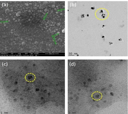
[48]. Fig. 3(a-d) explains the surface morphology
of the poly CR modified GCE using Field Emission
Scanning Electron Microscopy (FESEM). The
formation of small irregular spherical particles
indicates that the glassy carbon has been modified
by nano poly CR. It is noteworthy to mention that
the entire surface area has been uniformly coated
by the polymeric film.
Electrocatalytic Behavior of Poly Cresol Red GCE
The electrocatalytic behavior of poly cresol red
was investigated with DPV in 0.1 M Phosphate
Buffer Solution (PBS) at pH 7.0 as shown in Fig. 4.
A weak response and slow electron transaction for
both UA and AA is observed on a bare electrode
moreover the bare GCE unable to separate both
AA and UA in the solution mixture, where as the
nano poly CR modified electrode shows a good
electrocatalytic oxidation toward both UA and AA
(as seen in Fig. 4), indicating that the nano poly CR
modified electrode can effectively separate both
UA and AA in the solution mixture with decrease
in the oxidation potential of UA to 0.26 V from
a broad range of potentials. Furthermore, the
oxidation peak current (I
pa) of UA at the nano poly
CR modified electrode is ten times higher than
that of the bare electrode and for AA it is three
times higher. These results confirm that the nano
poly CR on the surface of the bare electrode can
effectively accelerate the electrochemical redox
behavior of UA and AA.
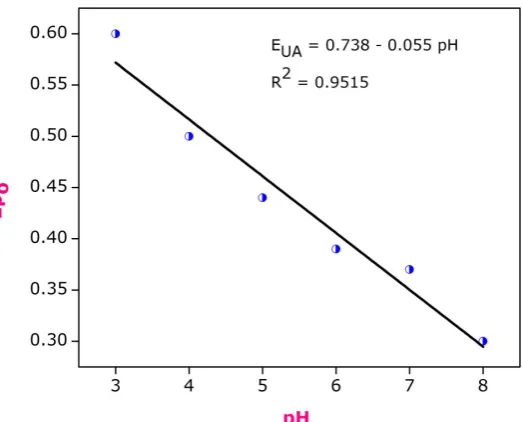
Effect of pH of UA in the Poly CR Modified GCE
The effect of pH on the peak current and peak
potential of the catalytic oxidation of UA was
investigated by cyclic voltammetry method. As a result
shown in Fig. 5, the peak potential for UA oxidation
Fig. 1. Cyclic voltammogram of poly cresol red158
C. Jayakumar et al. / Nano Poly Cresol Red over Glassy Carbon Electrode for Selective Determination of Uric acid
J Nanostruct 7(2): 155-164, Spring 2017
showed a linear variation with the increased value of
pH and it shifted to a more negative potential with a
slope of about –55 mV/pH, which suggested that the
total number of electrons and protons taking part in
the oxidation of UA was the same. As the oxidation
of UA is known to occur by a two-electron transfer
[49], thus the number of protons involved is also
predicted to be two (as shown in Fig. 6) Moreover
oxidation of UA was carried out in broad range of pH
ranging from 3-7. Since maximum peak current was
observed in pH 7.0, this was taken for the further
analysis Fig. 7 respectively.
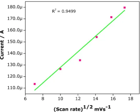
Effect of Scan Rate for UA in Poly Cresol Red GCE
Effect of scan rate was studied by cyclic voltammetry
method by placing 5mM UA in an electrochemical cell.
The scan rate has been varied from 50 mVs
-1to 300
mVs
-1. Fig. 8 and Fig. 9 show anodic peak current of UA
proportional with the square root of scan rate (ν
1/2)
with correlation coefficient 0.9797. This observation
suggests that oxidation process of UA in nano
poly CR modified electrode followed diffusion by
controlled process [50].
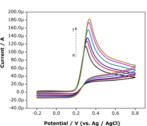
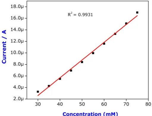
Concentration UA in the Presence of AA in Poly
Cresol Red GCE
The DPV method is normally used for the
determination of compounds because of its
high sensitivity [51]. It is well known that AA
is an electroactive molecule coexisting in a
biological system, which can also be oxidized in
the conventional solid electrode [52]. UA in the
presence of AA by nano poly CR modified GCE is
able to separate both 1 mM Uric acid and 1 mM AA
in the potential range of -0. 2 V, -0.8 V vs Ag/AgCl
Fig. 4. The DPV curves at different concentration
of UA at the modified electrode clearly show that
the anodic peak current increases linearly with an
159 J Nanostruct 7(2): 155-164, Spring 2017
C. Jayakumar et al. / Nano Poly Cresol Red over Glassy Carbon Electrode for Selective Determination of Uric acid
Fig. 4. DPV of 1mM UA and AA (a) bare GCE (b) nano poly CR GCE
Fig. 5. Plot of Ep0 (UA) Vs pH in poly CR GCE
160 J Nanostruct 7(2): 155-164, Spring 2017
increase in UA concentration from 30 to 575 µM
Fig. 10. The correlation coefficient of the various
concentrations of UA vs peak current is found to be
0.996 which is shown Fig. 11. Further the limit of
detection is 5 µM in the presence of 5 mM ascorbic
acid. All this result illustrates that the coexistence
of AA has no influence on UA determination.
Interference of Coexisting Compounds of Uric acid
in Real Samples
A part from the presence of AA, the other
reason for variation of UA oxidation peak current
is the presence of other interfering compounds.
For this a 300 µM of urea, 60 µM of oxalate, 60 µM
of glucose were added and tested in the presence
of 20 µM UA. The percentage of change in anodic
peak current is 5.6 %, 4.2 %, 4.5% for urea, oxalate
and glucose respectively. This results show that
the interfering compounds has no significant
effect in the detection of UA. In comparing other
interfering compound urea were tested in large
amount due to its large excess amount in urine.
The proposed method was validated by analysis
with the real samples. For this the human urines
Fig. 7. Plot of ipa (UA) Vs pH in poly CR GCEFig. 8. Effect of Scan rate: cyclic voltammograms of UA (5mM) in the nano poly CR modified GCE in 0.1mol L-1 PBS (pH-7.0) at various scan. The scan rate
C. Jayakumar et al. / Nano Poly Cresol Red over Glassy Carbon Electrode for Selective Determination of Uric acid
were collected from the healthy volunteers. The
human urine samples were filter and centrifuged
before the experiment. Then the samples were
diluted for 10 times with the phosphate buffer
solution of pH 7.0. DPV was carried out with the
diluted samples. The results are presented in the
Table 1.
Stability and Reproducibility of the Nano Poly CR
Modified GCE
The reproducibility of the developed sensor
has been measured by repetive scanning method
in PBS pH 7.0. For 10 determination of UA at poly
CR modified GCE the obtained relative strandard
deviation (R. S. D) is 4.8 % and 5.1 % this indicates
Fig. 9. Plots of ipa (UA) vs. square root of the scan rate in nano poly CRFig. 10. DPV of AA (5mM) at nano structured poly CR electrode in the presence
of different concentrations of L-dopa in µM: (a) 30 (b) 65 (c) 105 (d) 150 (e) 200
(f) 255 (g) 315 (h) 380 (i) 450 (j) 575
S.No Nature of sample Original value (μM) Spiked value (μM) Found (μM) Recovery (%)
1. Urine 22.2 10 30.8 95.6%
2. Urine 20.4 10 29.7 97.6%
162
C. Jayakumar et al. / Nano Poly Cresol Red over Glassy Carbon Electrode for Selective Determination of Uric acid
J Nanostruct 7(2): 155-164, Spring 2017
that the prepared polymeric film has excellent
ability to prevent the electrode from the surface
fouling. Further the long term stability of the
prepared modified GCE has been studied. For the
first 3 days there is no apparent current decrease
was observed in every day use and stored in 0.1 M
PBS (pH 7.0). after that 20 % of decreasing current
was observed on every day use. This shows the
presence of nano polymeric film over the surface
of the GCE.
CONCLUSION
The present study has demonstrated the
development of electrochemical detection of UA
that are based on the electro polymerization of
nano polymeric film. The surface morphology of
the poly CR has been examined by using FESEM
analysis. The results indicated that polymers
were homogeneously deposited on the surface
of the GCE with the particle size ranges from 30
nm - 65 nm. The electro polymerized nano poly
CR shows good electro catalytic effect towards
UA and AA in the solution mixture. Further it
shows significant enlargement in peak current
and a great decrease in the peak potential. The
reproducibility as well as the selectivity is good.
The selective detection of the UA in the presence
of AA is accomplished with the detection limit of
5.0 µM. Moreover the recovery percentage of
95.6 % and 97.6 % for UA determination in urine
samples indicates that the developed sensor is
very much reliable.
ACKNOWLEDGEMENT
Financial support from the University Grant
Commission (No.F: 42-382/2013 (SR)) is greatly
acknowledged.
CONFLICT OF INTEREST
The authors declare that there are no conflicts
of interest regarding the publication of this
manuscript.
REFERENCES
1. Abellan L, Vidal L, Rodríguez A.R, Berenguer M.A, Canals A, Morallon E. IDA microelec-trodes modified with Au-doped graphene oxide for the simultaneous determination of uric ac-id and ascorbic acid in urine samples. Electrochimi. Acta. 2017; 227: 275-284.
2. Li Y, Ran G,Yi W.J, Luo H.Q. A novel third generation uric acid biosensor using uricase electro-activated with ferrocene on a Nafion coated glassy carbon electrode. Microchim. Acta. 2012; 178: 115-121.
3. Abellán L.A, Ayan Varela M, Vidal L, Paredes J.I, Villar Rodil S, Canals A, Morallon E. Flavin mon-onucleotide-exfoliated graphene flakes as electrodes for the electrochemical determination of uric acid in the presence of ascorbic acid. J. Electroanal. Chem. 2016; 783: 41-48.
4. Pachla L.A, Reynolds D.L, Wright D.S, Kissinger P.T. Analytical methods for measuring uric acid in biological samples and food products. J. Assoc. Off. Anal. Chem. 1987: 70(1):1-14. 5. Wygaarden J, Kelly N.W. Gout and Hyperuricemica, Grune
and Stratton, New York, 1974: 1000.
6. Cunnigham S.K, Keaveny T.V. A two-stage enzymatic method for determination of uric acid and hypoxanthine/xanthine. Clin. Chim. Acta.1978; 86: 217.
C. Jayakumar et al. / Nano Poly Cresol Red over Glassy Carbon Electrode for Selective Determination of Uric acid
Zhang W.Y, Chen Y.C, Zhang F. Simultaneous determination of isoniazid, rifampicin, levofloxacin in mouse tissues and plasma by high performance liquid chromatography-tandem mass spectrometry. J. Chromatogr. B. Analyt. Technol. Biomed. Life Sci. 2010; 878: 2286-91.
8. Moussa L.A, Khassouani C.E, Soulaymani R, Jana M, Cassanas G, Alric R, Hue B. Therapeutic iso-niazid monitoring using a simple high-performance liquid chromatographic method with ultra-violet detection. J. Chromatogr. B. Analyt. Technol. Biomed. Life Sci. 2002; 766:181-7.
9. Karlaganis G, Peretti E, Lauterburg B.H. Analysis of isoniazid, acetylhydrazine and [15N2]acetylhydrazine in serum by capillary gas chromatography-ammonia chemical ionization mass spectrometry. J. Chromatogr. B: Biomed.Sci. Appl. 1987; 420: 171-177.
10. Liu J, Zhou W.H, You T.Y, Li F.L, Wanr E.K. Dong S.J. Detection of Hydrazine, Methylhydrazine, and Isoniazid by Capillary Electrophoresis with a Palladium-Modified Microdisk Array Electrode. Anal. Chem. 1996; 68: 3350-3353.
11. Safavi A, Karimi M.A, Nezhad M.R.H. Flow injection determination of isoniazid using N-bromosuccinimide- and N-chlorosuccinimide-luminol chemiluminescence systems. J. Pharm. Biomed. Anal. 2003; 30: 1499-1506.
12. Xiong Y, Zhou H.J, Zhang Z.J, He D.Y, He C. Electrochemiluminescence from Isoniazid Itself and Its Analytical Application. Spectrochim. Acta Part A: Mol. Biomol. Spectrosc. 2007; 66: 341-346.
13. Benetton S.A, Kedor Hackmann E.R.M, Santoro M.I.R.M, Borges V.M. Visible spectrophoto-metric and first-derivative UV spectrophotometric determination of rifampicin and isoniazid in pharmaceutical preparations. Talanta. 1998; 47: 639-643.
14. El Kommos M.E. Yanni A.S. Spectrophotometric determination of isoniazid using 6,7-dichloroquinoline-5,8-dione. Analyst. 1988; 113: 1091-1095.
15. Lapa R.A.S, Lima J.L.C, Santos J.L.M. Fluorimetric determination of isoniazid by oxidation with cerium(IV) in a multicommutated flow system. Anal. Chim. Acta. 2000; 419:17.
16. Majidi M.R, Jouyban A, Asadpour Zeynali K. Voltammetric behavior and determination of iso-niazid in pharmaceuticals by using overoxidized polypyrrole glassy carbon modified electrode. J. Electroanal. Chem. 2006; 589: 32-37.
17. Bergamini M.F, Santos D.P, Zanoni M.V.B. Electrochemical
behavior and voltammetric deter-mination of
pyrazinamide using a poly-histidine modified electrode. Bioelectrochemistry. 2010; 77: 133-138.
18. Chaney E.N, Baldwin R.P. Electrochemical determination of Adriamycin compounds in urine by preconcentration at carbon paste electrodes. Anal. Chem. 1982; 54: 2556-2560. 19. Goyal R.N, Gupta V.K, Oyama M, Bachheti N. Differential
pulse voltammetric determination of atenolol in pharmaceutical formulations and urine using nanogold modified indium tin oxide electrode. Electrochem. Commun. 2006; 8: 65-70.
20. Goyal R.N, Tyagi A, Bachheti N, Bishnoi S. The Electrocatalytic Activity of Bare Pyrolytic Graphite and Single Wall Carbon
Nanotube Modified Glassy Carbon Sensors Is Same for the Quantifica-tion of Bisoprolol Fumarate. Electrochim. Acta. 2008; 53: 2802-2808.
21. Zhang L, Lang Q, Shi Z. Electrochemical Synthesis of Three-Dimensional Polyaniline Network. J. Anal. Chem. 2010; 1: 102-112.
22. Brillians Revin S, Abraham John S. Highly sensitive determination of uric acid in the presence of major interferents using a conducting polymer film modified electrode. Bioelectrochemistry. 2012; 88: 22-29.
23. Claver J.B, Ortega I.F.D, Miron M.C.V, Vallvey L.F.C. Development of a cholesterol biosensor modified with carbon nanotube. Anal. Chim. Acta. 2011, 702, 254-261. 24. Yan H, Xiao H, Xie Q, Liu J, Sun L, Zhou Y, Zhang Y, Chao
L, Chen C, Yao S. Simultaneous electro-analysis of isoniazid and uric acid at poly(sulfosalicylic acid)/electroreduced carboxylated gra-phene modified glassy carbon electrode. Sens. Actuators B. 2015; 207: 167-176.
25. Wathoni N, Hasanah A.N, Gozali D, Wahyuni Y, Fauziah L.L. Determination of uric acid level by polyaniline and poly (allylamine): Based biosensor. J. Adv. Pharm. Technol. Res. 2014; 5: 13-6.
26. Ezhilarasi A, Judith Vijaya J, Kaviyarasu K, Maaza M, Ayeshamariam A, John Kennedy L. Green synthesis of NiO nanoparticles using Moringa oleifera extract and their biomedical applications: Cytotoxicity effect of nanoparticles against HT-29 cancer cells. J. Photochem. & Photobio. B: Bio. 2016; 164: 352-360.
27. Kaviyarasu K, Mariappan A, Neyvasagam K, Ayeshamariam A, Pandi P, Rajeshwara Palanichamy R, Gopinathan C, Genene T. Mola, Maaza M. Photocatalytic performance and antimicrobial ac-tivities of HAp-TiO2 nanocomposite thin films by sol-gel method. Sur. & Inter. 2017; 6: 247-255. 28. Kaviyarasu K, Geetha N, Kanimozhi K, Maria Magdalane C,
Sivaranjani S, Ayeshamariam A, Kennedy J, Maaza M. In vitro cytotoxicity effect and antibacterial performance of human lung epithelial cells A549 activity of zinc oxide doped TiO2 nanocrystals: investigation of bio-medical application by chemical method. Mat. Sci. Eng. C, 2017; 74: 325-333. 29. Kaviyarasu K, Kanimozhi K, Matinise N, Maria Magdalane
C, Kennedy J, Maaza M. Antiprolifera-tive effects on human lung cell lines A549 activity of cadmium selenide nanoparticles extracted from cytotoxic effects: Investigation of bio-electronic application. Mat. Sci. & Eng. C, 2017; 76: 1012-1025.
30. Maria Magdalane C, Kaviyarasu K, Judith Vijaya J, Siddhardha B, Jeyaraj B. Photocatalytic activi-ty of binary metal oxide nanocomposites of CeO2/CdO nanospheres: Investigation of optical and antimicrobial activity. J. Photochem. & Photobio. B: Bio. 2016; 163: 77-86.
31. Matinise N, Fuku X.G, Kaviyarasu K, Maaza M. ZnO nanoparticles via Moringa oleifera green synthesis: Physical properties & mechanism of formation. Appl. Sur. Sci. 2017; 406: 339-347.
164
C. Jayakumar et al. / Nano Poly Cresol Red over Glassy Carbon Electrode for Selective Determination of Uric acid
J Nanostruct 7(2): 155-164, Spring 2017
Nuc. Inst. & Meth. Phy. Res. Sec. B: Beam Inter. Mat. & Atoms, Accepted http://dx.doi.org/10.1016/j.nimb.2017.02.055. 33. Fuku X, Kaviyarasu K, Matinise N, Maaza M. Punicalagin
Green Functionalized Cu/Cu2O/ZnO/CuO Nanocomposite for Potential Electrochemical Transducer and Catalyst. Na-noscale Res. Lett. 2016; 11: 386.
34. Maria Magdalane C, Kaviyarasu K, Judith Vijaya J, Jayakumar C, Maaza M, Jeyaraj B. hydro-genation by UV–illuminated CeO2/CdO multilayered nanoplatelet arrays: Investigation of anti-fungal and antimicrobial activities. J. Photochem. & Photobio. B: Bio. 2017; 169: 110-123.
35. Kaviyarasu K, Devarajan P.A. A convenient route to synthesize hexagonal pillar shaped ZnO nanoneedles via CTAB surfactant. Adv. Mat. Lett. 2013; 4: 582-585.
36. Kaviyarasu K, Raja A, Prem Anand D. Structural elucidation and spectral characterizations of Co3O4 nanoflakes. Spectrochimica Acta Part A: Mol. & Biomol. Spec. 2013; 114: 586-591.
37. Kaviyarasu K, Sajan D, Prem Anand D. A rapid and versatile method for solvothermal synthesis of Sb2O3 nanocrystals under mild conditions. Appl. Nanosci. 2013; 3: 529-533. 38. Kaviyarasu K, Prem Anand D. Synthesis and characterization
studies of cadmium doped MgO nanocrystals for optoelectronics application. Der Pharma Chemica, Adv. Appl. Sci. Res. 2011; 2(6): 131-138.
39. Kaviyarasu K, Manikandan E, Kennedy J, Jayachandran M, Ladchumananandasivam R, Um-belino De Gomes U, Maaza M. Synthesis and characterization studies of NiO nanorods for en-hancing solar cell efficiency using photon upconversion materials. Cer. Int. 2016; 42: 8385-8394. 40. Kaviyarasu K, Prem Anand D, Stanly John Xavier S,
Augustine Thomas S, Selvakumar S. one pot synthesis and characterization of cesium doped SnO2 nanocrystals via a hydrothermal process. J. Mat. Sci. & Tech. 2012; 28: 15-20. 41. Jesudoss S.K, Judith Vijaya J, John Kennedy L, Iyyappa Rajan
P, Hamad. A. Al-Lohedan, Jothi Ramalingam R, Kaviyarasu K, Bououdina M. Studies on the efficient dual performance of Mn1–xNixFe2O4 spinel nanoparticles in photodegradation and antibacterial activity. J. Photochem. & Photobio. B: Bio. 2016; 165: 121-132.
42. Kaviyarasu K, Prem Anand D. Synthesis and characterization studies of cadmium doped MgO nanocrystals for optoelectronics
application. Adv. Appl. Sci. Res. 2011; 2(6): 131-138.
43. Kasinathan K, Kennedy J, Elayaperumal M, Henini M, Malik M. Photodegradation of organic pollutants RhB dye using UV simulated sunlight on ceria based TiO2 nanomaterials for antibac-terial applications. Sci. Rep. 2016; 6: 38064. 44. Kaviyarasu K, Sajan D, Selvakumar M.S, Augustine Thomas
S, Prem Anand D. A facile hydro-thermal route to synthesize novel PbI2 nanorods. J. Phy. & Chem. Sol. 2012; 73: 1396-1400.
45. Kaviyarasu K, Devarajan P.A, A convenient route to synthesize hexagonal pillar shaped ZnO nanoneedles via CTAB surfactant. Adv. Mater. Lett 2013; 4: 582-585. 46. Zhang S, Si Z, Yang Y. A highly selective photoelectrochemical
biosensor for uric acid based on core-shell Fe3O4@C nanoparticle and molecularly imprinted TiO2. Biosens. Bioelectron. 2015; 65: 115-120.
47. Ahmad R, Tripathy N, Jang N.K, Khang G, Hahn Y.B. Fabrication of highly sensitive uric acid bio-sensor based on directly grown ZnO nanosheets on electrode surface. Sens. Actuators B. 2015; 206: 146-151.
48. Amiri M, Imanzadeh H, Banaei A. Carbon nanoparticles with tosyl functional group for distin-guishing voltammetric peaks of ascorbic acid and uric acid. Mater. Sci. Eng. C. Mater. Biol. Appl. 2015; 47: 189-195.
49. Nematollahi S.M, Golabi J. Electrochemical Study of Bromide in the Presence of 1,3-Indandione. Application to the Electrochemical Synthesis of Bromo Derivatives of 1,3-Indandione. Electroanal. Chem. 1996; 405: 133-140. 50. Zare H.R, Nasirizadeh N, Ardakani M. Electrochemical
properties of a tetrabromo-p-benzoquinone modified carbon paste electrode. Application to the simultaneous determina-tion of ascorbic acid, dopamine and uric acid. Electroanal. Chem. 2005; 577: 25-33.
51. Atta N.F, Kady M.F.E. Novel poly(3-methylthiophene)/Pd, Pt nanoparticle sensor: Synthesis, characterization and its application to the simultaneous analysis of dopamine and ascorbic acid in biological fluids. Sens. Actuators B. 2010; 145: 299-310.