Conditional Hfq Association with Small Noncoding RNAs in
Pseudomonas aeruginosa
Revealed through Comparative UV
Cross-Linking Immunoprecipitation Followed by
High-Throughput Sequencing
Kotaro Chihara,
a,bThorsten Bischler,
cLars Barquist,
d,eVivian A. Monzon,
fNaohiro Noda,
a,bJörg Vogel,
d,gSatoshi Tsuneda
aaDepartment of Life Science and Medical Bioscience, Waseda University, Tokyo, Japan
bBiomedical Research Institute, National Institute of Advanced Industrial Science and Technology, Ibaraki, Japan
cCore Unit Systems Medicine, University Hospital of Würzburg, Würzburg, Germany
dHelmholtz Institute for RNA-based Infection Research, Helmholtz Center for Infection Research, Würzburg, Germany
eFaculty of Medicine, University of Würzburg, Würzburg, Germany
fEuropean Molecular Biology Laboratory, European Bioinformatics Institute, Hinxton, United Kingdom
gInstitute of Molecular Infection Biology, University of Würzburg, Würzburg, Germany
ABSTRACT
Bacterial small noncoding RNAs (sRNAs) play posttranscriptional
regula-tory roles in cellular responses to changing environmental cues and in adaptation to
harsh conditions. Generally, the RNA-binding protein Hfq helps sRNAs associate with
target mRNAs to modulate their translation and to modify global RNA pools
de-pending on physiological state. Here, a combination of
in vivo
UV cross-linking
im-munoprecipitation followed by high-throughput sequencing (CLIP-seq) and total
RNA-seq showed that Hfq interacts with different regions of the
Pseudomonas
aeruginosa
transcriptome under planktonic versus biofilm conditions. In the present
approach,
P. aeruginosa
Hfq preferentially interacted with repeats of the AAN triplet
motif at mRNA 5
=
untranslated regions (UTRs) and sRNAs and U-rich sequences at
rho-independent terminators. Further transcriptome analysis suggested that the
as-sociation of sRNAs with Hfq is primarily a function of their expression levels, strongly
supporting the notion that the pool of Hfq-associated RNAs is equilibrated by RNA
concentration-driven cycling on and off Hfq. Overall, our combinatorial CLIP-seq and
total RNA-seq approach highlights conditional sRNA associations with Hfq as a novel
aspect of posttranscriptional regulation in
P. aeruginosa
.
IMPORTANCE
The Gram-negative bacterium
P. aeruginosa
is ubiquitously distributed in
diverse environments and can cause severe biofilm-related infections in at-risk
individu-als. Although the presence of a large number of putative sRNAs and widely conserved
RNA chaperones in this bacterium implies the importance of posttranscriptional
regula-tory networks for environmental fluctuations, limited information is available regarding
the global role of RNA chaperones such as Hfq in the
P. aeruginosa
transcriptome,
espe-cially under different environmental conditions. Here, we characterize Hfq-dependent
differences in gene expression and biological processes in two physiological states: the
planktonic and biofilm forms. A combinatorial comparative CLIP-seq and total RNA-seq
approach uncovered condition-dependent association of RNAs with Hfq
in vivo
and
ex-pands the potential direct regulatory targets of Hfq in the
P. aeruginosa
transcriptome.
KEYWORDS
CLIP-seq, Hfq,
Pseudomonas aeruginosa
, biofilms, noncoding RNA
T
o thrive in fluctuating environments, bacteria must adapt to environmental threats
such as temperature fluctuations, nutrient/oxygen limitations, and antibiotic
expo-sure. In this context, regulatory noncoding RNAs (ncRNAs) have recently been
impli-CitationChihara K, Bischler T, Barquist L, Monzon VA, Noda N, Vogel J, Tsuneda S. 2019. Conditional Hfq association with small noncoding RNAs inPseudomonas aeruginosa
revealed through comparative UV cross-linking immunoprecipitation followed by high-throughput sequencing. mSystems 4:e00590-19.https://doi.org/10.1128/ mSystems.00590-19.
EditorMark J. Mandel, University of Wisconsin-Madison
Copyright© 2019 Chihara et al. This is an open-access article distributed under the terms of theCreative Commons Attribution 4.0 International license.
Address correspondence to Satoshi Tsuneda, [email protected].
Received16 September 2019
Accepted13 November 2019
Published
Molecular Biology and Physiology
3 December 2019
on September 8, 2020 by guest
http://msystems.asm.org/
cated in posttranscriptional regulation of diverse cellular processes, including
metab-olism, stress response, and virulence (1). Among the ncRNAs, small noncoding RNAs
(sRNAs) are transcribed distal to their target RNAs. Regulation is generally accomplished
through incomplete base pair formation, owing to which sRNAs often need
RNA-binding proteins (RBPs) to facilitate base pairing with their target RNAs (2). Hfq is one
of the most extensively studied RBPs among Gram-negative bacteria (3). The primary
mode of action of Hfq is through acceleration of sRNA-mRNA annealing (4, 5) and
subsequent RNA stabilization or degradation (6–8) though alternative regulatory
mech-anisms have also been described (9–11).
Pseudomonas aeruginosa
is a notorious bacterium as an opportunistic
biofilm-forming pathogen of burn wounds, medical devices, and the lungs of
immunocom-promised individuals (12). This bacterium can grow in a wide range of environments
other than the human body, its high adaptability potentially resulting from its large
genome and unusually high proportion of transcriptional and posttranscriptional
reg-ulators (13–15). Although 680 ncRNAs have been putatively detected thus far in
P.
aeruginosa
PAO1 and PA14 genomes (16–18), only a few sRNAs have been
experimen-tally validated (19). For example, PrrF1/PrrF2 (PrrF1/2) are expressed in an iron
acqui-sition regulatory factor Fur-dependent manner (20) and contribute to translational
regulation of iron-dependent proteins, serving a similar functional role to the
Enterobacteriaceae
sRNA RyhB (21).
P. aeruginosa
expresses the RNA chaperone Hfq;
however, its C-terminal domain (CTD) is truncated in comparison to CTDs of the
model Hfq proteins of
Escherichia coli
and
Salmonella
(22).
P. aeruginosa
Hfq exerts
pleiotropic effects: the production of virulence factors, quorum sensing, and
mo-tility are impaired in the Δ
hfq
strain (23, 24). Additionally, the catabolite repression
control protein Crc represses the function involved in utilization of less preferred
carbon sources, and the Hfq/Crc-binding sRNA CrcZ binds reciprocally and is
cross-regulated with Hfq in
P. aeruginosa
(25–27). Recently, Kambara et al. have
shown that Hfq binds hundreds of nascent transcripts cotranscriptionally, often in
concert with Crc (28).
New technologies based on high-throughput sequencing are increasingly
pro-viding insight into the functions of RBPs and their associated sRNAs (29, 30). In
particular,
in vivo
UV cross-linking immunoprecipitation followed by
high-throughput sequencing (CLIP-seq) can detect transcriptome-wide binding partners
of RBPs and identifies common binding sites down to single-nucleotide resolution
(31–34). Moreover,
in vivo
preferential ligation of RNAs has begun to unravel the
sRNA interactome (35–38). While these large-scale approaches have provided
global insights into individual RBP-binding RNAs, the mechanism and functions of
Hfq-mediated regulation of the
P. aeruginosa
transcriptome remain unclear,
espe-cially under different environmental conditions.
In this study, we performed simultaneous CLIP-seq (32, 34) and total RNA-seq to
understand the molecular mode of action and physiological effects of Hfq under two
medically and scientifically relevant conditions: planktonic and biofilm growth. Our
comparative approach highlights competitive sRNA regulation depending on
expres-sion and allows us to reassess the key functions of Hfq in
P. aeruginosa
.
RESULTS
Identification of different RNA interactions between planktonic and biofilm
forms.
To investigate the interactions of Hfq with target RNAs under two pervasive
conditions, i.e., planktonic and biofilm forms, we employed comparative CLIP-seq of
the
P. aeruginosa
PAO1
hfq
::3
⫻
FLAG strain. In this strain, growth rate, colony
morphology, and pigment production remained unimpaired (see Fig. S1 in the
supplemental material). UV irradiation to induce RNA-protein cross-linking was
carried out on early stationary planktonic cultures (optical density at 600 nm
[OD
600] of 2.0), and suspensions of colony biofilms formed on a cellulose membrane
on Luria-Bertani (LB) agar for 48 h. Autoradiography and Western blot analyses
indicated that UV cross-linking and anti-FLAG coimmunoprecipitation with
strin-Chihara et al.
on September 8, 2020 by guest
http://msystems.asm.org/
gent washing successfully enriched Hfq-RNA complex under both physiological
conditions (Fig. 1A and Fig. S2).
We performed next-generation sequencing for both cross-linked and
non-cross-linked samples in three biological replicates, subsequently seeing good correlations
within each experimental condition (Fig. S3). Peak calling using the tool PEAKachu
(
https://github.com/tbischler/PEAKachu
; see Materials and Methods) identified 991
putative Hfq-binding sites as peaks (average peak length
⫾
standard deviation,
44.2
⫾
14.9 nucleotides [nt]) with significant enrichment in cross-linked samples
throughout the
P. aeruginosa
PAO1 genome (Fig. 1B; also Fig. S4 and Table S1). We
identified 187 overlapping peaks between planktonic and biofilm conditions, where
overlapping peaks were defined as having both the start and stop positions within
40 nt of each other (Fig. 1C). Significant peaks were classified on the basis of RNA
classes (Fig. 1D). We generated an untranslated region (UTR) annotation in accordance
with previously reported transcription start site (TSS) data from differential RNA-seq (39)
and terminators predicted via TransTermHP (40) via the pipeline ANNOgesic (41), along
with manual curation of sRNAs from size selection sRNA-seq conducted previously (16,
17). Under both conditions, the majority of the peaks were classified into mRNAs (5
=
UTR, coding DNA sequence [CDS], and 3
=
UTR) and sRNAs, with fewer peaks for
remaining unannotated intergenic regions.
To determine the metabolic pathways in which Hfq-binding RNAs are enriched,
DAVID enrichment analysis was performed for the peaks, with the exception of the
sRNAs and intergenic regions, under planktonic and biofilm conditions with a modified
Fisher’s exact
P
value threshold of
⬍
0.1 (Table S2) (42). In the planktonic growth, genes
related to carbon metabolism, such as the tricarboxylic acid (TCA) cycle, glycolysis, and
gluconeogenesis, were enriched, consistent with the interaction between carbon
ca-tabolite repression control protein Crc and Hfq (Fig. 1E, left) (27, 28). In contrast, genes
related to carbon metabolism and two-component systems were enriched under the
biofilm condition (Fig. 1E, right). Intriguingly, aminoacyl-tRNA biosynthesis was
specif-ically highly enriched under the biofilm condition. Together, comparative CLIP-seq
analysis between planktonic and biofilm forms identified different RNAs associated with
Hfq and dynamic regulations of biological processes.
Sequence and structural motifs from binding sites on mRNAs.
Although
P.
aeruginosa
Hfq is a functional homologue of
E. coli
Hfq, only the N terminus is highly
conserved (22). Initially, to investigate whether
P. aeruginosa
Hfq, whose CTD is
truncated compared to that of
E. coli
Hfq, also interacts with mRNAs in similar regions
to those described for enterobacteria (31, 32), the peak density of Hfq peaks along all
detected mRNAs was determined via meta-gene analysis using start or stop codons as
reference points. Strong peak densities were observed around both start and stop
codons, showing that
P. aeruginosa
Hfq preferentially binds the 5
=
UTRs and 3
=
UTRs
(Fig. 2A and B). As examples, Hfq binds to the 5
=
UTR of
rhlI
, which is translationally
upregulated in late exponential phase (24), or the 3
=
UTR of
katA
, which putatively
functions as a sponge for PrrF1 sRNA (37) (Fig. 2C and D).
Next, 665 and 617 peaks derived from the 5
=
UTR and 3
=
UTR, respectively, in both
planktonic and biofilm data sets were used for MEME motif analysis. Top-ranked motifs
from MEME sequence analysis were predicted as five repeats of an AAN triplet at the
5
=
UTRs and GC-rich sequences, followed by U-rich tails at the 3
=
UTRs (Fig. 2E and F),
reminiscent of a Rho-independent terminator. In addition, the top-ranked structural
motif from CMfinder was predicted as a highly conserved stem-loop structure (Fig. 2G).
We also analyzed sequence and structural motifs in total peaks from planktonic and
biofilm conditions separately. These condition-specific analyses showed sequence and
structural motifs similar to those of the combined result (Fig. S5).
Sequence and structural motifs from binding sites on sRNAs.
Meta-gene analysis
for all detected sRNAs indicated that Hfq may preferentially interact with the central
region of sRNAs (Fig. 3A). For instance, Hfq peaks in PrrF1/2 sRNAs were identified
toward the central region of the transcripts (Fig. 3B), in contrast with
Salmonella
Hfq
on September 8, 2020 by guest
http://msystems.asm.org/
FIG 1 Overview of cross-linking immunoprecipitation with high-throughput sequencing (CLIP-seq) analysis ofPseudomonas aeruginosaHfq under planktonic and biofilm conditions. (A) Autoradiogram and Western blotting of the CLIP-enriched Hfq-RNA complex under two physiological conditions. XL⫹, cross-linking; XL-, non-cross-linking; P, planktonic; B, biofilm. Biological replicates are shown in Fig. S1 in the supplemental material. (B) The distribution of Hfq peaks throughout the P. aeruginosaPAO1 genome. Peaks from planktonic and biofilm conditions are highlighted in blue and red, respectively. (C) The Venn diagram shows the number of detected peaks under planktonic and biofilm conditions. Two peaks from both conditions wherein both the start and stop positions are within 40 nt are regarded as the same peak. (D) Classification of Hfq peaks into RNA classes (5=UTR, CDS, 3=UTR, sRNA, tRNA, and intergenic peaks). The 5=UTRs and 3=UTRs were annotated from TSSs validated by differential RNA-seq (39) and terminators predicted by TransTermHP (40), as well as manual curation of sRNAs from size selection sRNA-seq conducted by previous researches (16, 17). (E) DAVID enrichment analysis of Hfq peaks from 502 and 180 mRNAs except for intergenic regions under planktonic and biofilm conditions, respectively. The results of KEGG pathway enrichment analysis are presented. Overall results are shown in Table S2.
Chihara et al.
on September 8, 2020 by guest
http://msystems.asm.org/
CLIP-seq conducted previously, wherein Hfq peaks were skewed toward sRNA 3
=
ends,
particularly toward Rho-independent terminators (32). Nonetheless, Hfq peaks were
observed at 3
=
ends for some sRNAs in our CLIP-seq analysis. For example, in the region
encoding the sRNA RgsA, which posttranscriptionally regulates mRNAs encoding Fis
and AcpP (43), a strong peak was detected at the 3
=
end (Fig. 3C).
Since Hfq preferentially interacts with GC-rich sequences followed by U-rich tails in
3
=
UTRs (Fig. 2F), we speculated that Hfq may interact with the same motif in sRNAs.
FIG 2 Pseudomonas aeruginosaHfq binds to AAN triplet repeats on 5=UTRs and rho-independent terminators. (A and B) Meta-gene analysis along mRNAs with start (A) and stop (B) codons as the reference points. (C and D) Read coverage at therhlI(C) andkatA(D) loci as representatives of Hfq peaks at the 5=UTR and 3=UTR, respectively. TSS (black arrows) and terminator (open circle) annotations were derived from references Gill et al. (39) and Kingsford et al. (40) Rep, replication; XL, cross-linking; P, planktonic; B, biofilm. (E and F) MEME sequence motif analysis for 665 and 617 peaks from the 5=UTR (E) and 3=UTR (F) shows the AAN triplet repeats and GC-rich sequences followed by U-rich tails, respectively. (G) CMfinder structural motif analysis of 991 peaks. The highest-scoring motif is shown.
on September 8, 2020 by guest
http://msystems.asm.org/
However, the top-ranked sequence motif in sRNAs was predicted as four repeats of the
AAN triplet (Fig. 3D), rather resembling the one detected in 5
=
UTR peaks (Fig. 2E).
Previously, Sonnleitner et al. reported that the sRNA CrcZ contains CA repeats, which
are a hallmark of Crc regulation (25); moreover, CrcZ binds to Hfq and conditionally
sequesters it to reduce Hfq-mediated repression of catabolic gene expression (26).
Therefore, we hypothesized that CrcZ may also associate with Hfq via CA repeats within
the AAN triplet. Three Hfq peaks in CrcZ were detected under the planktonic condition,
FIG 3 Pseudomonas aeruginosaHfq binds to the AAN triplet repeats and rho-independent terminators in sRNAs. (A) Meta-gene analysis along sRNAs. Length normalization was achieved through proportional binning in accordance with the different lengths of the sRNA sequences. (B and C) Read coverage at the sRNA PrrF1 (B) and RgsA (C) loci, as indicated, as representatives of Hfq peaks at the central region and the 3=terminus of sRNAs, respectively. Rep, replication; XL, cross-linking; P, planktonic; B, biofilm. (D) MEME sequence motif analysis for peaks from sRNAs shows the AAN triplet repeats. (E and F) Secondary structures of representative sRNAs CrcZ (E) and PhrS (F). Red letters indicate Hfq cross-linking sites under the planktonic condition. In CrcZ structures, the red arcs indicate previously shown CA repeat motifs (25). Secondary structures were predicted using mfold (69).
Chihara et al.
on September 8, 2020 by guest
http://msystems.asm.org/
two of which had AAN triplets in accordance with CA repeat regions (Fig. 3E). In
contrast, the general consensus motif for the interaction between Hfq and sRNAs in
enterobacteria, namely, the Rho-independent terminator, was also observed at some
cross-linking sites. In the region encoding the sRNA PhrS, two Hfq peaks were detected,
of which one contained the GC-rich stem-loop followed by a U-rich sequence at the 3
=
end (Fig. 3F).
In summary,
P. aeruginosa
Hfq with a truncated CTD is sufficient to bind to repeats
of the triplet AAN, which are present at 5
=
UTRs or some sRNAs, and GC-rich stem-loop
structures followed by U-rich sequences, which are classical Rho-independent
termi-nators.
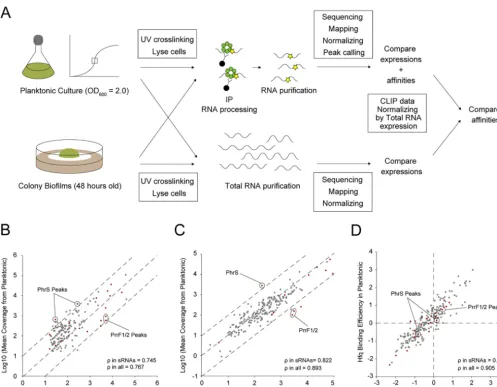
Disentanglement of preferential sRNA affinity to Hfq and expression.
A number
of sRNAs were associated with Hfq under both planktonic and biofilm conditions
though the Hfq peak intensities of these sRNAs differed between the two forms (Fig. 3B
and C, and Table S1). Since CLIP-seq peaks are a result of both RNA expression and RNA
affinity to Hfq under each condition, it is challenging to directly compare CLIP-seq
results between different conditions. To disentangle differences in gene expression
from those in protein binding, we carried out an
ad hoc
normalization of CLIP-seq using
RNA-seq-derived expression measurements (Fig. 4A).
We considered only overlapping peaks between the two conditions. First, read
coverage of cross-linked samples under the planktonic condition was divided by
coverage from the biofilm condition (“Compare expressions
⫹
affinities” denoted in
Fig. 4A, and Fig. 4B). Some sRNAs were differentially enriched in the Hfq CLIP-seq
between the planktonic and biofilm conditions. For instance, PhrS was more strongly
enriched under the planktonic condition (false discovery rate [FDR] of two PhrS
peaks, 5.12
⫻
10
⫺6, 2.63
⫻
10
⫺4) (Fig. 4B). Since this sRNA is upregulated from the early
stationary phase, depending on the anaerobic sensing factor Anr, and drives PQS/
pyocyanin synthesis by upregulating the transcriptional factor MvfR (44),
MvfR-regulating genes
pqsABCDE
,
phnAB
, and
phzS
were also transcriptionally upregulated
under the planktonic condition (Table S3). In addition, sRNAs P26, CrcZ, ErsA, and
NrsZ were also more enriched under the planktonic condition although the levels
of enrichment were not statistically significant. In contrast, PrrF1/2 were more strongly
enriched
under
the
biofilm
condition
(FDR
of
PrrF1/2
peaks, 2.31
⫻
10
⫺15,
6.78
⫻
10
⫺17) (Fig. 4B).
P. aeruginosa
overcomes iron limitation through pyoverdine/
pyochelin production during infection or biofilm development, and PrrF1/2 is in turn
associated with these pigments (20, 45, 46). In addition to PrrF1/2, sRNAs Ffs and SsrS
were also more enriched under the biofilm condition (FDR for Ffs peak, 2.76
⫻
10
⫺12;
FDR for two SsrS peaks, 3.77
⫻
10
⫺8and 2.59
⫻
10
⫺30).
To control for expression effects, we conducted total RNA-seq from the same
planktonic and biofilm cultures used in our CLIP experiments. Considering the
normal-ization biases upon rRNA depletion and the possibility of rRNA binding to Hfq, we did
not deplete rRNA. The three biological replicates displayed high correlations within
each condition (Fig. S6A and B). DESeq2 comparative analysis of 5,697 transcripts
detected via RNA-seq revealed that 478 and 589 RNAs were upregulated under
planktonic and biofilm conditions, respectively (FDR,
⬍
0.05; log
2fold change [FC],
⬎
1)
(“Compare expressions” denoted in Fig. 4A, and Fig. 4C; see also Fig. S6C and Table S3).
Here as well, PhrS and PrrF1/2 were upregulated exclusively in the planktonic and
biofilm forms, respectively (FDR of PhrS expression, 6.38
⫻
10
⫺34; FDR of PrrF1/2
ex-pression, 2.83
⫻
10
⫺29, 1.42
⫻
10
⫺33) (Fig. 4C). Overall, CLIP-seq and total RNA-seq
results inferred differential sRNA regulation between two phenotypic conditions.
To eliminate the effect of differential RNA expression from Hfq affinity, the fold
change in CLIP read coverages between planktonic and biofilm conditions were divided
by those of total RNA coverage (Fig. 4A, right, and Fig. 4D). We did not consider
non-cross-linked samples because the subjected RNAs were not suggested to be false
positives from calculation of both the log
2FC(cross-linked sample/non-cross-linked
sample) [FC(XL
⫹
/XL
⫺
)] and the FDR (47, 48). Figure 4D shows that differential sRNA
on September 8, 2020 by guest
http://msystems.asm.org/
association with Hfq was no longer seen after the normalization based on total
RNA-seq, implying that the Hfq-sRNA affinity remains constant across conditions. In
fact, while the correlation coefficient
of sRNAs from CLIP-seq and total RNA-seq
between conditions is relatively low (
in sRNAs, 0.745 and 0.822) (Fig. 4B and C), the
correlation in Hfq-binding efficiency is high (
in sRNAs, 0.922) (Fig. 4D). These results
suggest that the primary driver of differential sRNA association with Hfq is expression
rather than differences in relative affinities due to changing RNA pools. This lends
support to a model of RNA concentration-driven cycling of Hfq, known as the
associa-tive/active cycling model. In this model, free RNA transiently binds to the Hfq-RNA
complex, with frequent RNA exchange preventing Hfq availability from becoming a
limiting factor in regulation under most conditions (5).
DISCUSSION
Gram-negative
P. aeruginosa
is ubiquitously distributed in diverse environments.
Upon biofilm formation in the human body, this bacterium causes chronic
biofilm-FIG 4 Normalization and comparison of cross-linking immunoprecipitation with high-throughput sequencing of Hfq between planktonic and biofilm conditions. (A) A schematic representation of the comparison of RNA affinities to Hfq between planktonic and biofilm forms. Only overlapping peaks between two phenotypes were considered. Read coverages of cross-linked samples from planktonic conditions were divided by those from biofilms. Concomitantly, read coverages of total RNA from the planktonic condition were divided by those from biofilms. To remove the effect of RNA expression from peak information, fold changes of CLIP-normalized read coverages between planktonic and biofilm conditions were divided by those of total RNA-normalized coverage. (B to D) Mean coverages normalized according to size factors of overlapping peaks detected by CLIP-seq (B) and total RNA-seq (C) between planktonic and biofilm conditions. Dashed lines denote the reference diagonal. (D) Affinity comparison of Hfq normalized peaks on overlapping RNAs between planktonic and biofilm conditions. Dashed lines denote the reference bases. Peaks from sRNAs and others are highlighted in red and gray, respectively. Significantly enriched sRNAs in planktonic (PhrS) or biofilm (PrrF1/2) cultures are indicated with circles. The correlation coefficientwas calculated from planktonic versus biofilm.
Chihara et al.
on September 8, 2020 by guest
http://msystems.asm.org/
related infections in burn wounds, in
in vivo
-dwelling devices, and in the lungs of
immunocompromised patients with cystic fibrosis (12). Although posttranscriptional
regulatory networks that enable survival in dynamic environments are known to be
driven by sRNAs and associated RNA chaperones such as Hfq, RsmA, Crc, and the newly
identified ProQ (2), limited information is available regarding the global role of RNA
chaperones in the
P. aeruginosa
transcriptome, especially under different
environmen-tal conditions. CLIP-seq analysis, which helps decipher RNA-RBP interactions with RNA
targets, is well suited to explore regulatory functions of RBPs depending on a cell’s
physiological state (31–33).
In this study, we performed comparative CLIP-seq analysis between early stationary
planktonic cultures and colony biofilms, thus elucidating condition-specific RNA
regu-lation by Hfq. Consequently, 991 peaks with significant enrichment in cross-linked
samples were identified throughout the
P. aeruginosa
PAO1 genome although fewer
peaks were observed under the biofilm condition than under the planktonic condition
(Fig. 1A to D). RNA abundance decreases under biofilm conditions, with a concomitant
reduction in bacterial growth (49). Nevertheless, biofilm-specific Hfq-RNA interactions
were observed even at a low peak number (Fig. 1C to E). Previously, Pusic et al. reported
that CrcZ is the most abundant sRNA bound to Hfq in
P. aeruginosa
PA14 anoxic biofilm
and indirectly impacts biofilm formation (50). On the other hand, our CLIP-seq result
shows that CrcZ binds to Hfq under the planktonic condition rather than the biofilm
condition (see Table S1 in the supplemental material). The difference in Hfq-binding
sRNAs between two studies may come from the difference in the biofilm conditions.
Pusic et al. formed the anoxic biofilm in polypropylene tubes supplemented with
synthetic cystic fibrosis sputum medium, and this condition may stimulate CrcZ
ex-pression through the anaerobic sensing factor Anr. In contrast, in the current study,
colony biofilms were formed on LB agar aerobically, and this condition may repress
CrcZ expression due to the availability of a preferred carbon source. The present effort,
as a result, illuminated multifaceted roles of Hfq underlying the dynamic regulations of
biological processes between different physiological conditions, including different
sorts of biofilms.
Since
P. aeruginosa
Hfq can successfully complement its
E. coli
ortholog, it is a
functional homologue of
E. coli
Hfq (51). The association of
P. aeruginosa
Hfq with RNA
was investigated on the basis of sequence/structure motifs and the general distribution
of detected peaks (Fig. 2). Meta-gene analysis of peak distribution among mRNAs
mirrored previously described Hfq-binding preferences in
Salmonella
(32), indicating
that Hfq peaks are highly enriched at both the 5
=
UTR and 3
=
UTR (Fig. 2A to D). In
addition, sequence and structural motif analyses revealed that five AAN triplet
se-quence repeats at the 5
=
UTRs and the stem-loop structure, followed by the U-rich tail
at the 3
=
UTRs are preferential binding sites for Hfq (Fig. 2E to G). The sequence motif
from the 3
=
UTRs corresponds to Rho-independent terminators previously identified by
Salmonella
Hfq CLIP-seq (32). On the other hand, while the results derived from the 5
=
UTRs are congruent with previous reports regarding the distal surface of
E. coli
Hfq-binding A-rich sequences
in vitro
(52), this is the first evidence of AAN triplet
sequence repeats in mRNAs binding Hfq
in vivo
.
P. aeruginosa
Hfq lacks a long CTD, unlike
E. coli
and
Salmonella
Hfq. The mechanism
of action of the CTD in Hfq remains controversial; however, the CTD of
E. coli
Hfq is
required for the release of annealed mRNA-sRNA double-stranded conjugates from the
rim surface (53, 54). Since a truncated CTD wraps over the rim of
P. aeruginosa
Hfq and
since there is no association with the proximal and distal surfaces (53, 54), the present
results can be interpreted that AAN triplets at the 5
=
UTRs and GC-rich stem-loop
followed by U-rich tails at the 3
=
UTRs are common binding sites across bacterial orders,
at least between
Enterobacteriales
and
Pseudomonadales
, regardless of the length of the
CTDs. Although lack of a CTD strengthens the RNA association on rim surface, a UA-rich
consensus motif for the rim surface was indiscernible from our CLIP-seq, suggesting
that a truncated CTD may be enough to release mRNA-sRNA conjugates (55).
Interestingly, the peak distributions among sRNAs were relatively broad (Fig. 3A),
on September 8, 2020 by guest
http://msystems.asm.org/
and motif analysis revealed that four AAN triplet repeats in sRNAs are preferential
binding sites for Hfq (Fig. 3D). In enterobacteria, sRNAs have been divided into so-called
class I and class II sRNAs based on their sequence conservation and mode of interaction
with Hfq, among which class II sRNAs contain AAN motifs in the stretched sequence, as
well as U-rich tails at the 3
=
ends, and are more stable than class I sRNAs (6). Although
the majority of
E. coli
sRNAs are class I sRNAs, the prediction of AAN triplet repeats
implies a higher relative abundance of the class II sRNAs in the
P. aeruginosa
transcrip-tome. Interestingly, the CTD of
E. coli
Hfq has been suggested to offer a binding
advantage specifically to class II sRNAs (53). Why does
P. aeruginosa
, which has a short
Hfq CTD compared with that of
E. coli
, seem to preferentially express class II sRNAs? One
answer may come from recent structural analysis of the association of Crc as the fourth
partner with the sRNA-Hfq-mRNA ternary regulatory complex. Crc itself does not
interact with sRNAs through Rho-independent terminators; however, it does enhance
the association between Hfq and A-rich target sequences via an Hfq (distal
surface)-RNA-Crc sandwich conformation (27, 56), perhaps thus potentially selecting for the AAN
triplet motif. It may be interesting to consider the possibility that sRNAs with AAN
motifs tend to regulate mRNAs with U-rich motifs. However, Schu et al. reported that
a chimeric sRNA ChiX-RyhB in which the 5
=
end of ChiX with an AAN motif was fused
to RyhB with a U-rich motif reduces the negative regulation of RyhB target mRNA
sodB
with AAN motif and that another chimeric sRNA, ChiXΔAAN-RyhB-ChiX, which lacks an
AAN motif in the ChiX region, restores the negative regulation to
sodB
in
E. coli
(6). This
suggests that sRNA cannot regulate its target when each binding site to Hfq competes.
Therefore, a class II sRNA with both an AAN motif and U-rich motif may not regulate a
target with a U-rich motif as long as the binding of U-rich sequences in both the sRNA
and the target RNA to proximal surface of Hfq competes.
CLIP-seq converts read counts obtained from Illumina sequencing to peaks. Because
obtained peaks are products of both RNA expression and RNA affinity, it is difficult to
directly compare CLIP-seq results between different phenotypes or conditions. To
circumvent this issue, we carried out an
ad hoc
normalization of CLIP-seq results based
on RNA expression (Fig. 4). Disentanglement from background expression suggested
that the association of certain sRNAs to Hfq is primarily altered based on their
differential expression (Fig. 4A to C), a fact reminiscent of the associative/active cycling
model of Hfq-RNA interactions (5). Generally, several mechanisms may underlie
changes in RNA association with Hfq, depending on physiological states. First, since
hfq
expression levels did not significantly differ between planktonic and biofilm conditions
(Table S3), competition among sRNAs for invariable binding sites on Hfq may have
occurred. Second, sRNA association can be altered depending on the variations in
target RNA expression in each environment. This target-centric view is reminiscent of
condition-specific base paring between miRNA and microRNA response elements in
higher eukaryotes (57). Finally, the turnover rate of complex formation potentially
affects the RNA-Hfq association. Interactions between RBP and RNA were previously
reported to last 2 to 3 min
in vivo
; however, they were robust
in vitro
, with a half-life of
⬎
100 min, owing to turnover of complex formation depending on the levels of free
RNAs (5). In other words, while RNA with a low abundance can temporarily bind RBP,
it is replaced with highly abundant RNA. Additionally, another RBP, Crc, enhances Hfq
association with AAN triplet repeats in
P. aeruginosa
(56). Therefore, the Hfq
interac-tome may be strengthened with RNAs, wherein the interaction with AAN triplets is
rigid. While the current study cannot unravel which mechanisms in RNA association
with Hfq are preferential
in vivo
, our combinatorial approach shows that the affinity of
an individual sRNA does not change and that the association with Hfq is just dependent
on expression differences. Although one of the causes which make the understanding
of the mechanism in RNA association with Hfq
in vivo
difficult is lacking in complete list
of sRNA-Hfq-mRNA interactions, recent progress in the study about sRNA-mRNA
inter-actome is encouraging using
in vivo
preferential ligation of RNAs (36). We could find a
clue of a molecular mechanism of action of Hfq by comparing Hfq-binding efficiency
Chihara et al.
on September 8, 2020 by guest
http://msystems.asm.org/
among the different conditions provided in the present study with comprehensive
sRNA interactome studies.
Finally, we have identified more than 100 putative sRNAs bound to Hfq in either or
both of the planktonic and biofilm forms with manual curation of sRNAs from size
selection sRNA-seq conducted previously (16, 17) (Table S1). The evidence that these
sRNAs bind to Hfq suggests that they may be functional, not just RNA degradation
products. Further experiments will be needed to understand the mechanism of action
of the novel Hfq-binding sRNAs in
P. aeruginosa
. Altogether, the present combinatorial
comparative CLIP-seq and total RNA-seq approach uncovered condition-dependent
association of sRNAs with Hfq
in vivo
and dramatically expanded the potential direct
regulatory targets of Hfq in the
P. aeruginosa
transcriptome.
MATERIALS AND METHODS
Strains, plasmids, and growth conditions.Strains, plasmids and oligonucleotides used here are enlisted in Table S4 in the supplemental material. All experiments were performed usingP. aeruginosa
PAO1 or its derived strains. Each strain was cultured at 37°C in Luria-Bertani (LB) medium.
Construction of FLAG-tagged Hfq-harboring andhfqdeletion strains.PAO1hfq::3⫻FLAG and Δhfqstrains were constructed through conjugative transfer of appropriate plasmids and homologous recombination between the chromosome and plasmid DNA as previously described (58). pG19hfq:: 3⫻FLAG was constructed by cloning PCR products at HindIII/BamHI sites 1 kb upstream of thehfqstop codon in the PAO1 chromosome, three copies of a FLAG tag, and 1 kb downstream of thehfqstop codon in the PAO1 chromosome into the pG19II backbone. pG19Δhfqwas constructed by cloning PCR products at HindIII/XbaI sites 500 bp upstream of thehfqstart codon and 500 bp downstream of thehfqstop codon in the PAO1 chromosome into the pG19II backbone.
UV cross-linking, immunoprecipitation, and RNA purification. For the planktonic condition, 200-ml bacterial cultures of three replicates were maintained up to an OD600of 2.0 in LB medium. For the biofilm condition, an overnight culture was diluted 100-fold, and 10l of the aliquot was seeded on a cellulose membrane (HATF02500; Millipore) placed on the LB agar. Before the cellulose membrane was placed on LB agar, it was UV irradiated on both sides for 10 min each for surface sterilization. Colony biofilms were incubated statically for 48 h at 37°C. Membranes with colony biofilms were aseptically transferred to fresh LB agar every 24 h. Because the OD600could not be determined for colony biofilms, we measured the weight of pellets from both planktonic (OD600of 2.0) and 48-h-incubated colony biofilms and determined the number of necessary colony biofilms for CLIP experiments. After 48 h of incubation, 36 colony biofilms from the same biological replicate were scraped with a sterilized loop into 200 ml of LB medium. UV cross-linking and immunoprecipitation were performed as previously described (32, 34). Briefly, half of the culture from each condition was irradiated at 800 mJ of UV light at 254 nm. After UV cross-linking, the sample, as well as non-cross-linked control samples, was centrifuged for 20 min at 4,700 rpm at 4°C. Cell pellets were lysed in a Retch mill at 30 Hz for 10 min with 1 ml of 0.1-mm glass beads and 800l of NP-T buffer (50 mM NaH2PO4, 300 mM NaCl, 0.05% Tween 20, pH 8.0). NP-T buffer supplemented with 8 M urea was added to each supernatant at an equal volume and incubated for 5 min at 65°C with agitation at 900 rpm. Anti-FLAG magnetic beads were washed three times with 500l of NP-T buffer, resuspended in 125l of NP-T buffer, and treated with a 120-l suspension of urea-treated samples. After 1 h of incubation at 4°C, samples were washed twice with 500l of high-salt buffer (50 mM NaH2PO4, 1 M NaCl, 0.05% Tween 20, pH 8.0), followed by two washes with 500l of NP-T buffer. Beads were resuspended in Benzonase mix (500 units of Benzonase nuclease [E1014; Sigma-Aldrich] in NP-T buffer with 1 mM MgCl2) and incubated for 10 min at 37°C with agitation at 900 rpm. After one wash with high-salt buffer and two washes with calf intestinal alkaline phosphatase (CIP) buffer (100 mM NaCl, 50 mM Tris-HCl, pH 7.4, 10 mM MgCl2), beads were resuspended in 200l of CIP mix (20 units of CIP [M0290; NEB] in CIP buffer) and incubated for 30 min at 37°C with agitation at 800 rpm. After one wash with high-salt buffer and two washes with polynucleotide kinase (PNK) buffer (50 mM Tris-HCl, pH 7.4, 10 mM MgCl2, 0.1 mM spermidine), beads were resuspended in 100l of PNK mix (10 units of T4 PNK [EK0032; ThermoFisher Scientific], 10Ci of [␥-32P]ATP in PNK buffer) and incubated for 30 min at 37°C, followed by addition of 10l of 1 mM nonradioactive ATP and incubation for 5 min at 37°C. After two washes with NP-T buffer, beads were resuspended in 30l of protein loading buffer and incubated for 5 min at 95°C. Beads were magnetized and supernatants were transferred to fresh tubes.
Five-microliter aliquots and the rest of the supernatants were loaded and separated via SDS-polyacrylamide gel electrophoresis (12% resolving gel) for 1.5 h at 340 mA, followed by Western blot analysis and RNA extraction. After electrophoresis, proteins were electrotransferred onto a polyvinylidene difluoride membrane, which was blocked in 1⫻TBS-T buffer (20 mM Tris, 150 mM NaCl, 0.1% Tween 20, pH 7.6) with 10% skim milk for 45 min. Thereafter, the membrane was probed overnight at 4°C with monoclonal anti-FLAG (31430, 1:1,000; ThermoFisher Scientific) antibody diluted in 1⫻TBS-T buffer containing 3% bovine serum albumin, washed three time for 5 min each in 1⫻TBS-T buffer, probed for 1 h with anti-mouse horseradish peroxidase (HRP) antibody (F1804, 1:10,000; Sigma-Aldrich ) diluted in 1⫻TBS-T buffer containing 3% bovine serum albumin, and washed three times for 5 min each time in 1⫻TBS-T buffer. Chemiluminescent signals were detected using Image Quant LAS 4000 (GE Healthcare). After RNA electrophoresis, the RNA was transferred from the gel to Protran membrane (10600016; GE Healthcare). The membrane was placed in a cassette with a phosphor screen and exposed overnight. The
on September 8, 2020 by guest
http://msystems.asm.org/
autoradiogram was printed on clear paper and aligned with the membrane, and bands were cut out. Each membrane piece was cut into smaller pieces and placed in 2 ml of LoBind tubes with 400l of proteinase K (PK) solution (1.3 mg/ml PK [EO0491; ThermoFisher Scientific], 10 units of RNase inhibitor [10777019; Invitrogen] in 2⫻PK buffer [100 mM Tris-HCl, pH 7.9, 10 mM EDTA, 1% SDS]), followed by incubation for 1 h at 37°C with agitation at 1,000 rpm and then by incubation for 1 h at 37°C with agitation at 1,000 rpm with 100l of PK buffer with 9 M urea. Thereafter, 450l of supernatants from proteinase K-treated membranes was mixed with an equal volume of phenol-chloroform-isoamyl alcohol in phase-lock gel tubes and incubated for 5 min at 30°C with agitation at 1,000 rpm. Each mixture was centrifuged for 12 min at 13,000 rpm at 4°C, and 400l of the aqueous phase was precipitated with 3 volumes of ice-cold ethanol, 1/30 volume of 3 M sodium acetate (NaOAc; pH 5.2), and 1l of GlycoBlue (AM9515; Invitrogen) for 2 h at –20°C. The precipitated pellet was washed with 80% ethanol, briefly dried for 5 min at 20°C, and resuspended in 11l of sterilized water.
Total RNA purification.Two-hundred microliters of stop solution (95% [vol/vol] ethanol and 5% [vol/vol] water-saturated phenol, pH⬎7.0) was added into 1-ml aliquots of each cross-linked culture and immediately incubated at – 80°C. The frozen culture was defrosted on ice and centrifuged for 20 min at 4,500 rpm at 4°C. Total RNA was extracted via the hot phenol extraction method. Briefly, the culture containing the stop solution in the tube was resuspended in a mixture containing 600l of Tris-EDTA (TE) buffer, 0.5 mg/ml lysozyme (pH 8.0), and 60l of 10% (wt/vol) SDS. The tube was placed in a water bath for 5 min at 65°C, followed by addition of 66l of NaOAc (pH 5.2). Thereafter, 750l of phenol was added into the tube and mixed via tube inversion every 30 s during incubation for 5 min at 65°C. The tube was centrifuged for 15 min at 13,000 rpm at 4°C. The aqueous phase was transferred to a phase-lock gel tube, and 750l of chloroform was added. After samples were mixed via tube inversion, the mixture was centrifuged for 12 min at 13,000 rpm at 4°C. The aqueous phase was precipitated with 2 volumes of ice-cold ethanol and 1/30 volume of 3 M NaOAc (pH 6.5) for 2 h at –20°C. The precipitated pellet was washed with ice-cold 75% ethanol, briefly dried for 5 min at 20°C, and resuspended in 50l of sterilized water. Thereafter, 10 and 40g (vol/wt) of RNA were extracted from biofilm and planktonic cultures, respectively, in 39.5l of sterilized water. A total of 10.5l of DNase solution (5 units of DNase I and 5 units of RNase inhibitor in DNase buffer) was added to each sample and incubated for 30 min at 37°C, and 100l of sterilized water was added. Thereafter, 150l of the total mixture was transferred to a phase-lock gel tube with 150l of phenol-chloroform-isoamyl alcohol. After the samples was mixed via tube inversion, the mixture was centrifuged for 15 min at 13,000 rpm at 4°C. The aqueous phase was precipitated with 2.5 volumes of ice-cold ethanol and 1/30 volume of 3 M NaOAc (pH 6.5) for 2 h at –20°C. The pellet was washed with ice-cold 75% ethanol, briefly dried for 5 min at 20°C, and resuspended in 40l of sterilized water. RNA quantity and quality were determined via a NanoDrop spectrophotometer and electrophoresis on a 1% (wt/vol) agarose gel, respectively.
cDNA library preparation and sequencing.A cDNA library of the CLIP samples was prepared using an NEBNext Multiplex Small RNA Library Prep Set for Illumina (E7300; NEB) in accordance with the manufacturers’ instructions with certain modifications. First, a 3=SR adapter and 5=SR adapter (Illumina) were diluted 10-fold with nuclease-free water before use. Second, each reagent was prepared at half the volume instructed by the manufacturer. Third, 2.5l of each purified RNA was used for cDNA library preparation. PCR amplification was performed for cDNA under the following conditions: 18, 20, and 22 cycles for initial amplification and 23 cycles during the final amplification. cDNA libraries were quantified using a Qubit double-stranded DNA (dsDNA) assay (Q32854; Invitrogen) and Agilent 2100 Bioanalyzer dsDNA assay (Agilent). High-throughput sequencing was performed at Core Unit Systems Medicine, University of Würzburg, Würzburg, Germany. Twelve cDNA libraries from CLIP were pooled on an Illumina NextSeq 500 mid-output flow cell and sequenced in paired-end mode (2⫻75 cycles). For total RNA sequencing, high-throughput sequencing was performed at Vertis Biotechnologie AG, Freising, Germany. cDNA libraries were prepared from total RNA and pooled on an Illumina NextSeq 500 mid-output flow cell and sequenced in single-end mode (1⫻75 cycles).
Sequence processing and mapping.For CLIP-seq, to ensure high sequence quality, read 1 (R1) and read 2 (R2) files containing the Illumina paired-end reads in FASTQ format were quality and adapter trimmed via Cutadapt (59), version 1.15/1.16, using a cutoff Phred score of 20 in NextSeq mode, and reads without any remaining bases were discarded (command line parameters: --nextseq-trim⫽20 -m 1 -a AGATCGGAAGAGCACACGTCTGAACTCCAGTCAC -A GATCGTCGGACTGTAGAACTCTGAACGTGTAGAT CTCGGTGGTCGCCGTATCATT). To eliminate putative PCR duplicates, paired-end reads were collapsed using FastUniq (60). After trimming, we applied the pipeline READemption (61), version 0.4.5, to align all reads longer than 11 nt to the P. aeruginosaPAO1 chromosome (NCBI accession no.NC_002516.2) reference genome using segemehl (62), version 0.2.0, with an accuracy cutoff of 80%. From the results, only those reads mapping uniquely to one genomic position were considered for all subsequent analyses.
For RNA-seq, to ensure high sequence quality, Illumina reads were quality and adapter trimmed via Cutadapt (59), version 1.15, using a cutoff Phred score of 20 in NextSeq mode, and reads without any remaining bases were discarded (command line parameters: --nextseq-trim⫽20 -m 1 -a AGATCGGAAGAG CACACGTCTGAACTCCAGTCAC). After sequences were trimmed, we applied the pipeline READemption (61), version 0.4.5, to align all reads longer than 11 nt to the P. aeruginosaPAO1 (NCBI accession no.
NC_002516.2) reference genome using segemehl, version 0.2.0 (62), with an accuracy cutoff of 95%.
Transcript and UTR annotations. The transcript and subsequently the UTR annotations were generated for the planktonic and biofilm RNA-seq data with the pipeline ANNOgesic (41), version 0.7.33. Therefore, the annotation reference file forP. aeruginosaPAO1 (NCBI accession no.NC_002516.2), the reported TSS based on differential RNA-seq data (39), and the terminators detected by ANNOgesic (41)
Chihara et al.
on September 8, 2020 by guest
http://msystems.asm.org/
via TransTermHP (40) were used. The transcripts were adjusted based on the reference genome annotation positions. The 3=UTRs were annotated based on the transcripts as well as terminators, and the 5=UTR annotations were based on the TSS and transcripts. Other parameters were set to default.
For all analyses related to annotated genomic features such as CDSs and tRNAs, gene annotations from PseudoCAP (http://www.pseudomonas.com) were considered.
Read count normalization and peak calling based on CLIP-seq.Read counts per position were exploratorily analyzed to isolate the core of positions present in cross-linked and non-cross-linked library pairs, as described previously (34). The areas with low read counts were filtered from both cross-linked and non-cross-linked libraries using a standard deviation of 6 or less as an index. After the difference in read counts between the two libraries was plotted, the size factor was calculated using the DESeq normalization procedure from the high-count positions in both libraries across all replicates (63).
We applied PEAKachu, version 0.1.0 (https://github.com/tbischler/PEAKachu), for the peak calling in a similar way as described previously (34). First, BAM files for the respective pairs of cross-linked and non-cross-linked libraries were used to run in paired-end (-P) and paired-replicate (-r) mode. The maximum fragment size (-M) was set to 50, and annotations generated as GFF format (see above) were used to map overlapping features to the called peaks. Normalization was performed in the ‘manual’ mode using previously determined size factors (see above). Other parameters were set to default. Second, the boundary of initial peaks was set through block definition computed by the blockbuster algorithm (64) based on pooled read alignments from all cross-linked libraries using default parameters. Third, the PEAKachu tool runs DESeq2 (65) to analyze the significance of peak enrichment in the cross-linked libraries relative to levels in the non-cross-linked libraries with parameter values as follows: mad-multiplier (-m), 1.0; fold change (-f), 1.0; and adjustedPvalue (-Q), 0.05. Finally, PEAKachu was used for each replicon and strand to generate normalized coverage plots for the facilitation of data visual-ization.
Differential expression analysis based on total RNA-seq.We applied READemption (61) to assess the overlap of read alignments for each library to the same annotations used in the CLIP-seq analysis on the sense strand. Each read with a minimum overlap of 1 nt was counted with a value based on the number of locations where the read was mapped. If the read overlapped more than one annotation, the respective value was counted once for each overlapping region. The resulting read counts were subjected to differential expression analysis of planktonic versus biofilm samples via DESeq2 (65), version 1.18.1. Fold change shrinkage was applied by setting the parameter ‘betaPrior⫽TRUE’.
Analysis of sequence and structural motifs.The sequences of peaks from planktonic, biofilm, and combined conditions were used to perform MEME sequence motif analysis (66). Minimum and maximum motif widths were set at 6 and 50, respectively, while other parameters were set to default.
Structural motifs of the sequences of peak regions extended by an additional 10 nt upstream and downstream from planktonic, biofilm, and combined conditions were analyzed using CMfinder, version 0.2.1 (67). The minimum length of single stem-loop candidates was set as 20, while other parameters were set to default. Each analyzed motif was visualized using R2R (68).
Statistical and other analysis.Descriptive statistical analyses for peak overlapping between two conditions, peak distributions across theP. aeruginosagenome, and peak classification among RNA classes were performed using Microsoft Excel. A peak density plot was constructed using Python 3. Genes identified via CLIP-seq analysis were functionally characterized using the PseudoCAP annotation (http:// www.pseudomonas.com). KEGG enrichment analysis was performed from genes identified via CLIP-seq analysis using DAVID (https://david.ncifcrf.gov/summary.jsp). The default parameters and databases were used, and multiple testing adjustments were performed using the Benjamini-Hochberg method.
Data availability.Raw sequencing reads in FASTQ format are available in NCBI’s Gene Expression Omnibus (GEO [https://www.ncbi.nlm.nih.gov/geo]) under accession numberGSE136112.
SUPPLEMENTAL MATERIAL
Supplemental material for this article may be found at
https://doi.org/10.1128/
mSystems.00590-19
.
FIG S1
, TIF file, 1.3 MB.
FIG S2
, TIF file, 2.6 MB.
FIG S3
, TIF file, 1.7 MB.
FIG S4
, TIF file, 1.5 MB.
FIG S5
, TIF file, 0.9 MB.
FIG S6
, TIF file, 1.7 MB.
TABLE S1
, XLSX file, 0.2 MB.
TABLE S2
, XLSX file, 0.05 MB.
TABLE S3
, XLSX file, 1.5 MB.
TABLE S4
, DOCX file, 0.03 MB.
ACKNOWLEDGMENTS
We thank Jens Hör and Tetsushi Suyama for assistance with the registration of this
study in Würzburg University and in the National Institute of Advanced Industrial
Science and Technology, respectively. We also thank Charlotte Michaux (Imperial
on September 8, 2020 by guest
http://msystems.asm.org/
College London) for technical assistance with CLIP experiments, Kana Morinaga,
Ma-sanori Toyofuku, and Nobuhiko Nomura (Tsukuba University) for generously providing
the necessary plasmids and
E. coli
S17-1 strain, Sung-Huan Yu (Max Planck Institute of
Biochemistry) for his help with ANNOgesic, and Takaya Yamazoe (Tsukuba University)
for constructing scripts to compare peak positions between planktonic and biofilm
forms.
This study was supported by Grant-in-Aid for JSPS Research Fellow grant number
17J10663 and by the project Z-6 of the Interdisciplinary Center for Clinical Research of
the University Hospital Würzburg.
REFERENCES
1. Hör J, Gorski SA, Vogel J. 2018. Bacterial RNA biology on a genome scale. Mol Cell 70:785–799.https://doi.org/10.1016/j.molcel.2017.12.023. 2. Holmqvist E, Vogel J. 2018. RNA-binding proteins in bacteria. Nat Rev
Microbiol 16:601– 615.https://doi.org/10.1038/s41579-018-0049-5. 3. Chao Y, Vogel J. 2010. The role of Hfq in bacterial pathogens. Curr Opin
Microbiol 13:24 –33.https://doi.org/10.1016/j.mib.2010.01.001. 4. Lease RA, Woodson SA. 2004. Cycling of the Sm-like protein Hfq on the
DsrA small regulatory RNA. J Mol Biol 344:1211–1223.https://doi.org/10 .1016/j.jmb.2004.10.006.
5. Fender A, Elf J, Hampel K, Zimmermann B, Wagner EG. 2010. RNAs actively cycle on the Sm-like protein Hfq. Genes Dev 24:2621–2626.
https://doi.org/10.1101/gad.591310.
6. Schu DJ, Zhang A, Gottesman S, Storz G. 2015. Alternative Hfq-sRNA interaction modes dictate alternative mRNA recognition. EMBO J 34: 2557–2573.https://doi.org/10.15252/embj.201591569.
7. Morita T, Aiba H. 2011. RNase E action at a distance: degradation of target mRNAs mediated by an Hfq-binding small RNA in bacteria. Genes Dev 25:294 –298.https://doi.org/10.1101/gad.2030311.
8. De Lay N, Schu DJ, Gottesman S. 2013. Bacterial small RNA-based negative regulation: Hfq and its accomplices. J Biol Chem 288: 7996 – 8003.https://doi.org/10.1074/jbc.R112.441386.
9. Lee T, Feig AL. 2008. The RNA binding protein Hfq interacts specifically with tRNAs. RNA 14:514 –523.https://doi.org/10.1261/rna.531408. 10. Andrade JM, Santos RF, Chelysheva I, Ignatova Z, Arraiano CM. 2018. The
RNA-binding protein Hfq is important for ribosome biogenesis and affects translation fidelity. EMBO J 37:e97631.https://doi.org/10.15252/ embj.201797631.
11. Chen J, Gottesman S. 2017. Hfq links translation repression to stress-induced mutagenesis in E. coli. Genes Dev 31:1382–1395.https://doi .org/10.1101/gad.302547.117.
12. Mulcahy LR, Isabella VM, Lewis K. 2014. Pseudomonas aeruginosa bio-films in disease. Microb Ecol 68:1–12.https://doi.org/10.1007/s00248 -013-0297-x.
13. Stover CK, Pham XQ, Erwin AL, Mizoguchi SD, Warrener P, Hickey MJ, Brinkman FS, Hufnagle WO, Kowalik DJ, Lagrou M, Garber RL, Goltry L, Tolentino E, Westbrock-Wadman S, Yuan Y, Brody LL, Coulter SN, Folger KR, Kas A, Larbig K, Lim R, Smith K, Spencer D, Wong GK, Wu Z, Paulsen IT, Reizer J, Saier MH, Hancock RE, Lory S, Olson MV. 2000. Complete genome sequence of Pseudomonas aeruginosa PAO1, an opportunistic pathogen. Nature 406:959 –964.https://doi.org/10.1038/35023079. 14. Galán-Vásquez E, Luna B, Martínez-Antonio A. 2011. The regulatory
network of Pseudomonas aeruginosa. Microb Inform Exp 1:3.https://doi .org/10.1186/2042-5783-1-3.
15. Grenga L, Little RH, Malone JG. 2017. Quick change: post-transcriptional regulation in Pseudomonas. FEMS Microbiol Lett 364:fnx125.https://doi .org/10.1093/femsle/fnx125.
16. Ferrara S, Brugnoli M, De Bonis A, Righetti F, Delvillani F, Deho G, Horner D, Briani F, Bertoni G. 2012. Comparative profiling of Pseudomonas aeruginosa strains reveals differential expression of novel unique and conserved small RNAs. PLoS One 7:e36553. https://doi.org/10.1371/ journal.pone.0036553.
17. Gomez-Lozano M, Marvig RL, Molin S, Long KS. 2012. Genome-wide identification of novel small RNAs in Pseudomonas aeruginosa. En-viron Microbiol 14:2006 –2016. https://doi.org/10.1111/j.1462-2920 .2012.02759.x.
18. Wurtzel O, Yoder-Himes DR, Han K, Dandekar AA, Edelheit S, Greenberg EP, Sorek R, Lory S. 2012. The single-nucleotide resolution transcriptome
of Pseudomonas aeruginosa grown in body temperature. PLoS Pathog 8:e1002945.https://doi.org/10.1371/journal.ppat.1002945.
19. Sonnleitner E, Sorger-Domenigg T, Madej MJ, Findeiss S, Hackermüller J, Hüttenhofer A, Stadler PF, Bläsi U, Moll I. 2008. Detection of small RNAs in Pseudomonas aeruginosa by RNomics and structure-based bioinfor-matic tools. Microbiology 154:3175–3187.https://doi.org/10.1099/mic.0 .2008/019703-0.
20. Wilderman PJ, Sowa NA, FitzGerald DJ, FitzGerald PC, Gottesman S, Ochsner UA, Vasil ML. 2004. Identification of tandem duplicate regula-tory small RNAs in Pseudomonas aeruginosa involved in iron homeo-stasis. Proc Natl Acad Sci U S A 101:9792–9797.https://doi.org/10.1073/ pnas.0403423101.
21. Masse E, Gottesman S. 2002. A small RNA regulates the expression of genes involved in iron metabolism in Escherichia coli. Proc Natl Acad Sci U S A 99:4620 – 4625.https://doi.org/10.1073/pnas.032066599. 22. Nikulin A, Stolboushkina E, Perederina A, Vassilieva I, Blaesi U, Moll I,
Kachalova G, Yokoyama S, Vassylyev D, Garber M, Nikonov S. 2005. Structure of Pseudomonas aeruginosa Hfq protein. Acta Crystallogr D Biol Crystallogr 61:141–146. https://doi.org/10.1107/S09074449 04030008.
23. Sonnleitner E, Hagens S, Rosenau F, Wilhelm S, Habel A, Jäger KE, Bläsi U. 2003. Reduced virulence of a hfq mutant of Pseudomonas aeruginosa O1. Microb Pathog 35:217–228. https://doi.org/10.1016/S0882-4010 (03)00149-9.
24. Sonnleitner E, Schuster M, Sorger-Domenigg T, Greenberg EP, Bläsi U. 2006. Hfq-dependent alterations of the transcriptome profile and effects on quorum sensing in Pseudomonas aeruginosa. Mol Microbiol 59: 1542–1558.https://doi.org/10.1111/j.1365-2958.2006.05032.x. 25. Sonnleitner E, Abdou L, Haas D. 2009. Small RNA as global regulator of
carbon catabolite repression in Pseudomonas aeruginosa. Proc Natl Acad Sci U S A 106:21866 –21871. https://doi.org/10.1073/pnas.pnas .0910308106.
26. Sonnleitner E, Blasi U. 2014. Regulation of Hfq by the RNA CrcZ in Pseudomonas aeruginosa carbon catabolite repression. PLoS Genet 10: e1004440.https://doi.org/10.1371/journal.pgen.1004440.
27. Sonnleitner E, Wulf A, Campagne S, Pei XY, Wolfinger MT, Forlani G, Prindl K, Abdou L, Resch A, Allain FH, Luisi BF, Urlaub H, Blasi U. 2018. Interplay between the catabolite repression control protein Crc, Hfq and RNA in Hfq-dependent translational regulation in Pseudomonas aerugi-nosa. Nucleic Acids Res 46:1470 –1485. https://doi.org/10.1093/nar/ gkx1245.
28. Kambara TK, Ramsey KM, Dove SL. 2018. Pervasive targeting of nas-cent transcripts by Hfq. Cell Rep 23:1543–1552. https://doi.org/10 .1016/j.celrep.2018.03.134.
29. Barquist L, Vogel J. 2015. Accelerating discovery and functional analysis of small RNAs with new technologies. Annu Rev Genet 49:367–394.
https://doi.org/10.1146/annurev-genet-112414-054804.
30. Saliba AE, Santos CS, Vogel J. 2017. New RNA-seq approaches for the study of bacterial pathogens. Curr Opin Microbiol 35:78 – 87.https://doi .org/10.1016/j.mib.2017.01.001.
31. Tree JJ, Granneman S, McAteer SP, Tollervey D, Gally DL. 2014. Identifi-cation of bacteriophage-encoded anti-sRNAs in pathogenic Escherichia coli. Mol Cell 55:199 –213.https://doi.org/10.1016/j.molcel.2014.05.006. 32. Holmqvist E, Wright PR, Li L, Bischler T, Barquist L, Reinhardt R, Backofen
R, Vogel J. 2016. Global RNA recognition patterns of post-transcriptional regulators Hfq and CsrA revealed by UV crosslinking in vivo. EMBO J 35:991–1011.https://doi.org/10.15252/embj.201593360.
33. Potts AH, Vakulskas CA, Pannuri A, Yakhnin H, Babitzke P, Romeo T. 2017. Chihara et al.
on September 8, 2020 by guest
http://msystems.asm.org/
Global role of the bacterial post-transcriptional regulator CsrA revealed by integrated transcriptomics. Nat Commun 8:1596.https://doi.org/10 .1038/s41467-017-01613-1.
34. Holmqvist E, Li L, Bischler T, Barquist L, Vogel J. 2018. Global maps of ProQ binding in vivo reveal target recognition via RNA structure and stability control at mRNA 3’ ends. Mol Cell 70:971–982.e6.https://doi .org/10.1016/j.molcel.2018.04.017.
35. Waters SA, McAteer SP, Kudla G, Pang I, Deshpande NP, Amos TG, Leong KW, Wilkins MR, Strugnell R, Gally DL, Tollervey D, Tree JJ. 2017. Small RNA interactome of pathogenic E. coli revealed through crosslinking of RNase E. EMBO J 36:374 –387.https://doi.org/10.15252/embj.201694639. 36. Melamed S, Peer A, Faigenbaum-Romm R, Gatt YE, Reiss N, Bar A, Altuvia Y, Argaman L, Margalit H. 2016. Global mapping of small RNA-target interactions in bacteria. Mol Cell 63:884 – 897.https://doi.org/10.1016/j .molcel.2016.07.026.
37. Han K, Tjaden B, Lory S. 2016. GRIL-seq provides a method for identifying direct targets of bacterial small regulatory RNA by in vivo proximity ligation. Nat Microbiol 2:16239.https://doi.org/10.1038/nmicrobiol.2016 .239.
38. Zhang YF, Han K, Chandler CE, Tjaden B, Ernst RK, Lory S. 2017. Probing the sRNA regulatory landscape of P aeruginosa: post-transcriptional control of determinants of pathogenicity and antibiotic susceptibility. Mol Microbiol 106:919 –937.https://doi.org/10.1111/mmi.13857. 39. Gill EE, Chan LS, Winsor GL, Dobson N, Lo R, Ho Sui SJ, Dhillon BK, Taylor
PK, Shrestha R, Spencer C, Hancock REW, Unrau PJ, Brinkman F. 2018. High-throughput detection of RNA processing in bacteria. BMC Genom-ics 19:223.https://doi.org/10.1186/s12864-018-4538-8.
40. Kingsford CL, Ayanbule K, Salzberg SL. 2007. Rapid, accurate, computa-tional discovery of Rho-independent transcription terminators illumi-nates their relationship to DNA uptake. Genome Biol 8:R22.https://doi .org/10.1186/gb-2007-8-2-r22.
41. Yu SH, Vogel J, Förstner KU. 2018. ANNOgesic: a Swiss army knife for the RNA-seq based annotation of bacterial/archaeal genomes. GigaScience 7:giy096.https://doi.org/10.1093/gigascience/giy096.
42. Huang DW, Sherman BT, Tan Q, Collins JR, Alvord WG, Roayaei J, Stephens R, Baseler MW, Lane HC, Lempicki RA. 2007. The DAVID Gene Functional Classification Tool: a novel biological module-centric algo-rithm to functionally analyze large gene lists. Genome Biol 8:R183.
https://doi.org/10.1186/gb-2007-8-9-r183.
43. Lu P, Wang Y, Zhang Y, Hu Y, Thompson KM, Chen S. 2016. RpoS-dependent sRNA RgsA regulates Fis and AcpP in Pseudomonas aerugi-nosa. Mol Microbiol 102:244 –259.https://doi.org/10.1111/mmi.13458. 44. Sonnleitner E, Gonzalez N, Sorger-Domenigg T, Heeb S, Richter AS,
Backofen R, Williams P, Hüttenhofer A, Haas D, Bläsi U. 2011. The small RNA PhrS stimulates synthesis of the Pseudomonas aeruginosa quino-lone signal. Mol Microbiol 80:868 – 885.https://doi.org/10.1111/j.1365 -2958.2011.07620.x.
45. Banin E, Vasil ML, Greenberg EP. 2005. Iron and Pseudomonas aerugi-nosa biofilm formation. Proc Natl Acad Sci U S A 102:11076 –11081.
https://doi.org/10.1073/pnas.0504266102.
46. Nguyen AT, O’Neill MJ, Watts AM, Robson CL, Lamont IL, Wilks A, Oglesby-Sherrouse AG. 2014. Adaptation of iron homeostasis pathways by a Pseudomonas aeruginosa pyoverdine mutant in the cystic fibrosis lung. J Bacteriol 196:2265–2276.https://doi.org/10.1128/JB.01491-14. 47. Winther K, Tree JJ, Tollervey D, Gerdes K. 2016. VapCs of Mycobacterium
tuberculosis cleave RNAs essential for translation. Nucleic Acids Res 44:9860 –9871.https://doi.org/10.1093/nar/gkw781.
48. Winther KS, Roghanian M, Gerdes K. 2018. Activation of the stringent response by loading of RelA-tRNA complexes at the ribosomal A-site. Mol Cell 70:95–105.e4.https://doi.org/10.1016/j.molcel.2018.02.033. 49. Klumpp S, Zhang Z, Hwa T. 2009. Growth rate-dependent global effects
on gene expression in bacteria. Cell 139:1366 –1375.https://doi.org/10 .1016/j.cell.2009.12.001.
50. Pusic P, Tata M, Wolfinger MT, Sonnleitner E, Haussler S, Blasi U. 2016. Cross-regulation by CrcZ RNA controls anoxic biofilm formation in Pseudomonas aeruginosa. Sci Rep 6:39621.https://doi.org/10.1038/ srep39621.
51. Sonnleitner E, Moll I, Blasi U. 2002. Functional replacement of the Escherichia coli hfq gene by the homologue of Pseudomonas aerugi-nosa. Microbiology 148:883– 891.https://doi.org/10.1099/00221287-148 -3-883.
52. Link TM, Valentin-Hansen P, Brennan RG. 2009. Structure of Escherichia coli Hfq bound to polyriboadenylate RNA. Proc Natl Acad Sci U S A 106:19292–19297.https://doi.org/10.1073/pnas.0908744106.
53. Santiago-Frangos A, Kavita K, Schu DJ, Gottesman S, Woodson SA. 2016. C-terminal domain of the RNA chaperone Hfq drives sRNA competition and release of target RNA. Proc Natl Acad Sci U S A 113:E6089 –E6096.
https://doi.org/10.1073/pnas.1613053113.
54. Santiago-Frangos A, Jeliazkov JR, Gray JJ, Woodson SA. 2017. Acidic C-terminal domains autoregulate the RNA chaperone Hfq. Elife 6:e27049.https://doi.org/10.7554/eLife.27049.
55. Ishikawa H, Otaka H, Maki K, Morita T, Aiba H. 2012. The functional Hfq-binding module of bacterial sRNAs consists of a double or single hairpin preceded by a U-rich sequence and followed by a 3’ poly(U) tail. RNA 18:1062–1074.https://doi.org/10.1261/rna.031575.111.
56. Pei XY, Dendooven T, Sonnleitner E, Chen S, Blasi U, Luisi BF. 2019. Architectural principles for Hfq/Crc-mediated regulation of gene expres-sion. Elife 8:e43158.https://doi.org/10.7554/eLife.43158.
57. Bossi L, Figueroa-Bossi N. 2016. Competing endogenous RNAs: a target-centric view of small RNA regulation in bacteria. Nat Rev Microbiol 14:775–784.https://doi.org/10.1038/nrmicro.2016.129.
58. Hmelo LR, Borlee BR, Almblad H, Love ME, Randall TE, Tseng BS, Lin C, Irie Y, Storek KM, Yang JJ, Siehnel RJ, Howell PL, Singh PK, Tolker-Nielsen T, Parsek MR, Schweizer HP, Harrison JJ. 2015. Precision-engineering the Pseudomonas aeruginosa genome with two-step allelic exchange. Nat Protoc 10:1820 –1841.https://doi.org/10.1038/nprot.2015.115. 59. Martin M. 2011. Cutadapt removes adapter sequences from
high-throughput sequencing reads. EMBnet J 17:10 –12.https://doi.org/10 .14806/ej.17.1.200.
60. Xu H, Luo X, Qian J, Pang X, Song J, Qian G, Chen J, Chen S. 2012. FastUniq: a fast de novo duplicates removal tool for paired short reads. PLoS One 7:e52249.https://doi.org/10.1371/journal.pone.0052249. 61. Förstner KU, Vogel J, Sharma CM. 2014. READemption—a tool for the
computational analysis of deep-sequencing-based transcriptome data. Bioinformatics 30:3421–3423. https://doi.org/10.1093/bioinformatics/ btu533.
62. Hoffmann S, Otto C, Doose G, Tanzer A, Langenberger D, Christ S, Kunz M, Holdt LM, Teupser D, Hackermuller J, Stadler PF. 2014. A multi-split mapping algorithm for circular RNA, splicing, trans-splicing and fusion detection. Genome Biol 15:R34. https://doi.org/10.1186/gb-2014-15-2 -r34.
63. Anders S, Huber W. 2010. Differential expression analysis for sequence count data. Genome Biol 11:R106.https://doi.org/10.1186/gb-2010-11 -10-r106.
64. Langenberger D, Bermudez-Santana C, Hertel J, Hoffmann S, Khaitovich P, Stadler PF. 2009. Evidence for human microRNA-offset RNAs in small RNA sequencing data. Bioinformatics 25:2298 –2301.https://doi.org/10 .1093/bioinformatics/btp419.
65. Love MI, Huber W, Anders S. 2014. Moderated estimation of fold change and dispersion for RNA-seq data with DESeq2. Genome Biol 15:550.
https://doi.org/10.1186/s13059-014-0550-8.
66. Bailey TL, Johnson J, Grant CE, Noble WS. 2015. The MEME Suite. Nucleic Acids Res 43:W39 –W49.https://doi.org/10.1093/nar/gkv416.
67. Yao Z, Weinberg Z, Ruzzo WL. 2006. CMfinder–a covariance model based RNA motif finding algorithm. Bioinformatics 22:445– 452.https://doi.org/ 10.1093/bioinformatics/btk008.
68. Weinberg Z, Breaker RR. 2011. R2R–software to speed the depiction of aesthetic consensus RNA secondary structures. BMC Bioinformatics 12:3.
https://doi.org/10.1186/1471-2105-12-3.
69. Zuker M. 2003. Mfold web server for nucleic acid folding and hybridiza-tion predichybridiza-tion. Nucleic Acids Res 31:3406 –3415. https://doi.org/10 .1093/nar/gkg595.