INTRODUCTION
Tularemia caused by Francisella tularensis, an intracellu-lar obligate aerobic gram-negative coccobacillus, is a zoono-sis with natural focality distributed in geographical regions of the Northern hemisphere. Its actual medical and veterinarian importance has been stressed over the last decade by a pro-nounced activation of natural foci, accompanied by epidemic occurrence of the disease in humans, as well as by fear of bio-terrorism and misuse of F. tularensis as a potential biological weapon [1,2].
Tularemia in humans manifests in different ways depend-ing to a large extent on the mode of transmission: arthropod
bite, direct contact, ingestion or inhalation of the infectious agent. The clinical presentations of tularemia have been clas-sically divided into six classic forms: ulceroglandular, glandu-lar, oculoglanduglandu-lar, oropharyngeal, respiratory, and typhoidal tularemia [2-4]. However, overlapping of the different symp-toms is frequently observed. The occurrence of several dif-ferent clinical forms of tularemia that often initially presents with non-specific symptoms resembling influenza or other respiratory tract infections makes clinical diagnosis very diffi-cult. Laboratory diagnosis is essential for optimum treatment. Since the mortality rate is high in most severe cases, early diag-nosis of tularemia is crucial [3-6].
In Turkey, tularemia outbreaks have been described as early as 1936–1938, but tularemia was not reportable until 2004. Recently, multiple tularemia outbreaks in Turkey have been reported as it is now considered a re-emerging zoonotic disease in Turkey [7-9]. The only F. tularensis subspecies found in most of Eurasia, including Turkey, is holarctica. Genetic
*Corresponding author: Selçuk KILIÇ, M.D.,
Public Health Institution of Turkey (PHIT), National Tularemia Reference Laboratory, Refik Saydam Campus, Sağlık Mah. Adnan Saygun Cad. No:55, 06100 Sihhiye, Ankara, Turkey. E-mail: [email protected] Submitted: 22 November 2015 / Accepted: 21 December 2015
The use of matrix-assisted laser desorption
ionization-time of flight mass spectrometry in the
identification of
Francisella tularensis
Onur Karatuna1, Bekir Çelebi2, Simge Can3, Işın Akyar1, Selçuk Kiliç2*
1Department of Medical Microbiology, Acibadem University School of Medicine, Icerenkoy Mh, Istanbul, Turkey, 2National Tularemia
Reference Laboratory, Public Health Institution of Turkey, Refik Saydam Campus, Ankara, Turkey, 3Department of Microbiology, Acibadem
Labmed Medical Laboratories, Acibadem University, Icerenkoy Mh, Istanbul, Turkey
ABSTRACT
Francisella tularensis is the cause of the zoonotic disease tularemia and is classified among highly pathogenic bacteria (HPB) due to its low infection dose and potential for airborne transmission. In the case of HBP, there is a pressing need for rapid, accurate and reliable identification. Phenotypic identification of Francisella species is inappropriate for clinical microbiology laboratories because it is time-consuming, hazardous and subject to variable interpretation. Matrix-assisted laser desorption ionization-time of flight mass spectrometry (MALDI-TOF MS) was recently evaluated as a useful tool for the rapid identification of a variety of microorganisms. In this study, we evaluated the use of MALDI-TOF MS for the rapid identification of Francisella tularensis and differentiation of its subspecies. Using national collection of Francisella isolates from the National Tularemia Reference Laboratory (Public Health Institution of Turkey, Ankara), a total of 75 clinical isolates were investigated by species and subspecies-specific polymerase chain reaction (PCR) test and MALDI-TOF MS. All isolates were originally identified as F. tularen-sis subsp. holarctica according to region of difference 1 (RD1) subspecies-specific PCR results. For all isolates MALDI-TOF MS provided results in concordance with subspecies-specific PCR analysis. Although PCR-based methods are effective in identifying Francisella species, they are labor-intensive and take longer periods of time to obtain the results when compared with MALDI-TOF MS. MALDI-TOF MS appeared to be a rapid, reliable and cost-effective identification technique for Francisella spp. Shorter analysis time and low cost make this an appealing new option in microbiology laboratories.
KEY WORDS: Francisella tularensis; tularemia; identification; MALDI-TOF MS; PCR
diversity is low, probably because it’s a recently emerging pathogen [10]. Laboratory diagnosis is usually based on the detection of bacteria either by culture or by nucleic acid amplification techniques and serology. Despite the fastidious characteristics of F. tularensis which requires sulfhydral com-pounds (cysteine or cystine) for optimal growth and its highly infectious nature causing laboratory-acquired infections, cul-ture recovery and characterization remains the “gold standard” for laboratory confirmation of tularemia infections [4,5,11]. Conventional culture and biochemical tests, as well as molec-ular methods, have been used in clinical microbiology labora-tories for the identification of Francisella spp. [2,4-6]. Species and subspecies are subsequently determined by testing bio-chemical reactions/tests such as production of acid from car-bohydrates (maltose, d-glucose, lactose, sucrose, and glycerol), citrulline ureidase activity, H2S production in cysteine-supple-mented medium, and β-lactamase production. These proce-dures are inappropriate for clinical microbiology laboratories because they are time-consuming, difficult to perform, and hazardous [11,12]. In the last decade, to decrease the possibility of misidentification and obtain more rapid identification and confirmation, molecular methods such as polymerase chain reaction (PCR)-based diagnostics and 16/23S ribosomal RNA sequencing have been considered as alternative approaches to the phenotypic methods [2,11].
Over the last few years, matrix-assisted laser desorption ionization-time of flight mass spectrometry (MALDI-TOF MS) which enables identification of bacteria by comparing the mass spectra that correspond to protein profiles of bacterial cells to reference spectra stored in the database has been intro-duced as a fast, reliable and cost-effective technique for routine application in clinical microbiology laboratories [13-15]. Even though the performance of MALDI-TOF MS and phenotypic systems in identifying Francisella isolates have previously been reported [11], information on the performance of the modern methods is scarce in identifying clinical isolates of Francisella
species. Furthermore, challenging the MALDI-TOF MS with isolates from different geographical origins is needed to assess its usefulness as a universal method.
The aim of this study was to compare the performance of MALDI-TOF MS with subspecies-specific PCR method for the identification of Francisella species from Turkey.
MATERIALS AND METHODS
Bacterial isolates and reference strains
A total of 75 clinical Francisella strains isolated from humans were enrolled into the study. F. tularensis subsp. holarctica LVS (NCTC 10857) and F. tularensis subsp. tularensis SCHU S4 (FSC237) were included as reference strains. The isolates were carefully selected to ensure geographical diversity among the
isolates and collected from different regions of Turkey during the period October 2009 and March 2012. All isolates were stored at −86°C and were subcultured on cysteine heart agar supplemented with 9% heated (chocolatised) sheep blood (CHAB) plates.
PCR method
DNA was extracted from pure cultures of F. tularensis
strains grown on CHAB plates by using a commercial kit based on silica-gel-membrane technology (QIAamp DNA mini kit; QIAGEN GmbH, Hilden, Germany). Briefly, a loop-ful of bacteria were harvested and transferred to a 1.5 mL microcentrifuge tube, bacterial suspension was prepared by adding phosphate-buffered saline to a final volume of 200
primers. The primers used for the amplification of RD1 are shown in Table 1. The PCR steps included denaturation at 95°C for 5 min, followed by 40 cycles of denaturation at 95°C for 50 s, primer annealing at 60°C for 40 s, and primer extension at 72°C for 80 s. After the final extension step at 72°C for 7 min, each reaction mixture was subjected to gel electrophoresis in a 1.5% agarose gel for 1 h at 80 V, and the DNA fragments were detected with ethidium bromide staining. In this PCR assay F. tularensis subsp. holarctica strainsyield fragments of 924 bp, whereas F. tularensis subsp. tularensis strains yield fragments of 1522 bp which allowed differentiation at the sub-species level. In each PCR assay F. tularensis subsp. holarctica
LVS (NCTC 10857) and F. tularensis subsp. tularensis SCHU S4 (FSC237) reference strains were used as positive controls and a mixture containing water instead of bacterial DNA was used as negative control.
MALDI-TOF MS analysis
MALDI-TOF MS analyses in this study followed the pro-tocols described previously [19]. The strains which were incu-bated for 48 hours at 35°C on CHAB were transferred into 1.5 mL screw cap tubes, suspended with 75% ethanol and incu-bated for 30 min at room temperature. After vortexing they were centrifuged at 13,000 × g for 5 min. The supernatant was discarded and the pellet was left at room temperature for dry-ing. Afterwards, the pellet was mixed thoroughly with 50 µL of 70% aqueous formic acid. After addition of 50 µL of acetonitrile the mixture was centrifuged at 13,000 × g for 5 min and 1 µL of the microorganism extract supernatant was placed onto the polished steel MALDI target plate (Bruker Daltonics, Bremen, Germany) in duplicate and allowed to dry at room tempera-ture. Each sample was overlaid with 1 µL of matrix solution which consisted of saturated α-cyano-4-hydroxycinnamic acid in 50% acetonitrile-2.5% trifluoroacetic acid and the plate was air dried at room temperature. The plate was then loaded into a Bruker Autoflex III MALDI-TOF mass spectrometer (Bruker Daltonics, Bremen, Germany) and analysis was per-formed. The spectra were automatically recorded in the linear positive ion mode with delayed extraction at a laser frequency of 20 Hz within a mass range from 2.000 to 20.000 Da. For each spectrum 600 satisfactory shots in 100-shot steps from the sampling area of the target spot were obtained. Spectra were eligible for further analysis when the peaks had a reso-lution better than 400 intensity a.u. (arbitrary units). Each run
included a bacterial test standard with a characteristic pep-tide and protein profile, provided by Bruker for calibration, a negative extraction control (sterile water) and the reference QC strains(Escherichia coli ATCC 25922 and Staphylococcus aureus ATCC 29213). In case of failures the analyses were repeated using fresh colonies with the same method.
Data analysis
The mass spectra were evaluated with the MALDI Biotyper software version 3.0 (Bruker Daltonik, Bremen, Germany). The list of the best peaks of the spectrum are cre-ated automatically by the software after smoothing, normal-ization and baseline subtraction. The obtained spectra are analyzed by standard pattern matching algorithm, which com-pares the raw spectra with the spectra of the MALDI Biotyper Library (MBL) by using the standard settings. The results of MALDI Biotyper analysis are listed in a ranking table where the best match gives the identification result depending on its value. The results are expressed as log(score) values, which range from 0 (no spectra) to 3 (perfect match). Log(score) val-ues were interpreted as recommended by the manufacturer: score values of ≥1.7 generally indicate a relationship at the genus level, and values of ≥2.0 generally indicate relationships at the species level. The highest score is used for species iden-tification. However, at the time the study was performed there were no recorded reference F. tularensis strains in the standard MBL but only in a special “SR database” (security relevant database) which was not available in our laboratory. Therefore in-house generated spectral profiles of reference F. tularensis
strains, F. tularensis subsp. holarctica LVS (NCTC 10857) and
F. tularensis subsp. tularensis SCHU S4 (FSC237), were added to the MALDI Biotyper Support Library (MBSL) and used for the identification of 75 clinical Francisella tularensis isolates.
RESULTS
The PCR test targeting the tul4 gene which is common in
Francisellatularensis species was found positive in all isolates investigated in this study (Figure 1). For the determination of the subspecies of Francisella tularensis strains, the region of difference 1 (RD1) subspecies-specific PCR test was employed (Figure 2). All study isolates (n=75) yielded RD1 fragments of 924 bp that corresponds to the RD1 size of F. tularensis subsp.
holarctica. The mass spectra obtained by MALDI-TOF MS
TABLE 1. Oligunucleotide sequences used for the PCR amplification of tul4 and RD1 targets
Target Primer Oligonucleotide sequence (5’ to 3’) Fragment size (bp)
tul4 TUL4-435 GCTGTATCATCATTTAATAAACTGCTG 428 TUL4-863 TTGGGAAGCTTGTATCATGGCACT
for the test strains yielded log(score) values ranging between 1.704 and 2.357 (mean 1.897) for matching against the refer-ence strain F. tularensis subsp. holarctica LVS. Whereas the scores obtained for matching against the reference strain
F. tularensis subsp. tularensis SCHU S4 were found to be sub-stantially lower, ranging between 1.272 and 1.951 (mean 1.655). For all study isolates the Maldi Biotyper software yielded results for F. tularensis subsp. holarctica LVS as the first match organism which was followed by a second match with F. tula-rensis subsp. tularensis SCHU S4 with lower scores. Even though the obtained spectra visually looked very similar, the MALDI Biotyper software could correctly identify the study isolates as F. tularensis subsp. holarctica (sample spectral pro-files for the study isolate F. tularensis subsp. holarctica TUL S-013 and the two reference strains are shown in Figure 3). Even though the first match organism was F. tularensis subsp.
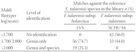
holarctica for all organisms, only 19 (25.3%) isolates received a score of >2.000 indicating reliable identification at the species level, the remaining 56 isolates (74.7%) received scores in the range 1.700 - 2.000 which translates as reliable identification at the genus level. Thus, using the cutoffs established by the manufacturer, MALDI-TOF MS-based identification of clini-cal F. tularensis isolates exhibited 100% concordance with the species-specific tul4 PCR assay, and 25.3% concordance with the RD1 PCR assay. The general performance of MALDI-TOF MS-based identification of clinical F. tularensis isolates is sum-marized in Table 2.
Regarding the duration of each method, the processing of the isolates for MALDI-TOF MS analysis took approximately 1 to 2 h per isolate or per batch but analysis itself took less than 10 min for a single isolate and around 0.5 to 1 h when multiple isolates are processed. For the PCR assay however, it took 4–6 h to complete the runs and analyse the PCR products. When the required hands-on time and the risk of contamination associated with PCR method are considered, MALDI-TOF MS-based identification was found to be easier to perform.
DISCUSSION
The current approach for the identification of Francisella
spp. is based mainly on standard slide agglutination and biochemical tests and molecular methods, including PCR, sequencing and hybridization assays using specific probes [4,5,11]. Conventional Francisella identification is both very time-consuming and sometimes inaccurate. Rapid and reliable identification of this agent is of major concern for opti-mal patient management and especially for the implementa-tion of effective measures for disease control [2,4].
In this study, the most relevant diagnostic methods avail-able in the field of clinical microbiology were compared for
the identification of Francisella. All isolates were identified correctly with both PCR and MALDI-TOF MS system. Species-specific PCR and Bruker MALDI-TOF MS identified all isolates belonging to F. tularensis correctly. This result is in accordance with the findings of Seibold et al., who reported a correct identification rate of 100% for Francisella spp. by MALDI-TOF MS [11].
F. tularensis subsp. holarctica is the only Francisella
TABLE 2. Performance of Bruker MALDI Biotyper software
for the identification of F. tularensis subsp. holarctica clinical isolates (n=75)
Maldi Biotyper log(score)
Level of identification
Matches against the reference
F.. tularensis species in the library n (%)
F. tularensis subsp.
holarctica F. tularensistularensis subsp.
LVS SCHU S4 <1.700 No identification 0 42 (56.0) 1.700-2.000 Genus only 56 (74.7) 33 (44.0) >2.000 Genus and species 19 (25.3) 0
FIGURE 2. The PCR amplification results for the Francisella
spe-cies using RD1 primers. Lane 1 and 2 are positive control strains F. tularensis subsp. tularensis SCHU S4 (FSC237) and F. tularensis subsp. holarctica LVS (NCTC 10857), respectively. Lane 3 is neg-ative control (water), lanes 4-8 are clinical Francisella strains. Molecular sizes in base pair (bp) are indicated at the left. (M: Molecular mass standard) In this PCR assay F. tularensis subsp. tularensis yielded a 1.522 bp amplicon, whereas the size of the amplicon for F. tularensis subsp. holarctica was 924 bp which allowed the discrimination of the two subspecies.
FIGURE 1. The PCR amplification results for the Francisella
subspecies which has been reported in the Eurasia region, including Turkey [8,9,20,21]. This has been linked to the low genetic diversity and relatively recent emergence of the patho-gen in the region [10]. The findings of our study, together with the other reports from Turkey [9,21] have identified F. tularen-sis subsp. holarctica as the only subspecies present in Turkey.
The ability of MALDI-TOF MS to correctly identify proteomic differences was shown by carrying out compara-tive proteome analysis of cellular extracts obtained from the
Francisella subsp. tularensis, mediaasiatica and holarctica
successfully and diagnostic markers and putative factors of virulence were identified [22]. The same concept was further proven by using whole-cell MALDI-TOF MS spectra which identified three specific biomarkers, namely the histone-like protein HU form B, the 10 kDa chaperonin Cpn10, and the 50S ribosomal protein L24 that were found to be relatively conserved amongst the Francisella genus but enabling the distinction of subspecies owing to slight differences in their sequences [23].
Regarding the duration of analysis of both methods, MALDI-TOF MS was found to be more rapid and practical. In our study, analysis procedure of MALDI-TOF MS was completed approximately in one and a half hours with the routinely used extraction method including the preparation step for a single isolate.
On the aspect of costs, the cost of MALDI-TOF MS-based identification is roughly one-eighth of that of phenotypic iden-tification and one-fifth of in-house PCR assay. In our labora-tory setting, we calculated the cost of each method as $ 2.0 with MALDI-TOF MS, $ 9.5 with PCR assay including tests for
tul4 and RD1, and $ 14-16 when following a conventional iden-tification algorithm. The PCR assay does not require expen-sive equipment and therefore, in those laboratories where advanced instruments such as MALDI-TOF MS are not yet
available and laboratories with small amounts of samples, the PCR assay used in the present study might still be a cost-effec-tive method in the rapid identification of F. tularensis.
The main disadvantage of MALDI-TOF MS-based iden-tification is that the lower limit of detection, when compared to PCR-based identification, is relatively high which requires using pure cultures grown on solid media. The analytical per-formance of the Bruker system was found to yield a detection limit of 9.0 × 10³ to 1.3 × 10⁵ bacteria per μL [24]. This high limit of detection renders direct testing of clinical samples generally not feasible. Among clinical specimens tested for feasibility, promising results were obtained by testing of positive blood cultures [25] and urine [26] owing to the high numbers of bac-teria present in these clinical specimens. For monomicrobial urine samples, bacterial counts ≥1 × 10⁷ bacteria/mL yielded successful results to allow direct identification from urine with 87.5% sensitivity [26]. Our study was conducted on isolates that were previously isolated from humans and stored in deep freezers, thus direct testing of clinical specimens by MALDI-TOF MS for the presence of Francisella spp. was not feasible in our set up. The evaluation study performed for the in-house PCR assay used in this study, however, revealed 100% sensi-tivity and specificity for human clinical specimens at a lower detection limit of 100 genomic equivalent [27].
Even though the studies conducted at single laborato-ries provide very promising results for the identification of
Francisella spp. by MALDI-TOF MS, the results of an inter-laboratory ring trial carried out with eleven laboratories in nine countries pointed out the need for a complete and comprehensive database with spectra from a broad strain col-lection for the accurate microbial identification [28]. In this trial F. tularensis subsp. holarctica along with 15 other bacte-ria were sent out to laboratories under blinded conditions. The initial MALDI-TOF MS analysis results obtained for the
FIGURE 3. Spectral profiles obtained by Bruker MALDI-TOF MS for the clinical F. tularensis subsp. holarctica isolate TUL S-013 (top) and
Francisella challenge strain yielded a score of 70% between the participating laboratories but when the mass spectra were collected at the study center (Robert Koch Institute, Berlin, Germany) and subsequently analysed using the own database of the institute, a score of 95% was obtained. The findings of the study indicate that for laboratories which are processing highly pathogenic bacteria on a regular basis, MALDI-TOF MS can serve as a rapid and reliable identification method, however the spectral databases should be supported with additional high-quality spectral entries to enable improved accuracy in identification.
CONCLUSION
In conclusion, identification of F. tularensis by MALDI-TOF MS exhibited good correlation with identification obtained by molecular methods. The PCR methods used in the present study are effective in identifying this fastidous microorganism. However, MALDI-TOF MS represents a rapid and reliable system that allows the identification of bacteria from colonies grown on agar culture plates in just a few minutes, with a very simple methodology and hardly any consumable costs. These advantages make MALDI-TOF MS a good candidate method for the identification of F. tularensis
and its subspecies.
ACKNOWLEDGEMENTS
This study was approved by the Acibadem University Medical Research Assessment Committee (ATADEK Decision No. 2012-36).
DECLARATION OF INTERESTS
The authors declare that there are no conflicts of interest.
REFERENCES
[1] Gurycova D. Epidemiologic characteristics of tularemia in Slovakia. Bratisl Lek Listy 2006;107(5):224.
[2] World Health Organization (WHO) Epidemic and Pandemic Alert and Response (2007) WHO guidelines on tularaemia. Geneva: WHO. http://www.who.int/csr/resources/publications/ WHO_CDS_EPR_2007_7.pdf. Accessed 22 November 2015. [3] Sjöstedt A. Tularemia: history, epidemiology, pathogen physiology,
and clinical manifestations. Ann N Y Acad Sci 2007;1105:1–29. http://dx.doi.org/10.1196/annals.1409.009.
[4] Tärnvik A, Chu MC. New approaches to diagnosis and ther-apy of tularemia. Ann N Y Acad Sci 2007;1105:378–404. http://dx.doi.org/10.1196/annals.1409.017.
[5] Splettstoesser WD, Tomaso H, Al Dahouk S, Neubauer H, Schuff-Werner P. Diagnostic procedures in tularaemia with special focus on molecular and immunological techniques. J Vet Med B Infect Dis Vet Public Health 2005;52:249–261. http://dx.doi.org/10.1111/j.1439-0450.2005.00863.x.
[6] Splettstoesser W, Guglielmo-Viret V, Seibold E, Thullier P.
Evaluation of an immunochromatographic test for rapid and reliable serodiagnosis of human tularemia and detection of
Francisella tularensis-specific antibodies in sera from differ-ent mammalian species. J Clin Microbiol 2010;48(5):1629–1634. http://dx.doi.org/10.1128/JCM.01475-09.
[7] Gürcan Ş. Epidemiology of tularemia. Balkan Med J 2014;31(1):3–10. http://dx.doi.org/10.5152/balkanmedj.2014.13117.
[8] Kılıç S. Tularemia: the pathogen and epidemiology. Turkiye Klinikleri J Inf Dis-Special Topics 2014;7(2):52–61.
[9] Kılıç S, Birdsell DN, Karagöz A, Çelebi B, Bakkaloğlu Z, Arıkan M, et al. Water as source of Francisella tularensis infec-tion in humans, Turkey. Emerg Infect Dis 2015;21(12):2213–2216. http://dx.doi.org/10.3201/eid2112.150634.
[10] Vogler AJ, Birdsell D, Wagner DM, Keim P. An opti-mized, multiplexed multi-locus variable-number tan-dem repeat analysis system for genotyping Francisella tularensis. Lett Appl Microbiol 2009;48(1):140–144. http://dx.doi.org/10.1111/j.1472-765X.2008.02484.x.
[11] Seibold E, Maier T, Kostrzewa M, Zeman E, Splettstoesser W. Identification of Francisella tularensis by whole-cell matrix-as-sisted laser desorption ionization–time of flight mass spectrome-try: fast, reliable, robust, and cost-effective differentiation on spe-cies and subspespe-cies levels. J Clin Microbiol 2010;48(4):1061–1069. http://dx.doi.org/10.1128/JCM.01953-09.
[12] Sjöstedt A. Francisellae. In; Garrity GB, Don J, Krieg NR, Staley JR (eds). Bergey's Manual of Systematic Bacteriology Volume 2: The Proteobacteria, Part B: The Gammaproteobacteria, 2nd edn. New York: Springer US; 2005, pp. 199–210.
[13] Carbonnelle E, Mesquita C, Bille E, Day N, Dauphin B, Beretti JL, et al. MALDI-TOF mass spectrometry tools for bacterial identification in clinical microbiology laboratory. Clin Biochem 2011;44:104–109. http://dx.doi.org/10.1016/j.clinbiochem.2010.06.017.
[14] Croxatto A, Prod'hom G, Greub G. Applications of MALDI-TOF mass spectrometry in clinical diagnos-tic microbiology. FEMS Microbiol Rev 2012;36:380–407. http://dx.doi.org/10.1111/j.1574-6976.2011.00298.x.
[15] Seng P, Rolain JM, Fournier PE, La Scola B, Drancourt M, Raoult D. MALDI-TOF-mass spectrometry applications in clinical microbiology. Future Microbiol 2010;5(11):1733–1754. http://dx.doi.org/10.2217/fmb.10.127.
[16] Sjöstedt A, Eriksson U, Berglund L, Tärnvik A. Detection of
Francisella tularensis in ulcers of patients with tularemia by PCR. J Clin Microbiol 1997;35(5):1045–1048.
[17] Broekhuijsen M, Larsson P, Johansson A, Byström M, Eriksson U, Larsson E, et al. Genome-wide DNA microar-ray analysis of Francisella tularensis strains demonstrates extensive genetic conservation within the species but identi-fies regions that are unique to the highly virulent F. tularen-sis subsp. tularensis. J Clin Microbiol 2003;41(7):2924–2931. http://dx.doi.org/10.1128/JCM.41.7.2924-2931.2003.
[18] Huber B, Escudero R, Busse HJ, Seibold E, Scholz HC, Anda P, et al. Description of Francisella hispaniensis sp. nov., isolated from human blood, reclassification of Francisella novicida (Larson et al. 1955) Olsufiev et al. 1959 as Francisella tularensis subsp.
novicida comb. nov. and emended description of the genus
Francisella. Int J Syst Evol Microbiol 2010;60(Pt 8):1887–1896. http://dx.doi.org/10.1099/ijs.0.015941-0.
[19] Mellmann A, Cloud J, Maier T, Keckevoet U, Ramminger I, Iwen P, et al. Evaluation of matrix-assisted laser desorption ion-ization–time-of-flight mass spectrometry in comparison to 16S rRNA gene sequencing for species identification of non-fermenting bacteria. J Clin Microbiol 2008;46(6):1946–1954. http://dx.doi.org/10.1128/JCM.00157-08.
[20] Kılıç S, Çelebi B, Acar B, Ataş M. In vitro suscep-tibility of isolates of Francisella tularensis from Turkey. Scand J Infect Dis 2013;45(5):337–341. http://dx.doi.org/10.3109/00365548.2012.751125.
http://dx.doi.org/10.3201/eid2101.141087.
[22] Hubálek M, Hernychová L, Brychta M, Lenčo J, Zechovská J, Stulík J. Comparative proteome analysis of cellular proteins extracted from highly virulent Francisella tularensis ssp. tula-rensis and less virulent F. tularensis ssp. holarctica and F. tula-rensis ssp. mediaasiatica. Proteomics 2004;4:3048–3060. http://dx.doi.org/10.1002/pmic.200400939.
[23] Durighello E, Bellanger L, Ezan E, Armengaud J. Proteogenomic biomarkers for identification of Francisella species and sub-species by MALDI-TOF mass spectrometry. Anal Chem 2014;86(19):9394–9398. http://dx.doi.org/10.1021/ac501840g. [24] U.S. Food and Drug Administration. 510(k) Substantial Equivalence
Determination Decision Summary for MALDI Biotyper CA System. http://www.accessdata.fda.gov/cdrh_docs/reviews/K130831.pdf. Accessed 18 December 2015.
[25] Nonnemann B, Tvede M, Bjarnsholt T. Identification of pathogenic microorganisms directly from positive blood
vials by matrix-assisted laser desorption/ionization time of flight mass spectrometry. APMIS 2013;121(9):871–877. http://dx.doi.org/10.1111/apm.12050
[26] March Roselló GA, Gutiérrez Rodríguez MP, de Lejarazu Leonardo RO, Orduña Domingo A, Bratos Pérez MA. Procedure for microbial identification based on matrix-assisted laser desorption/ionization-time of flight mass spectrometry from screening-positive urine samples. APMIS 2014;122(9):790–795. http://dx.doi.org/10.1111/apm.12208.
[27] Çelebi B, Kılıç S, Yeşilyurt M, Acar B. Evaluation of a newly-devel-oped ready-to-use commercial PCR kit for the molecular diagnosis of Francisella tularensis. Mikrobiyol Bul 2014;48(1):135–142. [28] Lasch P, Wahab T, Weil S, Pályi B, Tomaso H, Zange S, et al.