R E S E A R C H A R T I C L E
Open Access
Low prevalence of
‘
classical
’
microscopic colitis
but evidence of microscopic inflammation in
Asian Irritable Bowel Syndrome patients with
diarrhoea
Ida Hilmi
1*, Juanda Leo Hartono
1, Jayalakshmi Pailoor
2, Sanjiv Mahadeva
1and Khean-Lee Goh
1Abstract
Background:There is increasing evidence for the role of microscopic inflammation in patients with IBS. We aimed to examine the prevalence of microscopic colitis and inflammation in Malaysian IBS patients with diarrhoea (IBS-D).
Methods:Consecutive patients who met the Rome III criteria for IBS-D and asymptomatic controls were
prospectively recruited. Colonoscopy was performed in all study subjects and systematic biopsies taken from all segments of the colon. The diagnosis of lymphocytic colitis and collagenous colitis was made using previously defined criteria. Patients with post infectious IBS were excluded.
Results:120 subjects (74 IBS-D, 46 controls) were recruited during the study period. In the IBS-D group, the colonoscopic (macroscopic) findings were as follows; normal findings n = 58 (78.4%), diverticula disease n = 5 (6.8%), diminutive polyps n = 9 (12.2%) and haemorrhoids n = 2(2.7%). No subject under the age of 40 had any significant findings. Microscopically, there was only one case (1.3%) with histology consistent with collagenous colitis. However, the IBS-D patients had a higher prevalence of moderate microscopic inflammation (n = 11, 14.9%) compared to controls (n = 1, 2.2%) (p = 0.005).
Conclusions:‘Classical’microscopic colitis is uncommon in Malaysian patients with IBS-D but a significant number of adults showed evidence of microscopic inflammation.
Background
Irritable Bowel Syndrome (IBS) is defined as a chronic ab-dominal discomfort associated with altered bowel habit. It is a common gastrointestinal disorder worldwide, with prevalence rates ranging from 2-22% in the West (depend-ing on criteria used) [1] and a rate of 15% in the Malaysian population, based on the Rome II criteria [2,3]. The Rome criteria, the most recent modification being the Rome III criteria [4], allows a positive diagnosis of IBS based on symptoms alone and additionally allows for subtype classi-fication depending on the predominant symptom. The exact pathogenesis of IBS is generally unknown but postu-lated mechanisms include alterations in gut motility, visceral hypersensitivity, bacterial flora, together with
psychological association [5-7]. Recently, there has been an increased interest in the role of microscopic inflamma-tion in patients with IBS [7-14].
‘Classical’ microscopic colitis is a histopathological diagnosis which is currently accepted as an umbrella term for either lymphocytic colitis or collagenous colitis. Both subtypes share similar histological features, apart from the fact that collagenous colitis has a significantly thickened sub-epithelial collagen layer. The annual inci-dence in western countries ranges from 1 to 12 per 100,000 and is a common finding (10-15%) in patients investigated for chronic diarrhoea [15]. It is a disease of the elderly with a strong female preponderance. The pathogenesis is not fully elucidated but autoimmunity, luminal antigens, drugs such as NSAIDs and bile salt malabsorption have been implicated [15]. The clinical symptoms include chronic diarrhoea, urgency, incontin-ence, anorexia, nausea, abdominal cramping, and mild * Correspondence:[email protected]
1
Gastroenterology Unit, Department of Medicine, Faculty of Medicine, University of Malaya, Kuala Lumpur, Malaysia
Full list of author information is available at the end of the article
weight loss. Not surprisingly, a significant number of pa-tients who have microscopic colitis also fulfill the Rome criteria for IBS [16-18]. Therefore, a subset of IBS pa-tients, in particular those with diarrhoea (IBS-D) who do not undergo colonoscopy and biopsies would have missed a diagnosis of microscopic colitis. This is clinic-ally relevant as there is effective treatment, in particular budesonide, which has been specifically licensed for this condition [15,19-22]. In addition, a diagnosis of micro-scopic colitis should prompt a search for associated autoimmune conditions such as coeliac disease.
As IBS represents a significant health problem in Malaysia, the aim of this study was to examine the prevalence of microscopic colitis in Malaysian patients with IBS-D. In view of the increasing evidence for the role of inflammation in patients with IBS, our secondary objective was to examine the histopathological features in these patients compared to controls.
Methods
This study was approved by the University of Malaya Medical Centre (UMMC) Ethics Committee. Consecutive patients who presented to the UMMC Endoscopy Unit, who met the Rome III criteria for IBS-D from May 2010 to May 2011, were prospectively recruited following in-formed consent. These patients were primarily referred from primary care physicians for further investigation, and none had undergone prior colonoscopy. In our clinical practice, a colonoscopy is usually requested after a nega-tive screen for organic causes of diarrhoea such as para-sitic infestation, thyroid disease, etc. However, screening for Coeliac Disease and Tropical Sprue are not routinely performed as they are rare in our population. The controls chosen during the same time period were asymptomatic subjects who had undergone colonoscopy for colorectal cancer screening or polyp surveillance. Subjects with the following features were excluded: prior gastrointestinal surgery, significant loss of weight, bloody stool, metabolic disorders such as thyrotoxicosis, corticosteroid usage in the past 4 weeks and those with a recent GI infection (i.e. indicative of post-infectious IBS).
Baseline demographic characteristics, detailed GI symp-toms, smoking history, alcohol intake, drug history and concomitant medical illness were documented. A single stool sample from each patient was sent for microscopy and culture.
Colonoscopy and biopsy
For all subjects, the bowel preparation used was polyethyl-ene glycol (Fortrans®). During colonoscopy, the macro-scopic findings were recorded. Two well-oriented biopsies were taken from each part of the colon (i.e. caecum, as-cending colon, transverse colon, desas-cending colon, sigmoid, and rectum) which appeared macroscopically normal. The
biopsies were fixed in 10% buffered formalin and sent for processing. The tissues were routinely processed for light microscopic examination. The paraffin embedded biopsy materials were sectioned and stained with the Hematoxylin and eosin stain. Additional stains such as Masson tri-chrome stains for collagen fibres were done when collage-nous colitis was suspected on H&E stains.
Histopathological assessment
Biopsy specimens were assessed by an experienced path-ologist who was blinded to the clinical indication for colonoscopy. Quantitative assessment of intraepithelial lymphocytes, intraepithelial neutrophils, subepithelial collagen thickness, and lamina propria infiltration (by lym-phocytes, plasma cells, and mononuclear cells) were docu-mented as well as the presence of crypt distortion and surface epithelial damage. The histological criteria for the diagnosis of lymphocytic colitis was as follows; increased intraepithelial lymphocytes (IEL) of 20 or more per 100 epithelial cell in conjunction with surface epithelial dam-age, normal collagen layer and normal crypt architecture. The histological criteria for collagenous colitis was abnor-mally thickened subepithelial collagen band of 10 μm or more, chronic inflammation including increased IEL and normal crypt architecture [23].
Results Demography
One hundred and twenty subjects, 74 who fulfilled the Rome III criteria and 46 controls were recruited. The demography of both cases (IBS-D patients) and controls are highlighted in Table 1. Among IBS-D patients, the male:female ratio was 1:1.4, and the median age was 51 years. The median stool frequency was 4 times per day (range 2–10), the median duration of symptoms was 12 months and nocturnal diarrhoea was seen in 3 (4%) subjects. In the control group, the male:female ratio was 1:0.9, and the median age was 62 years.
Colonoscopy findings
The colonoscopy (macroscopic) findings in the IBS-D group were as follows; normal findings n = 58 (78.4%), diverticula disease n = 5 (6.8%), diminutive polyps n = 9 (12.2%) and haemorrhoids n = 2(2.7%). No subject under the age of 40 had any significant findings. Colonoscopy findings in the control group were as follows: normal findings n = 27(58.7%), adenomas n = 15(32.6%) (one large rectal polyp 1 cm, the others <1 cm), diverticula disease n = 3(6.5%) and haemorrhoids n = 1(2.2%).
Histological findings
observed in the transverse, descending, sigmoid colon and rectum (Figure 1). There were no subjects who ful-filled the criteria for lymphocytic colitis.
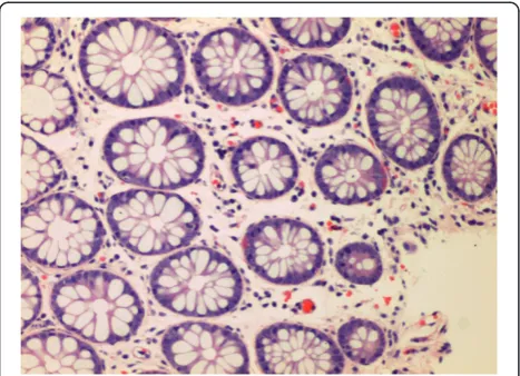
A further 11/74 (14.9%) IBS-D cases showed evidence of microscopic inflammation, with moderate lymphocytic and plasmacytic infiltration in the lamina propria. Eight cases had distal involvement but in four cases, inflamma-tion was seen only proximal to the splenic flexure only. In-filtration of an occasional crypt by neutrophils was seen in
five of the cases and scattered crypt abscesses in the trans-verse colon, sigmoid colon and rectum were present in one case. Moderate infiltration of the lamina propria only without crypt involvement was noted in the remaining five cases. An example of one of the cases is seen in Figure 2. The remaining 62 (83.7%) subjects with IBS-D did not show any evidence of significant inflammation throughout the colon (Figure 3).
Amongst those in the control group, moderate infiltra-tion of lymphocytes in the ascending and transverse colon, with no evidence of cryptitis, was found in 1/46 (2.2%) adult. This patient was an asymptomatic, 60-year-old Chinese male, who had undergone a colonoscopy for colo-rectal cancer screening. None of the other subjects in the control group showed any evidence of significant micro-scopic inflammation. Univariate analysis demonstrated that IBS-D subjects, compared to controls, had a greater prevalence of moderate to severe microscopic inflam-mation (14.9% vs 2.2%, OR 11.44, 95% CI = 1.49-240.69, p = 0.005).
Discussion
There are several relevant observations in our study. The primary objective was to look at the prevalence of micro-scopic colitis in patients who were diagnosed with IBS-D as defined by the Rome III criteria. From the study, it appears that classic microscopic colitis is very uncommon in our cohort of patients with IBS-D. However, a signifi-cant percentage of subjects with IBS-D had evidence of microscopic inflammation that did not fit the criteria for classical microscopic colitis. The most common abnor-malities seen were mixed chronic and acute inflammatory cells, lymphocytes, plasma cells and neutrophils; with or without cryptitis and crypt abscesses.
The categories of microscopic colitis were recently ex-panded by Falodia et al. into five subtypes: collagenous
Table 1 Summary of baseline characteristics, endoscopic and histological findings in patients with IBS-D and controls
IBS-D n(%) Controls n(%)
Total 74 Total 46
Gender Gender
Male 31(41.9%) Male 24(52.2%)
Female 43(58.1%) Female 22(47.8%)
Median age(years) 51(16–78) Median(range) 61(26–79)
Race Race
Malay 15(20.3%) Malay 8(17.4%)
Chinese 36(48.6%) Chinese 31(66%)
Indian 23(31.1%) Indian 7(14.9%)
Colonoscopic findings Colonoscopic findings
Normal 58 (78.4%) Normal 27(58.7%)
Polyps 9 (12.2%) Polyps 15(32.6%)
Diverticular disease 5 (6.8%) Diverticular disease 3(6.5%)
Haemorrhoids 2(2.7%) Haemorrhoids 1(2.2%)
Histological findings Histological findings
Normal 62(83.8%) Normal 0(97.8%)
Collagenous 1(1.4%) Collagenous colitis 0(0)
Lymphocytic colitis 0(0%) Lymphocytic colitis 0(0)
Non specific colitis 11(14.9%) Non specific colitis 1(2.2%)
Figure 1IBS-D patient with collagenous colitis.Note the thickened collagen band (arrow).
colitis, lymphocytic colitis, minimal change colitis (crypt architectural abnormality in the form of cryptitis and crypt dilatation in the absence of increase in intra-epithelial lymphocytes and subintra-epithelial collagenous band), microscopic colitis not otherwise specified (in-creased inflammatory cell infiltrates in the lamina propria in the absence of other abnormalities) and microscopic colitis with giant cells [24]. If we were to reclassify our patients according to the above categories, one patient had collagenous colitis, six patients had minimal change colitis and five patients had microsopic colitis not otherwise specified. However, the classifica-tion proposed by Falodia et al. has yet to be widely ac-cepted and the therapeutic studies carried out in the past only looked specifically at collagenous and lympho-cytic colitis. Inflammation was patchy throughout the colon and from the study, four cases showed inflamma-tion beyond the splenic flexure. This is similar to clas-sical microscopic colitis, where the diagnosis can be missed unless a complete colonoscopy with biopsies throughout the colon is conducted [15].
The role of inflammation in IBS has been of interest in recent times and evidence of low level inflammation has been demonstrated in both post infectious and non-post infectious IBS. Gwee et al. found increased expression of pro-inflammatory cytokine interleukin Iβ(IL-1β) mRNA in subjects with post-infectious IBS, which was not observed in adults without IBS [12]. Chadwick et al. [25,26] had divided the histological findings of IBS pa-tients into three groups; those with normal histology, those with evidence of microscopic inflammation and those who met criteria for lymphocytic colitis. With im-munostaining, it was found that all three groups had in-creased numbers of activated immunocompetent cells in the intestinal mucosa. Barbara et al. [27] reported that increased numbers of activated mast cells in close
proximity to intestinal innervation correlated with ab-dominal pain in IBS. In addition to this, there is evi-dence that patients with IBS-D have an increased expression of TNFα and IL-1β in the peripheral blood monocytes, not dissimilar to that found in patients with inflammatory bowel disease [8]. A study from Sri Lanka also showed low grade inflammation in IBS patients, simi-lar to Western studies but with an increase in eosinophils as well as the other chronic inflammatory cells [28].
An attractive concept is that IBS-D forms the mildest part of a disease spectrum, with idiopathic inflammatory bowel disease (in particular, ulcerative colitis) at the other end, with classical microscopic colitis in between [29,30]. There is a clear overlap in terms of their proposed patho-genesis including alterations in intestinal microbiota and bile salt malabsorption. However, the common pathways between the conditions remain poorly understood and the majority of IBS-D patients fail to demonstrate significant inflammation on histology.
In the light of Western studies demonstrating a signifi-cant overlap between classical microscopic colitis and IBS-D, as well as the increasing role of inflammation in IBS patients,‘to do or not to do’colonoscopy in patients with IBS-D remains as unclear as ever. Although the sin-gle case of collagenous colitis in our study was found in the‘typical’subject (elderly, female), non specific inflam-mation was seen in both age groups, both genders and across all three races. As the prevalence of collagenous and lymphocytic colitis appears to be low in our popula-tion, routine colonoscopy is not justified in young pa-tients who have been identified as IBS on symptom based criteria. This may change however, in light of promising data on the use of anti-inflammatory drugs such as mesalazine in IBS [31-34]. It is uncertain, how-ever, if only those with evidence of microscopic in-flammation will respond to anti-inflammatory therapy, thereby necessitating the use of routine colonoscopy in order to provide a more individualised approach to the management of these patients.
The low colonoscopic yield among IBS patients in this study is noteworthy. No macroscopic abnormalities were seen in subjects under the age of forty and it is also re-assuring to note that no significant pathology was identi-fied in the older adults. In fact, our findings were very similar to a large study carried out in USA where find-ings of polyps, diverticular disease and haemorrhoids in subjects with IBS were no different to that of asymptom-atic controls [35]. While it is certainly reasonable to still offer colonoscopy in older patients with IBS for colorec-tal cancer screening, the study emphasizes the import-ance of careful history taking in stratifying patients who do or do not require colonoscopy and those who require it urgently or otherwise. This is especially relevant in Malaysia, which is still considered a developing country,
with limited endoscopic facilities and only 106 registered gastroenterologists in a population of 25 million [36].
There are several limitations of the study. The study sample was small and patients were derived from sec-ondary care, which may not be representative of IBS pa-tients in the community. As IBS, like other functional GI diseases, is rarely life-threatening, medical consult-ation rates amongst sufferers are known to be low. CD3 staining for lymphocytes was not carried out which may have resulted in an under-diagnosis of lymphocytic col-itis [37]. Screening for Coeliac Disease, rare among our population but known to be associated with collagenous colitis, was not performed. However, the merits of this study lay in our strict adherence to the Rome III criteria and systematic sampling of colonic mucosal tissue, pro-viding an accurate estimation of inflammation in an Asian population with IBS-D.
Conclusions
In this prospectively conducted study, we have found a significant proportion of patients with IBS-D who have evidence of colonic inflammation on biopsies which may be part of a microscopic colitis spectrum. This further adds to the growing evidence for the role of inflamma-tion which will hopefully result in increasing the thera-peutic options for the management of this common but difficult to treat condition.
Competing interests
All authors declare no competing interests in the conduct of this study and preparation of the manuscript.
Authors’contributions
IH - research design, drafting of manuscript; JLH - research design, acquisition of data; JP - analysis and interpretation of results; SM - research design, acquisition of data, critical review of manuscript; GKL - critical review of manuscript. All authors read and approved the final manuscript.
Acknowledgements
This study was funded by a University of Malaya Research Grant (UMRG), No: RG027/09HTM.
Author details 1
Gastroenterology Unit, Department of Medicine, Faculty of Medicine, University of Malaya, Kuala Lumpur, Malaysia.2Department of Pathology, Faculty of Medicine, University of Malaya, Kuala Lumpur, Malaysia.
Received: 5 September 2012 Accepted: 3 May 2013 Published: 8 May 2013
References
1. Rey E, Talley NJ:Irritable bowel syndrome: novel views on the epidemiology and potential risk factors.Dig Liver Dis2009,41:772–780. 2. Rajendra S, Alahuddin S:Prevalence of irritable bowel syndrome in a
multi-ethnic Asian population.Aliment Pharmacol Ther2004,19:704–706. 3. Tan YM, Goh KL, Muhidayah R, Ooi CL, Salem O:Prevalence of irritable
bowel syndrome in young adult Malaysians: a survey among medical students.J Gastroenterol Hepatol2003,18:1412–1416.
4. Longstreth GF, Thompson WG, Chey WD, Houghton LA, Mearin F, Spiller RC:
Functional bowel disorders.Gastroenterology2006,130:1480–1491. 5. Barbara G, De Giorgio R, Stanghellini V, Cremon C, Salvioli B, Corinaldesi R:
New pathophysiological mechanisms in irritable bowel syndrome.
Aliment Pharmacol Ther2004,20(Suppl 2):1–9.
6. Talley NJ:Irritable bowel syndrome.Intern Med J2006,36:724–728. 7. Barbara G, Cremon C, Carini G, Bellacosa L, Zecchi L, De Giorgio R,
Corinaldesi R, Stanghellini V:The immune system in irritable bowel syndrome.J Neurogastroenterol Motil2011,17:349–359.
8. Liebregts T, Adam B, Bredack C, Roth A, Heinzel S, Lester S, Downie-Doyle S, Smith E, Drew P, Talley NJ,et al:Immune activation in patients with irritable bowel syndrome.Gastroenterology2007,132:913–920. 9. Spiller R, Campbell E:Post-infectious irritable bowel syndrome.Curr Opin
Gastroenterol2006,22:13–17.
10. Barbara G, De Giorgio R, Stanghellini V, Cremon C, Corinaldesi R:A role for inflammation in irritable bowel syndrome?Gut2002,51(Suppl 1):i41–i44. 11. Spiller R, Garsed K:Infection, inflammation, and the irritable bowel
syndrome.Dig Liver Dis2009,41:844–849.
12. Gwee KA:Post-Infectious Irritable Bowel Syndrome, an Inflammation-Immunological Model with Relevance for Other IBS and Functional Dyspepsia.J Neurogastroenterol Motil2010,16:30–34.
13. Akiho H, Ihara E, Nakamura K:Low-grade inflammation plays a pivotal role in gastrointestinal dysfunction in irritable bowel syndrome.
World J Gastrointest Pathophysiol2010,1:97–105.
14. Ford AC, Talley NJ:Mucosal inflammation as a potential etiological factor in irritable bowel syndrome: a systematic review.J Gastroenterol2011,
46:421–431.
15. Pardi DS, Kelly CP:Microscopic colitis.Gastroenterology2011,140:1155–1165. 16. Limsui D, Pardi DS, Camilleri M, Loftus EV Jr, Kammer PP, Tremaine WJ,
Sandborn WJ:Symptomatic overlap between irritable bowel syndrome and microscopic colitis.Inflamm Bowel Dis2007,13:175–181.
17. Carmona-Sanchez R, Carrera-Alvarez MA, Perez-Aguilar RM:Prevalence of microscopic colitis in patients with irritable bowel syndrome with diarrhea predominance.Rev Gastroenterol Mex2011,76:39–45. 18. Madisch A, Bethke B, Stolte M, Miehlke S:Is there an association of
microscopic colitis and irritable bowel syndrome–a subgroup analysis of placebo-controlled trials.World J Gastroenterol2005,11:6409.
19. Baert F, Schmit A, D’Haens G, Dedeurwaerdere F, Louis E, Cabooter M, De Vos M, Fontaine F, Naegels S, Schurmans P,et al:Budesonide in collagenous colitis: a double-blind placebo-controlled trial with histologic follow-up.Gastroenterology2002,122:20–25.
20. Miehlke S, Heymer P, Bethke B, Bastlein E, Meier E, Bartram HP, Wilhelms G, Lehn N, Dorta G, DeLarive J,et al:Budesonide treatment for collagenous colitis: a randomized, double-blind, placebo-controlled, multicenter trial.
Gastroenterology2002,123:978–984.
21. Miehlke S, Madisch A, Bethke B, Morgner A, Kuhlisch E, Henker C, Vogel G, Andersen M, Meier E, Baretton G,et al:Oral budesonide for maintenance treatment of collagenous colitis: a randomized, double-blind, placebo-controlled trial.Gastroenterology2008,135:1510–1516.
22. Miehlke S, Madisch A, Karimi D, Wonschik S, Kuhlisch E, Beckmann R, Morgner A, Mueller R, Greinwald R, Seitz G,et al:Budesonide is effective in treating lymphocytic colitis: a randomized double-blind placebo-controlled study.Gastroenterology2009,136:2092–2100.
23. Lazenby AJ, Yardley JH, Giardiello FM, Jessurun J, Bayless TM:Lymphocytic (“microscopic”) colitis: a comparative histopathologic study with particular reference to collagenous colitis.Hum Pathol1989,20:18–28. 24. Falodia S, Makharia GK, Sateesh J, Deo V, Tevatia MS, Gupta SD:Spectrum
of microscopic colitis in a tertiary care centre in India.Trop Gastroenterol
2007,28:121–125.
25. Chadwick VS, Chen W, Shu D, Paulus B, Bethwaite P, Tie A, Wilson I:
Activation of the mucosal immune system in irritable bowel syndrome.
Gastroenterology2002,122:1778–1783.
26. Gwee KA, Collins SM, Read NW, Rajnakova A, Deng Y, Graham JC, McKendrick MW, Moochhala SM:Increased rectal mucosal expression of interleukin 1beta in recently acquired post-infectious irritable bowel syndrome.Gut2003,52:523–526.
27. Barbara G, Stanghellini V, De Giorgio R, Cremon C, Cottrell GS, Santini D, Pasquinelli G, Morselli-Labate AM, Grady EF, Bunnett NW,et al:Activated mast cells in proximity to colonic nerves correlate with abdominal pain in irritable bowel syndrome.Gastroenterology2004,126:693–702. 28. De Silva AP, Nandasiri SD, Hewavisenthi J, Manamperi A, Ariyasinghe MP,
Dassanayake AS, Jewell DP, de Silva HJ:Subclinical mucosal inflammation in diarrhea-predominant irritable bowel syndrome (IBS) in a tropical setting.Scand J Gastroenterol2012,47:619–624.
30. Mearin F, Perello A, Balboa A:[Irritable bowel syndrome and inflammatory bowel disease: Is there a connection?].Gastroenterol Hepatol2009,
32:364–372.
31. Dorofeyev AE, Kiriyan EA, Vasilenko IV, Rassokhina OA, Elin AF:Clinical, endoscopical and morphological efficacy of mesalazine in patients with irritable bowel syndrome.Clin Exp Gastroenterol2011,4:141–153. 32. Hanevik K, Dizdar V, Langeland N, Eide GE, Hausken T:Tolerability and
effect of mesalazine in postinfectious irritable bowel syndrome.
Aliment Pharmacol Ther2011,34:259–260.
33. Bafutto M, Almeida JR, Leite NV, Oliveira EC, Gabriel-Neto S, Rezende-Filho J:
Treatment of postinfectious irritable bowel syndrome and noninfective irritable bowel syndrome with mesalazine.Arq Gastroenterol2011,
48:36–40.
34. Corinaldesi R, Stanghellini V, Cremon C, Gargano L, Cogliandro RF, De Giorgio R, Bartesaghi G, Canovi B, Barbara G:Effect of mesalazine on mucosal immune biomarkers in irritable bowel syndrome: a randomized controlled proof-of-concept study.Aliment Pharmacol Ther2009,
30:245–252.
35. Chey WD, Nojkov B, Rubenstein JH, Dobhan RR, Greenson JK, Cash BD:The yield of colonoscopy in patients with non-constipated irritable bowel syndrome: results from a prospective, controlled US trial.Am J Gastroenterol2010,105:859–865.
36. Malaysia National Specialist Register.[www.nsr.org.my/]
37. Mohamed N, Marais M, Bezuidenhout J:Microscopic colitis as a missed cause of chronic diarrhea.World J Gastroenterol2011,17:1996–2002.
doi:10.1186/1471-230X-13-80
Cite this article as:Hilmiet al.:Low prevalence of‘classical’microscopic colitis but evidence of microscopic inflammation in Asian Irritable Bowel Syndrome patients with diarrhoea.BMC Gastroenterology2013
13:80.
Submit your next manuscript to BioMed Central and take full advantage of:
• Convenient online submission
• Thorough peer review
• No space constraints or color figure charges
• Immediate publication on acceptance
• Inclusion in PubMed, CAS, Scopus and Google Scholar
• Research which is freely available for redistribution