R + C Factors and Sacro Occipital Technique
Orthopedic Blocking: a pilot study using pre and
post VAS assessment
Charles L. Blum, DC*
*Private Practice
1752 Ocean Park Boulevard, Santa Monica, California 90405 Tel: 310-392-9795 Affiliation: Sacro Occipital Technique Organization – USA – Director of Research
Disclaimer: No finanical benefit was accrued by the submitting of this study or will be attained for publication. No patient was withheld any care by consenting to be a participant in this study.
Sources of Support: There were no grants, financial support, or any secondary financial gain to the author or patients in this study. © JCCA 2015
Introduction: The concept of a systematic or predictive relationship between distant vertebral levels
distinct from accumulative functional compensatory mechanisms, such as in scoliosis, has been perpetuated within chiropractic technique systems based on clinical observation and experience. This study seeks to
investigate this relationship between the cervical and lumbar vertebrae.
Methods: Patients (experimental group n=26 and control group n=12) were selected from the patient base of one office, and were limited to patients that had sensitivity at specific cervical reflex points. Using a pre and post outcome measurement and sacro occipital technique R + C protocols, the related lumbar vertebra was adjusted in the direction indicated by the cervical vertebral sensitivity.
Results: Statistical analysis revealed there was a statistically significant difference between pre- and post-VAS measurements and found that the notable difference in mean change in VAS scores were statistically
significantly different between the experimental and control groups (p < .001).
Conclusion: The findings of this study suggest that further research into cervical and lumbar vertebra interrelationships, and the efficacy of orthopedic block
Introduction : Le concept d’une relation systématique ou prédictive entre les niveaux distants vertébraux distincte des mécanismes compensatoires fonctionnels cumulatifs, comme en cas de scoliose, a été perpétué dans les systèmes de techniques chiropratiques fondés sur des observations cliniques et sur l’expérience. Cette étude cherche à examiner la relation entre les vertèbres cervicales et lombaires.
Introduction:
Since the early 20th century, some within the chiropractic
profession have posited that there is a functional relation-ship between the lumbar and cervical vertebrae and have incorporated this concept into methods of evaluation and treatment.1 This concept has empirically been accepted
by many chiropractors for decades and was based on the work of Robert W. Lovett.2 Walther notes that, “The spine
appears to function with a specific harmonious movement as an individual walks, runs, and otherwise performs daily activities… The vertebra working in conjunction with each other, such as the 1st lumbar and 5th cervical, are
known as Lovett Brothers.”3
As the name Sacro Occipital Technique (SOT) implies, DeJarnette a chiropractor and osteopath, found a similar relationship between the sacrum and occiput, as well as between the cervical and lumbar vertebrae.4 He described
that a relationship exists between the atlas and the 5th
lum-bar vertebra, axis and the 4th lumbar vertebra and so forth,
following that pattern all the way to the mid thoracic re-gion. He called this relationship R + C (resistance and contraction) factors and found that each vertebrae within a pair affected one another (Figure 1).
This concept of a systematic or predictive relation-ship between distant vertebral levels distinct from ac-cumulative, functional, compensatory or adaptive mech-anisms, such as occur frequently in idiopathic scoliosis for example, has been perpetuated based on observation and clinical experience without published report of any systematic study. This pilot study seeks to investigate this relationship between the cervical and lumbar verte-brae.
Methods
Patients
Patients were selected from the patient base of one office (Table 1) and were limited to those who had sensitivity at specific cervical reflex points.
treatment, may be warranted. Further studies are needed to confirm whether a causal relationship exists between lumbar manipulation and decreased cervical spine sensitivity.
(JCCA 2015; 59(2):134-142)
k e y w o r d s : chiropractic, spine, cervical vertebra,
lumbar vertebra, Sacro Occipital Technique, Lovett Brother, R+C Factors
ainsi que l’efficacité du traitement de bloc orthopédique, peuvent être justifiées. D’autres études sont nécessaires pour confirmer s’il existe une relation de cause à effet entre la manipulation lombaire et la baisse de sensibilité de la colonne cervicale.
(JCCA 2015; 59(2):134-142)
m o t s c l é s : chiropratique, colonne, vertèbre
cervicale, vertèbre lombaire, technique sacro occipitale, Lovett Brother, facteurs R+C
Figure 1.
R+C Factors relate to how sensitivity at the cervical vertebrae may relate to position of lumbar vertebrae. A relationship is illustrated suggesting that sensitivity at the cervical transverse process relates to an anterior
rotation relating to the ipsilateral lumbar vertebrae’s transverse process. Also sensitivity at the lateral cervical
Inclusion
To determine whether patients qualified for study inclu-sion, patients were palpated for tenderness at the cervical reflex points which are located at the temporal bone styl-oid processes (adjacent to C1), lateral spinous processes (C2-C7) and lateral tips of the transverse processes (C1-C7). Palpation over these bony landmarks constitutes part of the normative examination for SOT practitioners. Sensitivity to palpation over these reflex points is thought to indicate the need for lumbar ipsilateral decompression or rotational adjustment.4 During the normative
examina-tion, patients were asked if they felt pain or tenderness in response to approximately 1-2 pounds of digital pressure (estimated subjectively without instrumentation).
R + C palpation can be performed in the standing, sit-ting, supine or prone position. These cervical reflex points were determined to be positively tender or painful when the patient reported that they felt significant discomfort upon palpation, and that this discomfort was localized to the point being contacted (over any of the above-men-tioned landmarks).
Excluded from this study were young children or any patients who had difficulty with communciation or under-standing questions (due to cognitive issues or language barriers).
Comparison groups
A non-randomized allocation method was used to en-roll participants into two groups: an experimental group, where an adjustment was delivered after initial palpation and a baseline measurement of pain intensity, and a con-trol group in which patients were not treated but were simply rechecked for palpatory tenderness after a brief waiting period of 3-5 minutes. Sequential patients were first enrolled into the experimental group and then into the control group.
Study maneuvers
Diagnostic Procedure
R + C Factors are reflex indicators at the cervical verte-brae used to identify vertebral rotation and lateral flexion inferiorities of the lumbar spine.4 Each lumbar vertebra is
purported to have a corresponding “Lovett Brother” indi-cator in the cervical spine. The cervical vertebral reflexes can be found in any posture, but will be most prominent
when in the position that patients experience their greatest amount of low back discomfort.
Locations for orthopedic blocking and treatment
Sacro occipital technique (SOT) recommends the use of pelvic blocks and sustained pressure at the lumbar verte-bra to allow the nervous system time to accommodate, the local connective tissue to remodel, and the lumbar ver-tebrae to experience gentle rotation or lifting pressures.4
The rationale for these procedures has been described in previous literature.5-8
Generally the Lovett Brother relationships found be-tween the cervical and lumbar vertebrae are as follows: cervical lateral transverse process sensitivity indicates ipsilateral lumbar transverse process anterior rotation; cervical lateral spinous process sensitivity relates to ipsi-lateral lumbar transverse process inferiority; and, given the absence of a spinous process at C1, the temporal styl-oid process is used to determine inferiority of the ipsilat-eral L5 transverse process.9
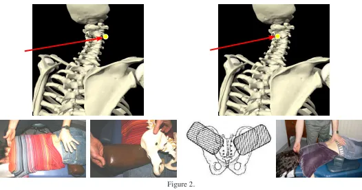
Orthopedic Blocking: R + C Factors [See Figure 2] • Treatment of vertebral inferiorities/compression One method to treat inferiorities can be accomplished by placing the pelvic blocks under the ASISs bilateral-ly, with the blocks facing 45 degrees caudalward, with the patient lying prone. The angle of the blocks and their position tends to create a distractive affect on the lumbar and lumbosacral regions. The goal is to create a focused decompression at the level of lumbar vertebral segment malalignment, as directed by (i.e. related to) the corres-ponding location cervical reflex point sensitivity.
• Treatment of vertebral rotations
More typically, the related lumbar vertebra was ad-justed opposite to the direction indicated by the cervic-al vertebrcervic-al sensitivity (according to R + C protocol).4 A
will appear to increase. This increased muscular tone is theorized to help rotate the vertebra into its correct pos-ition or possibly, by effecting the crossed extensor/flexor reflex, relaxing the muscles holding the lumbar vertebra in rotation.4
Furthermore, with the block in position, the spinous, lamina or mamillary processes can be contacted to rotate the vertebra opposite to the direction indicated by the cer-vical vertebra transverse process sensitivity. For example, with a right C2 transverse process sensitivity, the block would be placed under a prone patient on the left side be-tween the ASIS and greater trochanter, and the right side
of the L4 spinous process would be contacted, on the right side, with pressure directed from right to left.
Treating chiropractor
The treating chiropractor was in practice for over 30 years, and has taught and utilized sacro-occipital tech-nique methods for that period of time, and the patients in the study were all from his practice.
Outcome
A validated instrument, the Visual Analogue Scale (VAS) (Figure 2),10,11 was utilized to measure patient
self-re-Figure 2.
Orthopedic Block Placement
ported pain or sensitivity levels. The VAS has been used in studies measuring quality of life,12 pain and trauma,13
risk assessments regarding risk proneness,14 and in both
young and old patient populations.15 In this study, the
VAS was used to measure pain/sensitivity to palpation over cervical reflex points. Both pre- and post-treatment scales employed a 10 cm line, anchored by the terms, “no pain” at one extreme, and “unbearable” (pain) at the other extreme. The patients were asked to mark the line at a position that corresponded to their current level of pain. The distance between “no pain” and each patient’s current pain intensity marking was measured on a 1-to-100 mm scale.
Timing of measurements
Baseline VAS measurements were recorded at study entry prior to any lumbar manipulation (experimental) or waiting period (control). Post-intervention VAS measure-ments were recorded immediately after manipulation (ex-perimental) or a 3-to-5 minute waiting period (control). During the basline assessment, a note was made on each patient’s study form documenting the point of vertebral contact and sensitivity (VAS measurement). To improve the reliability of the reflex point assessments, patients was asked to help confirm (to the best of their abilities) that the points of contact were consistent between the baseline and post-intervention evaluations.
The attempt to find a sham and provide a control for manual treatment is often fraught with difficulty.16
There-fore, in lieu of using a direct sham intervention, patients in the control group were simply left untreated during a brief waiting period. However, all selection criteria, and manu-al pmanu-alpation and assessment maneuvers were standardized between the two groups. As with the experimental group, only patients who had a positive response to cervical re-flex palpation were asked to be part of the control group. Between the two VAS administrations, patients histories, ranges of motion, and results of palpation to elicit sensi-tivy, as well as other tests were performed. In the control group, lumbar manipulative therapy was eventually ad-ministered as indicated by the normative examination, but only after a brief waiting period and repeated administra-tion of the VAS.
Statistical Methods
Data were assessed for normality using
Kolmog-orov-Smirnov and Shapiro-Wilk tests. Results of these statistical tests were used to determine whether we could use parametric or nonparametric testing. To test for be-tween group differences in the pre-test VAS scores an in-dependent t-test analysis was used. Pre-test and post-test VAS scores were compared and tested using an independ-ent t-test and confirmed using the Mann-Whitney statis-tic. An analysis of covariance was performed to control for baseline differences in VAS scores between groups.
Ethics
All patients signed a consent form which explicitly in-formed them that while participating in the study their normal care would not be affected apart from possibly taking a few minutes longer to complete their scheduled visit. Institutional review board approval for this study was received from Cleveland Chiropractic College.
Results:
Two children, ages 2 and 4 years old, were excluded. An-other 4 patients were excluded because no sensitivity was noticed to palpation. This left 26 patients in the treatment group and 12 patients in the control group.
the VAS, was significantly greater in the experimental group (p < .001). Non-parametric test (Mann-Whitney U) was also significant (p < 0.001), confirming statistical significance. After controlling for baseline differences in VAS between groups a statistically significant difference (p= 0.001) was found between pre and post VAS meas-urement between the treatment and control groups using an ANCOVA test.
Discussion:
While the Lovett Brother concept has been accepted by some within the field of chiropractic, there is a clear lack of research evidence to support it’s clinical existence. Why or how could localized points on cervical vertebrae be related to positions of the lumbar vertebra?
Some biologically plausible theories for the R + C Factors and it’s clinical treatment via blocking have been proposed.1 These theories vary from myofascial17-25 and
myological26 interrelationships, referred pain patterns27-29,
facilitating tonic neck reflexes involving intersegmental spinal pathways,30 as well as postural righting
mechan-isms involving the cervical spine as well as propriocep-tive and plantar mechanoreceptors31. Optic feedback or
gaze has been found to be associated with cervical32 and
lumbar33 spine accommodation. Dental occlusion has also
been found to affect gaze and body posture.34 However, in
addition to gaze or optic feedback, there have also been some studies that have found vestibular, labyrinth and plantar mechanosensor relationships between posture and proprioceptive influences.35-38
It is possible that the R + C reflex points constitute complex neurological and myological interrelationships that are related to multifactorial influences. Some clinical findings have suggested that while lumbar vertebral dys-function could affect its cervical component, it is possible that cervical vertebral dysfunction could also affect its lumbar component.32
Various studies have found a clinical relationship be-tween the lumbosacral spine and cervicocranial regions. Chinappi and Getzoff found that integrated dental ortho-pedic and craniochiropractic care ameliorated the lumbo-sacral pain and improved head, jaw, neck and back func-tion.39 Kessinger in another study noted a relationship
between the effect of treatment to the cervical spine and improvement of lumbar spine ranges of motion.40 Giggey
and Tepe in their study showed a statistically significant improvement in cervical isometric extensor strength fol-low orthopedic lumbosacral block placement.41
While some degree of biological plausiblity may be present to support a relationship between the lumbar and cervical regions, what is challenging to understand is how thumb pressure for minutes to a lumbar vertebra along with pelvic block placement, could have a relative change in cervical vertebral sensitivity within seconds. A study by Meier et al found that thumb pressure to the lumbar Table 1:
Number and sex distribution per group
Experimental Group n Mean age
Male 15 46
Female 11 39
Control Group
Male 4 45
Female 9 47
Table 2:
Mean change in pre and post VAS per Experimental and Control Groups
Mean Pre-test
VAS Measurement
Mean Post-test
VAS Measurement
Mean Change
in VAS Experimental
Group (n=26) 40.7 (SD 21.1) 14.4 –26.4
Control Group
(n=12) 32.5 (SD 15.7) 31.6 –0.9
t-test
(unadjusted) p=.189 p<.001 p < .001*
ANCOVA
(adjusted) ηpp=.0012=.292**
* From parametric as well as non-parametric t-test for independent samples
spinous processes had relatively immediate responses in the cortex and cerebellum.42 Theoretically this change in
cortical and cerebellar activity could simultaneously af-fect cervical spine sensitization and accommodative func-tion. Also Jonckheerea et al described an electrophysio-logical phenomenon running up and down the spine that is elicited by pressure to the spine at specific points. They suggested that this electrophysiological phenomenon is associated with a standing spinal wave found in both normal and quadriplegic subjects. They suggest that “a standing spinal wave demonstrates that the neuronal cir-cuitry is embedded in the spine”,43 which combined with
the Meire et al’s findings, may help us better understand this complex lumbar and cervical vertebra relationship. Even though this study focused on the use of ortho-pedic pelvic block placement and evaluating levels of pain sensation at related cervical veretbrae, other stud-ies might also be performed utilizing other chiropractic treatment methods, such as diversified adjusting methods. Using the cervical vertebrae sensitivity as described in this study could function as a pre- and post-assessment tool to help guide the direction or vector for the diversi-fied adjustment to the lumbar spine, as well as help deter-mine if the adjustment was sufficient to help reduce the cervical veretebra sensitivity. There may be an advantage to using pelvic blocks and a sustained thumb or finger contact to the lumbar spine because the lumbar osseous landmark (spinous or transverse process) is maintained continuously, minimizing fascial glide off of the lumbar contact. Sometimes a change of sensitivity at the cervical spine may be affected by a change of vector at the lumbar vertebra contact, and this could be utilized as a pre-as-sessment with either block or diversified techniques. Re-ducing cervical vertebrae sensitivity by directing force to specific lumbar vertebra and in specific vectors may help localize the optimal vertebrae and direction of correction, regardless of the technique utilized. Also theoretically, by adjusting the lumbar spine using the cervical indicators as guides, this may help improve function and relieve pain in the cervical spine with patients in whom cervical spine adjusting is contraindicated.
While this study appears to indicate that a relationship between pressure to the lumbar spine or its position may have an affect on related cervical spine sentitivity, what is important to determine is whether this has clinical signifi-cance. Currently there is limited low level evidence that
supports this clinical relationship. Therefore future stud-ies investigating lumbar and cervical vertebral reflex rela-tionships will need to determine whether this relationship is clinically meaningful, let alone causal in nature.1,5,8,9,44
Limitations:
Beyond the obvious limitations of a poorly controlled study such as this, many questions arise as to what else may potentially explain the observed association between lumbar manipulation and reduced sensitivity in the cer-vical spine. More importantly, are non-validated clinic-al procedures such as those used in the current study--developed decades ago no less--clinically meaningful? Furthermore, are there other clinical indictors for mon-itoring actual anatomical positions of the vertebra that can be measured through or corroborated by radiographs and MRI? While clinical anecdotal reports on SOT proced-ures have suggested improved levels of patient reported pain, increased functionality, and reduction of disc herni-ation, sufficient study into this method of diagnosis and treatment has not been adequately performed to allow for definitively strong conclusions to date. The findings of this pilot study suggest that further research into cervical and lumbar vertebral interrelationships, as well as ortho-pedic block placement and treatment, may be warranted to determine the genuine reliability, validity, and efficacy of this particular system technique.
Conclusion:
This pilot study has attempted to investigate a relation-ship between SOT’s R + C Factors method of diagnosis and lumbar spine dynamics. Following orthopedic block placement and pressure to the lumbar spine, the cervic-al spine reflex areas were contacted to determine if there was a change to treatment. In most cases there was a lessening to pain upon palpation following treatment as compared to the control group with findings reaching sta-tistical significance. However, this study was affected by several methodological limitations and important threats to validity. The results of this must be interepreted ex-tremely cautiously, but represent a valuable beginning for future studies.
References
1. Blum C. Lovett Brothers: The relationship between the cervical and lumbar vertebra. J Vertebral Subluxation Res. 2004;6(1):1-3.
2. Lovett RW. Lateral Curvature of the Spine and Round Shoulders. P. Blakiston’s Son & Co: Philadelphia, PA, 1913:1-192.
3. Walther DS. Applied Kinesiology: Volume I, Basic Procedures and Muscle Testing, Systems DC, Pueblo, CO, 1981:67.
4. Monk R. SOT Manual. Sacro Occipital Technique Organization – USA: Sparta, NC, 2006:152-154.
5. Blum C. Sacro Occipital Technique pelvic block treatment for severe herniated discs: a case study. J Chiropractic Education. Spr 2004;18(1):38-9.
6. Blum CL, Esposito V, Esposito C, Orthopedic block placement and its affect on the lumbosacral spine and discs: three case studies with pre and post MRIs. J Chiropractic Education. Spr 2003;17(1):48. 7. Blum CL. Chiropractic and pilates therapy for the
treatment of adult scoliosis. J Manip Physiol Thera. 2002; 25(4:E3.
8. Blum CL. Incongruent sacro-occipital technique examination findings: Two unusual case histories. J Chiropr Edu. Spr 2002;16(1):67.
9. Shaneyfelt D, Blum CL, Benner CD. Styloid process sensitivity in a patient with low back pain and radicular syndrome: A case report. World Congress on Low Back & Pelvic Pain Conference. Dubai, Oct. 2013.
10. Jensen MP, Karoly P. Self-report scales and procedures for assessing pain in adults. In: Turk DC, Melzack R, eds. Handbook of Pain Assessment, New York, NY: Churchill Livingstone, 1983:635.
11. Price DD, McGrath PA, Rafii A, Buckingham B. The validation of visual analogue scales as ratio scale measures for chronic and experimental pain. Pain. 1983;17L45-6. 12. de Boer AG, van Lanschot JJ, Stalmeier PF,
van Sandick JW, Hulscher JB, de Haes JC, Sprangers MA. Is a single-item visual analogue scale as valid, reliable and responsive as multi-item scales in measuring quality of life? Qual Life Res. 2004 Mar;13(2):311-20.
13. Knop C, Oeser M, Bastian L, Lange U, Zdichavsky M, Blauth M. [Development and validation of the Visual Analogue Scale (VAS) Spine Score] [Article in German] Unfallchirurg. 2001 Jun;104(6):488-97.
14. Sicard B, Jouve E, Blin O, Mathieu C [Construction and validation of visual analogue scale for risk assessment] [Article in French] Encephale. 1999 Nov-Dec;25(6):622-9. 15. Tiplady B, Jackson SH, Maskrey VM, Swift CG. Validity
and sensitivity of visual analogue scales in young and older healthy subjects. Age Ageing. 1998;27(1):63-6. 16. Hawk C, Long CR, Rowell RM, Gudavalli MR, Jedlicka J.
A randomized trial investigating a chiropractic manual placebo: a novel design using standardized forces in the delivery of active and control treatments. J Altern Complement Med. 2005;11(1):109-17.
17. Farmer JA, Blum CL. Dural Port Therapy. J Chiropr Med. Spr 2002;1(2):1-8.
18. Shinomiya K, Dawson J, Spengler DM, Konrad P, Blumenkopf B. An analysis of the posterior epidural ligament role on the cervical spinal cord. Spine. 1996;21(18):2081-2088.
capitis posterior minor muscle and the dura mater. Spine. 1995;20(23):2484-2486.
20. Mitchell B, Humphreys BK, O’Sullivan E. Attachments of the ligamentum nuchae to cervical posterior spinal dura and the lateral part of the occipital bone. J Manip Physiol Thera. 1998;21(3):145-8.
21. Barbaix E, Girardin MD, Hoppner JP, Van Roy P, Claris JP. Anterior sacrodural attachments Trolard’s ligaments revisited. Manual Therapy. 2000;1(2):88-91.
22. Pontell ME, Scali F, Enix DE, Battaglia PJ, Marshall E. Histological examination of the human obliquus capitis inferior myodural bridge. Ann Anat. 2013;195(6):522-6. 23. Scali F, Pontell ME, Enix DE, Marshall E. Histological analysis of the rectus capitis posterior major’s myodural bridge. Spine J. 2013;13(5):558-63.
24. Bashline SD, Bilott JR, Ellis JP. Meningovertebral ligaments and their putative significance in low back pain. J Manip Physiol Thera. 1996;19(9):592-6.
25. Kershner DE, Binhammer RT. Lumbar intrathecal ligaments. Clin Anat. 2002 Mar;15(2):82-7.
26. Sutherland WG, Contributions of Thought, Meridian, Idaho: Sutherland Cranial Teaching Foundation, 1967. 27. Dvorak J, Dvorak V. Manual Medicine: Diagnostics,
Thieme Medical Publisher, Inc.: New York, NY, 1990: 53,253.
28. Kellgren JH. Observations of referred pain arising from muscles. Clin Sci. 1939; 3:175.
29. Kellgren JH. On the distribution of pain arising from deep somatic structures with charts of segmental pain areas. Clin Sci. 1939;3:35.
30. Lewis T, Kellgren JH. Observations relating to referred pain viscero-motor reflexes and other associated phenomena. Clin Sci. 1939;4:47.
31. Morningstar MW, Pettibon BR, Schlappi H, Schlappi M, Ireland TV. Reflex control of the spine and posture: a review of the literature from a chiropractic perspective. Chiropr Osteo. 2005.9;13:16.
32. Nansel D, Waldorf T, Cooperstein R. Effect of cervical spinal adjustments on lumbar paraspinal muscle tone: evidence for facilitation of intersegmental tonic neck reflexes. J Manip Physiol Thera. 1993;16(2):91-5. 33. Bonney RA, Corlett EN. Head posture and loading of the
cervical spine. Appl Ergon. 2002 Sep;33(5):415-7.
34. Loder RT. The sagittal profile of the cervical and lumbosacral spine in Scheuermann thoracic kyphosis. J Spinal Disord. 2001;14(3):226-31.
35. Gangloff P, Louis JP, Perrin PP. Dental occlusion modifies gaze and posture stabilization in human subjects. Neurosci Lett. 2000 Nov 3;293(3):203-6.
36. Ivanenko YP, Grasso R, Lacquaniti F. Effect of gaze on postural responses to neck proprioceptive and vestibular stimulation in humans. J Physiol. 1999 Aug 15;519 Pt 1:301-14.
37. Yasuda T, Nakagawa T, Inoue H, Iwamoto M, Inokuchi A. The role of the labyrinth, proprioception and plantar mechanosensors in the maintenance of an upright posture. Eur Arch Otorhinolaryngol. 1999;256 Suppl 1:S27-32. 38. Allum JH, Bloem BR, Carpenter MG, Hulliger M,
Hadders-Algra M. Proprioceptive control of posture: a review of new concepts. Gait Posture. 1998 Dec 1;8(3):214-242.
39. Chinappi AS, Getzoff H. Chiropractic/dental co-treatment of lumbosacral pain with temporomandibular joint involvement. J Manip Physiol Thera. 1996 Nov; 19(9):607-12.
40. Kessinger RC. The influence of upper cervical specific chiropractic care on lumbar range of motion. Chiropr Res J. 2000 Fall;7(2):80.
41. Giggey K, Tepe R. A pilot study to determine the effects of a supine sacroiliac orthopedic blocking procedure on cervical spine extensor isometric strength. J Chiropr Med. 2009 Jun;8(2):56-61.
42. Meier ML, Hotz-Boendermaker S, Boendermaker B, Luechinger R, Humphreys BK. Neural responses of posterior to anterior movement on lumbar vertebrae: a functional magnetic resonance imaging study. J Manip Physiol Thera. 2014; 37(1):32–41.
43. Jonckheerea E, Lohsoonthorn P, Musuvathy S, Mahajan V, Stefanovic M. On a standing wave Central Pattern
Generator and the coherence problem. Biomedical Signal Processing and Control. Oct 2010;5(4):336-347.