P O S T E R S E S S I O N
The use of the first generation Burch-Schneider
anti-protrusion cage in complex cases of acetabular
deficit in revision hip prosthetic surgery
V. Bellotti, D. Marino’
Department of Orthopaedic Surgery, University Federico II (Naples, IT)
Introduction We describe the use of the first generation Burch-Schneider anti-protrusion cage in revision hip prosthetic surgery in a series of 30 patients, and analyze the results at 8 years follow-up.
Material and methodsA series of 30 patients with severe acetabular deficit at the time of revision surgery of hip prosthetic implants were treated with the implantation of a Burch-Schneider Reinforcement Cage (17 women and 13 men). The mean age at surgery was 73 years (range 54–86 years). We operated the first 12 patients screwing the disalflange of the cage in the ischiatic bone. We then moved to a different way to secure the distal flange to the ischiatic bone, by inserting it into the bone to reach a better primary mechanical stability. Patients were evaluated retrospectively with a mean follow-up of 8 years (range 5–13 years).
ResultsIn one case with a deep infection, revision of the anti-pro-trusion cage was required. The cages showed a survival rate of 92% at mean follow-up. Clinical evaluation of the surviving patients showed a mean Harris Hip Score of 75 points (range 30–92). Radiological evaluation revealed a major rate of loosening in the group in which the distal flange was screwed to the ischiatic bone.
ConclusionsAlthough we present a small series, and other studies are required, the Burch-Schneider cage is a good tool in case of complex acetabular deficiency in revision hip prosthetic surgery, with regard to medium-term implant survival rate. Due to better mechanical results in our series, we emphasize the positioning of the distal flange into the ischiatic bone, without screwing it on the ischiatic bone, according to the literature.
Suggested readings
1. Symeonides PP, Petsatodes GE, Pournaras JD, Kapetanos GA, Christodoulou AG, Marougiannis DJ (2009) The Effectiveness of the Burch-Schneider antiprotrusio cage for acetabular bone deficiency: five to twenty-one years’ follow-up. J Arthroplasty 24(2):168–174 2. Schlegel UJ, Bitsch RG, Pritsch M, Aldinger PR, Mau H, Breusch SJ
(2008) Acetabular reinforcement rings in revision total hip arthro-plasty: midterm results in 298 cases Orthopade 37(9):904–913
Are autologous growth factors (PRP) really useful
in prosthetic integration? DEXA study in CFP stems
R. Cecchinato1, R. Gogue2, U. Mezzadri3, G. Fraschini3, P. Sirtori3
1Scuola di Specializzazione in Ortopedia e Traumatologia (Milan, IT); 2Scuola di Specializzazione in Ortopedia e Traumatologia (Perugia, IT); 3IRCCS San Raffaele (Milan, IT)
Scientific international literature agrees in assuming that ‘‘paraphys-iological’’ periprosthetic bone resorption that happens in the first months after total hip arthroplasty (THA) is an indirect index of osteointegration of the prosthetic components. With the aim of improving the quantity and the quality of the bone mass in host femurs, pharmacological products (bisphosphonates, Teriparatide) and autologous factors (PRP, stem cells) with osteoinductive prop-erties are more often used in orthopaedic surgery.
Several studies show the efficacy of substances as bisphosphonates in reducing periprosthetic bone loss, while there are no randomized studies on the real effects of local autologous factors.
We then decided to perform a randomized trial to evaluate the periprosthetic bone mass content with DEXA scan (Hologic QDR 4500 with metal removal software) on CFP prosthesised subjects using one of these autologous osteoinductive factors; Cascade’s
Platelet Rich Plasma (PRP), a product made of growth factors derived from platelets, one of the most widely used in orthopaedic surgery and easily obtainable during the surgical act, had been chosen.
Intraoperative apposition of PRP in 10 subjects (CFP + PRP Group) in the femoral calcar region did not significally change pBMD in respect to the control group, demonstrating the inefficacy of this product in contrasting periprosthetic bone resorption in our model. We can then assume that the biological osteoinductive action of PRP is not able to antagonize the biomechanical osteoresorptive force due to stress shielding.
Paracetamol and tramadol combination
for postoperative pain treatment in day-care knee
arthroscopy patients: a preliminary study
A. De Ponti1, E. Colnaghi2, F. Deni2, G.F. Fraschini1
1Orthopaedic Department and2Anaesthesiology Department, IRCCS
Ospedale San Raffaele (Milan, IT)
Objective The aim of our study was to evaluate the analgesic effectiveness and safety profile of Paracetamol 325 mg and Tramadol 37.5 mg in the treatment of postoperative pain in day hospital patients.
side effects was recorded and patient satisfaction was graded on a 4 points scale.
Results Fifty patients were studied. Patients satisfaction was high (grade 3 and 4) for 75%. The majority of patients scored a low score for pain at rest (83% below 3) and at movement (70% below 5). The main complaint was sickness which was present in 15% of patients and required discontinuation of therapy in 5%.
DiscussionThe combination of Paracetamol 325 mg with Tramadol 37.5 mg, seems to be an effective and safe therapy for postoperative pain management in day hospital patients undergoing knee arthros-copy. A double blind randomized study comparing Paracetamol and Tramadol association with Diclofenac is in progress to better eluci-date this topic.
Relationship between cutting errors and learning curve
in computer-assisted total knee replacement
A. Manzotti, P. Cerveri, E. De Momi, C. Pullen, N. Confalonieri
1st Orthopedic Department, C.T.O. Hospital, Via Bignami 1 (Milan, IT)
Computer-assisted total knee replacement (TKR) has been shown to improve radiographic alignment. Continuous feedback from the navigation system allows accurate adjustment of the bone cuts, thus reducing errors. The aim of this study was to determine the impact of experience both with computer navigation and knee replacement surgery on the frequency of errors in intraoperative bone cuts and implant alignment. Three homogeneous patient groups undergoing computer assisted TKR were included in the study. Each group was treated by one of three surgeons with varying experience in computer-aided and knee replacement surgery. Surgeon A had extensive experience in knee replacement and computer-assisted surgery. Sur-geon B was an experienced knee replacement surSur-geon. A general orthopaedic surgeon with limited knee replacement surgery experi-ence performed all surgeries in group C. The cutting errors and the number of re-cuts were determined intraoperatively. The complica-tions and mean surgical time were collected for each group. The postoperative frontal femoral component angle, frontal tibial com-ponent angle, hip-knee-ankle angle and comcom-ponent slopes were evaluated. The results showed that the number of cutting errors were lowest for TKR performed by the surgeon with experience in navi-gation. This difference was statistically significant when compared to the general orthopaedic surgeon. A statistically significant superior result was achieved in final mechanical axis alignment for the surgeon experienced in computer-guided surgery compared to the other two groups (179.3 degrees compared to 178.9 degrees and 178.1 degrees). However, the total number of outliers was similar, with no statisti-cally significant differences among the three surgeons. Experience with navigation significantly reduced the surgical time.
Metallosis as a complication of knee arthroplasty
A. Schiavone Panni, M. Tartarone, M. Vasso, S. Cerciello, A. Palombi, G. Maccauro
Department of Science for Health, University of Molise (Campobasso, IT)
Metallosis represents a rare and serious complication in knee pros-thetic surgery. It is caused by infiltration and accumulation of metallic
debris in the periprosthetic structures (bones and soft tissues), deriving from the friction among metallic prosthetic surfaces [1]. In knee arthroplasty, this event occurs as the result of tibial or metal-backed patellar polyethylene wear, which causes the direct contact between metallic components. The exact incidence of metallosis is unknown; it has been reported an incidence of 5.3% on 418 hip arthroplasties [2]. Metallosis complicates more frequently hip than knee replacements, although some authors reported a higher incidence rate in knee arthroplasty. The metal debris induce a high release of cytokines from the inflammatory cells, making a revision surgery necessary whenever osteolysis and prosthetic loosening occur [3]. We report two cases of severe metallosis that occurred respectively in a male patient who underwent unicompartimental knee prostheses 12 years before, and in a female patient with a total knee arthroplasty implanted 14 years before. In the first case, metallosis was caused by friction between femoral and tibial prosthetic metal components resulting from a full thickness polyethylene insert wear. In the second case, the consumption of the patellar plastic portion has led to friction between the metal-backed patellar component and the metallic femoral trochlea. In both patients, a single-stage revision was necessary, with rapid pain disap-pearance and a complete functional recovery of the knee joint.
References
1. Sanchis-Alfonso V (2007) Severe metallosis after unicompart-mental knee arthroplasty. Knee Surg Sports Traumatol Arthrosc 15:361–364
2. Chang JD, Lee SS, Hur M, Seo EM, Chung YK, Lee CJ (2005) Revisione total hip arthroplasty in hip joints with metallosis: a single-center experience with 31 cases. J Arthroplasty 20:568– 573
3. Matsushita I, Morita Y, Gejo R, Kimura T (2007) Severe bone defects and reduced mineralization caused by massive metallosis after total knee arthroplasty: histopathologic and bone morpho-metric findings. Mod Rheumatol 17:507–510
Surgical management of patello-femoral chondral
defects
A. Schiavone Panni, M. Tartarone, S. Cerciello, M. Vasso, C. Mazzotta, D. Santaiti
Department of Science for Health, University of Molise (Campobasso, IT)
restoration. They include autologous chondrocyte transplantation, osteocondral transplantation, Amic and Chondrogide. This last tech-nique consists in a type 1 and 3 collagen membrane which is formed by a solid and a porous layers. The former is superficial and acts as a barrier to avoid the mesenchimal cells passage into the joint space; the latter is deep and facilitate stem cells passage and adhesion. This membrane protects and stabilizes the blood plug, acts as a matrix for new cartilage formation in order to treat medium size chondral defects and above all is a single step procedure. In our Department 8 patients affected by medium size patello-femoral chondral defects were treated with this technique. Subjective results although the short follow-up (average 1 year) are encouraging. Thus we believe that chondrocytes represent a good option in the treatment of cartilage defects.
Myositis complicated by necrotising fascitis: a lesson
learned
F. Chiodini1, M. Berlusconi1, L. Di Mento1, D. Marchettini1, I. Scarabello1, M. Zago2, H. Kurihara2
1Trauma Surgery II and2Emergency and Trauma Unit,
Istituto Clinico Humanitas (Rozzano, IT)
Streptococcal myositis is a rare and often fatal infection caused by group A beta haemolytic streptococcus. The infection onset is in the muscle without the formation of an abscess and propagation to the subcutaneous tissues is delayed. We report a case of a young healthy male who presented with a myositis of the adductor loggia of the right thigh. The delay in the diagnosis and cure lead to an extension of the infection to the soft tissues of the lover limb, trunk and upper limb in a necrotising fascitis fashion and to an aggressive surgical debride-ment that resulted to be ineffective. The patient underwent closely to a disarticulation of the lower limb and aggressive antibiotic therapy that controlled the infection. The onset of a cerebral abscess with a transient left haemiplegia was also treated with antibiotics alone. A retrospective analysis of the case is presented along with a reinter-pretation of the exams taken in order to identify all the factors that could have leaded to an earlier diagnosis and a more effective treatment.
Reduction of surgical site infections after decolonization
of
S. Aureus
carriers in orthopaedic surgical team
M. Portigliatti Barbos1, S. Pecoraro1, W. Picco2, V. Veglio2, B. Mognetti1
1Department of Clinical and Biological Sciences, University of Turin
(Turin, IT);
2Fondazione Pinna Pintor (Turin, IT)
Literature and international guidelines identify antibiotic prophylaxis as a useful way to prevent surgical infection. Other recommendations to be considered are environmental treatment of the operative theatre, sterilization of surgical instruments and implants, asepsis and surgical technique. On the other hand, decolonization ofS. Aureuscarriers in surgical team or in patients is given no weight neither in the critical practice nor in literature.
The goal of this research is to verify the usefulness of decolonization of surgical team members in order to reduce the risk of surgical site infections (SSIs).
In order to establish the real frequency ofS. Aureus nasal carriers between the orthopaedic team members operating within 2 m from surgical site, we performed swab of both nares and oropharynx on 126 workers at the operating theatre who consented to cooperate to the study. 29 (23%) were found carriers ofS. Aureusand treated with mupirocin ointment (Bactroban) for 5 days. The treatment was repeated 2 months later.
Retrospective study on 1,000 consecutive patients operated without nasal decolonization of the surgical team showed 6%SSIs. Of the 300 cases considered after nasal decolonization, none developed a SSI.
In accordance with published statistic, our work confirms the presence of 23% S. Aureus carriers in surgical teams, thus emphasizing the importance of decolonization treatment not also for patients but— probably even more strikingly—for health workers of the operating theatre.
Though aware that more data need to be collected, this work might address to the development of preventive protocols and guidelines in order to introduce this kind of prophylaxis to reduce SSIs.
The percutaneous intramedullary proximo-distal
osteosynthesis in metacarpal fractures
F. Fanfani, R. De Vitis, G. Taccardo, A. Militerno, A. Pagliei, A. Tulli, F. Catalano
Complesso Integrato Columbus, U.C.S.C. (Rome, IT)
IntroductionAlso recognizing the validity of the bloodless treatment for many fracture’s type, in metacarpal fractures is frequently indi-cated mini-invasive surgical treatment to reduce the times of immobilization and to reduce or to abolish the following rehabilitation.
There are a lot of surgical approaches, techniques and materials of osteosynthesis used in literature.
All the techniques fundamentally have the following problems: 1. The opening of the fracture from some osteosynthesis (as the
plate) exposes to the generic risks of the open surgical treatment, essentially consisting in the risk of infection, delay of consoli-dation or nonunion, rigidity for iatrogenic adhesion of the extensor’s tendons.
2. The articular osteosynthesis (through the MF joint) trans or iuxta-articolar through the metacarpal head constantly involves serious problems of rigidity for capsulo-ligamentous damage or, how-ever, for adhesion.
3. The intramedullary proximo-distal osteosynthesis, sometimes described in literature, has to our notice the defect to use nails with an extremely low diameter that do not allow a stable osteosynthesis.
Over 15 years ago, we have felt the necessity to use a mini-invasive closed intramedullary proximo-distal osteosynthesis that obviated to the problems of the other methodics.
fracture. Two wires of the diameter of 1.5 mm are generally used. The choice of the diameter and of the number of wires to be used is, however, varying in relationship to the diameter of the medullary channel and to the type of fracture. Once performed the osteosyn-thesis, in the site of introduction the wires are refolded to straight angle and allowed to escape from the skin. No immobilization is applied and the patient is invited to move the fingers without restrictions in the immediate postoperative. Wire’s removal happens around the 40day.
ResultsIn the last 15 years we have treated more than 2000 metacarpal fractures with this technique. All the fractures healed. We have not observed therefore any case of nonunion. Complications have not been observed in the immediate postoperative, neither injury to extensor tendons, although about in the 10% of patients a superficial cutaneous infection at the point of introduction was present, anyway this occur-rence did not caused the early removal of the mean of synthesis.
Conclusions In our 15 year-experience, the closed intramedullary proximo-distal osteosynthesis with Kirschner’s wires represents the best surgical approach to the greatest part of the metacarpal fractures, in particular, this surgical technique has shown greater advantages in the prevention of nonunion and rigidity, and in the reduction or abolition of post-surgical rehabilitation.
Suggested readings
1. Ozer K, Gillani S, Williams A, Peterson SL, Morgan S (2008) Comparison of intramedullary nailing versus plate-screw fixation of extra-articular metacarpal fractures. J Hand Surg Am 33(10):1724–1731
2. Henry MH (2008) Fractures of the proximal phalanx and metacarpals in the hand: preferred methods of stabilization. J Am Acad Orthop Surg 16(10):586–595
3. Sohn RC, Jahng KH, Curtiss SB, Szabo RM (2008) Comparison of metacarpal plating methods. J Hand Surg Am 33(3):316–321
The intramedullary nailing with Rush’s nails
in forearm’s nonunions
R. De Vitis, F. Fanfani, G. Taccardo, S. Serpieri, A. Tulli, V. Cilli, F. Catalano
Complesso Integrato Columbus, U.C.S.C. (Rome, IT)
IntroductionThe intramedullary osteosynthesis is very often con-sidered a second choice, because, in the opinion of many authors, not all the types of nail guarantee the rotational stability. The intramed-ullary nailing of the forearm with Rush’s nails uses Rx-scopia and has different advantages: it is a mini-invasive technique with microin-cision and missed exposure of fracture, the extremely contained costs in comparison to the others technique, and, in skilled hands, the constant consolidation of fracture or nonunion.
Material and methodsThe nail of Rush are plunged in the radio by retrograde, in the ulna by anterograde and are nails, in biocompatible steel, of varying diameter between 2 and 5 mm, varying length, rel-atively flexible, with a curved extremity with a ‘‘curl’’, an important element in conferring a rotational stability to the synthesis. If possi-ble, the diameter and the length of the nail should be established before the operation. However, in ours technique the apparently disproportionate diameter of the nail, never inferior to 3.5 mm, con-fers a continuity cortical–cortical and it is a fundamental element for rotational stability of the synthesis. The relative flexibility of the nail of Rush allows such mean of synthesis to suit for the physiological bendings of the forearm’s bones. Although closed intramedullary
nailing is the best technique, in the most of cases, for the presence of the synthesis previously affixed, obliteration of the intramedullary channels and atrophy of the stumps of nonunion, shortness of one or both forearm’s bones with the necessity of a bone graft, a open (relatively closed) intramedullary nailing is needed. Because of the opening of the nonunion, it is necessary to proceed with the removal of the synthesis, the reconstitution of the medullary channels, and the restoration of the length of the two bones (where necessary, with a tricortical tubular bone graft withdrawn by the controlateral iliac crest). The patient, in any case, maintains an immobilization in BAM cast with elbow flexed to 90 and neutral prono-supination for 30 days.
Among 1996 and the 2007, we treated by using the technique of the intramedullary nailing with nails of Rush 55 pseudoarthrosis (12 biossee, 12 isolated of ulna, 19 isolated of radio) in 43 patients, 30 males and 13 females, with an age ranging among 20 and 81 years (average 38 years). In 50 pseudoarthrosis in 39 patients (90.9%) the fracture had been synthesized, originally, through plate and screws; in four pseudoarthrosis in three patients (7.3%) with intramedullary nail; in a case of a fracture of radio, then hesitated in pseudoarthrosis (1.8%), the patient had been treated in bloodless way through immobilization in BAM cast. In 10 cases (18.2%) the restoration of the length of the forearm’s bones was made necessary by an autol-ogous tricortical tubular bone graft withdrawn by the controlateral iliac crest and opportunely modeled. The time intervened by the fracture varied from 6 to 84 months (average 8.7 months). The fol-low-up period ranged between 8 and 85 months (7 years) with a mean follow-up of 18.5 months.
ResultsIn 54 pseudoarthrosis (98.2%) we had a complete recovery, only in one case (1.8%), a nonunion complicated by osteomyelitis, intramedullary nailing failed. The recovery time ranged between 5 and 12 months (average, 7 months). In some cases a following sur-gical treatment was necessary for the coexistent conditions with the nonunion or for complications. The clinical and functional results were judged excellent by 39 patients (90.7%), good by 3 patients (7%) and unsatisfactory by one patient (1.3%).
Conclusions The treatment of the nonunion mono or both of fore-arm’s bones results particularly difficult as shown by the existing casuistry in literature that reports an elevated incidence of failures. The more used surgical procedure in literature is bone graft and synthesis with plate and screws. In our opinion, the problems inherent this technique are spongiosization of the cortical bone, risks of re-fracture after the removal of synthesis, as well as a loss of the intraoperative performed compression with frequent risk of not con-solidation of the graft. The usual theoretical criticism to the intramedullary nailing with nail of Rush, as reported by many authors, is the scarce follow-up of the rotatory stability. In our experience, we observed an excellent rotatory stability. This probably can be explained by: (a) the use of high diameter nails involves a primary cortical-cortical taking (presplit); (b) the Rush’s nail is a synthesis able to adapt the physiological scoliosis of the two bones of forearm; (c) the’’curl’’ of the nail of Rush has a rotational stabilizing effect associated with the necessary initial immobilization.
Suggested readings
1. Eglseder WA (1998) Malunion/nonunion of radius and ulna shaft fractures. J South Orthop Assoc 7:58–64
2. Ring D, Allende C, Jafarnia K, Allende BT, Jupiter JB (2004) Ununited diaphyseal forearm fractures with segmental defects: plate fixation and autogenous cancellous bone-grafting. J Bone Joint Surg Am 86-A(11):2440–2445
Treatment of scaphoid nonunion: our experience
with mini-open
N. Galvano, M. Ferruzza, R. Lo Cascio, M. D’ Arienzo
Clinica Ortopedica, Palermo University (Palermo, IT)
The scaphoid nonunion develops into osteoarthritis of wrist. In Order to decreases the incidence of osteoarthritis is essential to get ana-tomical reduction and consolidation of the fracture. An incomplete consolidation of the isthmus of the scaphoid produces a cifotica deformation and variation in natomical proportions or carpal collapse. MTac and RMN are helpful in the evaluation of scaphoid collapse, nonunion and osteonecrosis.
In our Medical Department, during the 3-year-period 2006–2008, we treated nine cases of scaphoid nonunion by micro-incision and by applying a platelet gel on the fracture focus. Follow-up at 1, 3, 6, 12 months from surgery showed a complete recovery in all cases. The use of platelet gel in mini-open, in selected cases is a good alternative to traditional method.
Mini-subvastus approach for total knee arthroplasty:
5 years of experience
C. Zorzi, V. Madonna, F. Cortese, V. Condello, V. Iacono
Division of Orthopedics and Traumatology, Center for Arthroscopy and Knee Surgery, Hospital of S. Cuore-Don Calabria (Negrar, IT)
The application of minimally invasive surgery (MIS) for the total knee arthroplasty is increasingly common. From 2004 at the Hospital Sacro Cuore—Don Calabria Negrar (VR) we systematically used mini-subvastus approach for total knee arthroplasty.
The technique provides a central skin incision from the superior pole of the patella to the tibial tubercle, exposure of the medial retinaculum and mobilization of the vastus medialis muscle subcutaneously, incision of the medial retinaculum and blunt separation of the vastus medialis muscle from the intermuscolar septum, lateralization instead of eversion of the patella. In this way we get a good exposure of the articulation, without undermining the extensor apparatus and without cutting the superior-medial genicular artery. By preservation of the extensor mechanism, we obtain earlier return to active extension and flexion, less postoperative pain and reduced blood loss. Other advantages are: the conservation of superior-medial genicular artery, the maintenance of a normal patellar tracking with reduced use of the lateral release, a decreased use of analgesics, reduced hospitalization and an earlier straight leg raise (SLR). The relative contraindications are: obesity, previous standard parapatellar approach. The absolute contra-indications are, in our opinion, the severe valgus osteoarthritis with medial instability and flexion contractures in more than 15 degrees. We believe that this surgical approach, as demonstrated by the literature, offers some early advantages compared to standard parapatellar approach: reduced surgical times, reduced blood loss, faster and less painful functional recovery, shorter postoperative recovery and reduced hospitalization costs.
Suggested readings
1. Schroer WC, Diesfeld PJ, Reedy ME, LeMarr AR (2008) Mini-subvastus approach for total knee arthroplasty. J Arthroplasty 23(1):19–25
2. Schroer WC, Diesfeld PJ, Reedy ME, LeMarr AR (2007) Evaluation of complications associated with six hundred mini-subvastus total knee arthroplasties. JBJS Am 89(Suppl 3):76–81
3. Boerger TO, Aglietti P, Mondanelli N, Sensi L (2005) Mini-subvastus versus medial parapatellar approach in total knee arthroplasty. Clin Orthop Relat Res 440:82–87
Percutaneous repair of Achilles tendon:
a ten years follow-up
G. Scarfı`, C. Veneziani, R. Simoncini, N. Mondanelli
Department of Orthopaedics and Trauma, Florence University (Florence, IT)
The incidence of Achilles tendon ruptures is constantly increasing, probably as a result of the increment of recreational sport activities. Percutaneous tendon repair, described for the first time in 1977 by Ma and Griffith, minimize many of complications that accompany open surgical repairs and combine the advantages of surgical and non surgical management. Currently this technique is obtaining increasing popularity and is considered safe and effective in repairing ruptured Achilles tendons.
We use this procedure, modified, from 1997 and the purpose of this study is the evaluation of long term results of the first 20 cases operated between 1997 and 1999. At follow-up the control patients were evaluated subjectively with a questionnaire and clinically with a sensory assessment, measurement of calf circumference, ankle range of motion and a stress test with the patient on bilateral and unilateral tip toe. Moreover we performed bilateral ultrasonography examinations to control tendon healing and size and MRI evaluation for a more accurate study of tendon structure and thickness. The overall results were good. No re-ruptures. We confirm an increase of Achilles tendon thickness without structural impairments. At long term follow-up percutaneous repair proved tobe a simple, safe, reliable, low cost procedure with a high patient’s compliance.
Unicompartmental knee replacement with ZUK
prosthesis. Preliminary results
A. Schiavone Panni, M. Vasso, M. Tartarone, S. Cerciello, D. Santaiti, C. Mazzotta, A. Palombi
Department of Science for Health, University of Molise (Campobasso, IT)
IntroductionThe popularity of unicompartmental knee replacement has increased over the past 10 years, due to newer designs, improved instrumentation and surgical technique. Recent studies have described 10-year survivorship of 90%, which is comparable with that of total knee arthroplasty. Recent minimally invasive techniques are increasing the appeal of unicompartmental knee replacement to patients and surgeons. As a result, the classic indi-cations are being expanding to include younger patients with more active lifestyles. However, patients must be carefully selected: unicompartmental osteoarthritis or osteonecrosis, flexion contrac-ture less than 10, frontal deformity less than 10–15without need of ligament release, integrity of anterior cruciate ligament. Anterior pain and degenerative changes of patellofemoral joint may represent a determining factor in the decision for not proceeding to unicom-partmental replacement.
primitive osteoarthritis (51 patients) or osteonecrosis (19 patients). In 62 cases, the medial compartment was replaced, while the lateral compartment was replaced in remnant 8 patients. All patients received a ‘‘ZUK’’ (Zimmer, Warsaw, IN, USA) prosthesis, with a fixed-bearing metal-backed tibia. Maximum follow-up was 37 months. Plain radiographs and the Knee Society clinical and functional scor-ing were used to assess the outcome at each follow-up visit. All data were collected prospectively.
ResultsFor the 70 knees, the mean preoperative Knee Society knee and function scores were 46 and 54, respectively. Three patients abandoned the study, with the implants functioning well at the latest follow-up. At the time of the final follow-up, 67 patients had mean Knee Society knee and function scores of 80 and 94, respectively; all implants resulted intact and well functioning. Our patients pre-sented no polyethylene wear, osteolysis, component loosening or failure. We only reported an incomplete and less than 1 mm radiolucent line around the tibial component in two cases, without any evidence of component subsidence, suggesting a stable inter-face appearance.
ConclusionsThe purported advantages of unicompartmental knee arthroplasties include shorter operative time, less blood loss, quicker recovery, increased range of motion, and preservation of bone stock. Improved results obtained over the past 10 years, have been attributed to patient selection, improvement in surgical tech-nique, and better prosthetic design. Polyethylene wear and deterioration of the unreplaced compartments represent the most important causes of failure in modern unicompartmental knee implants.
Minimally plate osteosinthesis of humeral shaft
fractures
F. Chiodini, M. Berlusconi, L. Di Mento, D. Marchettini, I. Scarabello, M. Cavanna, J. Puchol
Traumatologia II, Istituto Clinico Humanitas (Rozzano, IT)
IntroductionMinimally Invasive Plate Osteosynthesis (MIPO) for humeral shaft fractures is still under debate but its popularity is rising. Most of the papers presented focus mainly on the safety of the technique and on the implant/radial nerve relationships. Less infor-mation is given about the indications and the results in relation to the type of fracture and patients and about the pitfalls of this demanding technique.
Material and methods Between 2006 and 2008 we operated 18 patients with humeral shaft fractures with MIPO. We mainly used 4.5 LCPs placed laterally on the humeral shaft through two incisions and after isolation of the radial and circumflex nerves. In some cases a third incision was made to reduce the fracture and insert a lag screw or to facilitate plate positioning. We retrospectively reviewed all the patients and registered the surgical time, complications, time to union and clinical results with the DASH and Constant scores.
ConclusionsWe conclude that MIPO of humeral shaft fractures is a demanding technique that requires a deep understanding of the frac-ture fixation principles and a good acquaintance with the techniques to achieve good results. We recommend MIPO technique for multi-fragmentary fractures. When dealing with a simple fracture we suggest to obtain a good fracture reduction, even through a minimal access to the fracture if needed. We do not recommend this procedure in obese patients and in not collaborative patients, although we believe that in selected cases the results can be good and easily predictable.
Comparative evaluation of the LCCK prosthesis
in primary and revision knee arthroplasty
F. Bellomo1, G. Teppex1, K. Zoccola1, S. Artiaco1, E.M. Fragkakis2, P. Bianchi2
1Department of Orthopaedics and Traumatology, Institute of
Orthopaedic Surgery Maria Adelaide, ASO CTO-Maria Adelaide (Turin, IT);
2Department of Orthopaedics, Traumatology, Rehabilitation, Plastic
and Reconstructive Sciences, II Orthopaedic Clinic, Second Univer-sity of Naples (Naples, IT)
IntroductionKnee instability after total joint arthroplasty is one of the most common cause of prosthetic failure. Knee ostheoarthritis with severe varus and valgus deformity and revision for previous prosthetic loosening are usually treated by means of constrained implants provided with modular components. In our study we com-paratively evaluated functional results in two groups of patients operated on by the LCCK prosthesis in case of primary and revision knee arthroplasty in order to detect the role of preoperative status on final clinical recovery.
Material and methodsStarting from 2004 to 2007, 30 LCCK total knee prosthesis were implanted in our Institute. All patients were included in the study. In 17 cases the prosthesis was applied as pri-mary implant in patients with severe varus and valgus knee deformity. In 13 patients the implant was used in knee prosthesis revision for aseptic (10 cases) or septic (3 cases) loosening. The average age of patients was 73 years (range 64–85 years) and the average follow-up was 23 months (range 12–48 months). Clinical and functional results have been evaluated with WOMAC and KSS score.
Results No case of implant failure was observed during periodical controls. The WOMAC and KSS scores showed good results partic-ularly in pain and articular function domains. We did not observe statistically significant differences among the two group of patients in which the LCCK prosthesis was used (primary vs. revision implant).
Discussion and conclusionsSevere knee deformities, instability and insufficient bone stock require a careful preoperative planning and, in some cases, the use of constrained prosthesis. In clinical practice the opportunity of using modular implants represents an essential element to fit the correct prosthesis in every patient. In our experience the LCCK knee prosthesis system respected preoperative expectations allowing to obtain favorable clinical results in both primary and revision knee arthroplasty.
Venous thromboembolism prophylaxis in orthopaedic
surgery: impact of regional guidelines
F. Venturini1, A. Scalvi2, D. Regis3, V. Biasi1, M. Torbol1, G. Scroccaro1
1Servizio di Farmacia, Azienda Ospedaliera di Verona (Verona, IT); 2Reparto di Ortopedia e Traumatologia, Ospedale Borgo Trento,
Azienda Ospedaliera di Verona (Verona, IT);
3Reparto di Ortopedia e Traumatologia, Ospedale Borgo Roma,
Azienda Ospedaliera di Verona (Verona, IT)
this study is to evaluate the impact of regional guidelines for VTE prophylaxis in orthopaedic surgery, with a particular focus on pre-scriptions for low molecular weight heparins (LMWHs) (e.g., patient eligibility, dose, duration). Secondary objective is to evaluate self reported therapy adherence at home.
Material and methodsProspective pre–post intervention study. Data on VTE use in orthopaedic surgery are collected before and after the implementation of regional guidelines in 21 hospitals in the Veneto Region, Italy. Guidelines dissemination is considered the interven-tion. The present study describes the results of prophylaxis use before guidelines dissemination.
Results Twenty-four orthopaedic wards participated to the study, accounting for 2,084 patients (50% women, 41.6% aged[65 years). Patients in the sample are mainly inpatients (72.2%,n=1,504) vs. day hospital patients (27.8%, n=580). Most frequent surgical interventions were: total hip replacement (9.1%), arthroscopy of the knee (7.6%), knee replacement 86.5%). Overall, 76.9% of the patients underwent at least one prophylactic measure, mainly with a LMWH. Nine-hundred and thirty-six patients underwent prophylaxis for sur-gical interventions with no clear indications on the most appropriate procedure from the published literature. Self reported therapy adherence was high: 85.6% of the interviewed patients reported to have used the drug for the whole prescribed period.
ConclusionsSeveral orthopaedic surgery interventions need clear indi-cations on what to prescribe for VTE prophylaxis. Next step of the project is to perform a literature review on these surgery interventions, and a risk evaluation based on patient’s and intervention’s characteristics.
The hallux valgus correction by distal metatarsus’
osteotomy (SERI Technique): results, complications
and observations concerning the surgical technique
D. Lazzara, A. Aquino, S. Piscini, G. Caruso, R Cervone, F. Del Prete, G. Lepri Alberti, S. Peggion, R. Virgili, A. Petrini
S.C. Ortopedia e Traumatologia, Ospedale Nuovo San Giovanni di Dio (Florence, IT)
Linear distal osteotomy with SERI (Simple, Effective, Rapid, Inex-pensive) technique (Boesch osteotomia, modified by Giannini) has recently imposed itself, thanks to all the advantages it undoubtedly offers, when compared to other surgical methods.
In particular, this technique well fits with the One Day Surgery, for the velocity of the surgical times and because fluoroscopy is not mandatory. In addition, the moderate pain and bleeding in the post-surgical period must be considered, as well as the possibility to immediately allow weight bearing using a Barouk’s shoe.
As concerning this technique, widely described in the literature, we evaluated several parameters such as position of the Kirschner (K) wire, lateral and plantar displacement, tilting of the head, level and direction of the osteotomy and contact between the osteotomic sur-faces trying to correlate them with the final result.
In our Center 117 operations of hallux valgus correction by means of linear distal osteotomy of first metatarsus were carried out by using the SERI technique in 115 patients (2 bilateral cases) between 2004 and 2008.
All patients underwent a follow-up ranging from 8 months and 5 years performed by the same surgeons. They were evaluated by a A.O.F.A.S clinical score and with the measurement of the pre- and post-surgical Rx parameters (period of osteotomy consolidation, IM and MF angles, PASA, DASA). In our case series we recorded complications in 5 patients; 2 cases of superficial infection, 1 case of accidental K wire removal by the patient (reoperated the same day to
reinsert the K wire), 1 case of Morton-like metatarsalgia at 2nd IM space, 1 case of consolidation delay (the patient recovered eventually, although with some delay).
Tibio-talo-calcaneal arthrodesis with a retrograde nail
M. Rossi, T. Binda, A. Fagetti, C. Montoli, F. D’Angelo
Dipartimento di Scienze Ortopediche e Traumatologiche, Universita` dell’Insubria (Varese, IT)
BackgroundThe tibio-talo-calcanear arthrodesis is an effective sur-gical treatment for the severe symptomatic degenerative pathology of the ankle and for the correction of malalignment the hind-foot. In literature several techniques and methods of synthesis are descri-bed. The use of second and third generation retrograde nailing, compared with the first generation, adds the possibility of compacting the surfaces to be joined and allows a trans-calcaneal access, which is a more respectful vascular-nervous structures approach.
Material and methods Between January 2000 and April 2007, 22 patients (9 men and 13 women) with an average age of 54 years (range 21–81) were treated with a tibio-talo-calcanear arthrodesis using a retrograde nail. The post-operative protocol included the use of a below-knee cast and a period of non-weight bearing for 4–6 weeks. In 7 cases, where autologous bone grafting was used, the protection of the joint was delayed for further 2 weeks. All the patients was clinically evaluated using both AOFAS and Mazur Score and radiologically assessed.
ResultsAt an average follow-up of 24.7 months (range 8–58) the rate of consolidation was 95%. Complications observed were 1 case of fracture at the apex of the nail, 1 case of perioperative infection. In 2 cases we found mild pain in the plantar region and in 2 cases sub-talar pain. The average AOFAS result was 64.2, the Mazur score was 61.83 with satisfaction in 85% of patients. The radiographic analysis showed a correct alignment in the frontal plan in all cases and only one case of non-union.
DiscussionThe arthrodesis with retrograde nail can be considered a relatively easy surgical technique, which provides maximum stability. It also proved to be useful in case of need of early weight-bearing. The success rate is, however, related to the proper indication of this type of intervention. A key factor for a good clinical outcome is to obtain a correct position with a neutral dorsi-flexion of the foot, an external rotation of 5110and 5of valgus deformity of the hindfoot. Contraindication are patient with a low compliance and all the cases in which there were considerable doubts about psychological accep-tance of the loss of joint motion.
Ankle arthrodesis: a comparison between retrograde
intramedullary nail and external fixation
M. Massobrio, G. Antonietti, P. Albanese, F. Necci
Dip. Scienze Apparato Locomotore, Universita` ‘‘La Sapienza’’ (Rome, IT)
Introduction We compared the indications and the results of the treatment of severe deformities of the tibiotarsic joint, performed either with the external fixator or with the retrograde nail.
created two groups of patients based upon the performed treatment: (1) patients (n=10) treated with a circular external fixator, mean age 42 years (min 17, max 72); (2) patients treated with a retrograde nail, mean age 61 years (min 47, max 74). The indication to the retrograde nail treatment as an alternative to the external fixation was given using the following criteria: (a) impossibility to put a prosthesis on the joint; (b) type of deformity; (c) previous arthrodesis surgery; (d) patient refusing E.F. treatment.
Results In both groups we achieved the deformity correction, the complete tibiotarsic joint fusion, and the recovery of the limb func-tionality (evaluated using a AOFAS chart). The residual ipometry was ranging between 1 and 2.5 cm. In all patients the tibial and talar articular surfaces were prepared (open surgery). In the group treated with an external fixator, the consolidating time was averaging 2.5 months (min 2 max 3.5). In the group of patients treated with a retrograde nail, the consolidating time was longer, 4 months (min 3 max 6), and it was related to the talus conditions, not to the deformity severity. In both groups, no patient underwent an iliac transplant.
Conclusions The external fixation allows the execution of the arthrodesis independently from the tibiotarsic bone stock. The treat-ment can be an immediate correction, a gradual one, or associated to a bone transport, if necessary. The external fixation is mostly indicated in complex deformities, with a severe lack of bone stock, and in reviews of previous arthrodesis. The retrograde nail allows an immediate correction of the deformity, but requires more residual bone stock of the talus. In case of an insufficient bone stock, the nail stability is precarious or risky. The arthrodesis and the deformity correction with a retrograde nail, may result into an ipometry of more than 3 cm, that will require further surgical techniques.
Suggested readings
1. Salem KH, Kinzl L, Schmelz A (2006) Ankle arthrodesis using Ilizarov ring fixators: a review of 22 cases. Foot Ankle Int 27:764–770
2. Niinimaki TT, Klemola TM, Leppilahti JI (2007) Tibiocalcaneal arthrodesis with a compressive retrograde intramedullary nail: a report of 34 consecutive patients. Foot Ankle Int 28:431–434
Peritalar release according to Simons for treatment
of congenital clubfoot: medium-term clinical
and X-ray results
L. Murena1, C. Ratti2, F. D’Angelo1, M.F. Surace1, E. Vulcano1, G. Zatti3
1Dipartimento di Scienze Orthopediche e Traumatologiche,
Universita` dell’Insubria (Varese, IT);
2Unita` di Ortopedia, Ospedale Sant’Anna (Como, IT);
3Dipartimento di Ortopedia e Traumatologia, Ospedale San Gerardo
(Monza, IT)
IntroductionThe objective of the present study is to evaluate med-ium-term results of the Simons procedure for the treatment of congenital clubfoot.
Material and methodsFifteen patients affected by III degree con-genital talipes equinovarus (TEV) were treated surgically. TEV was bilateral in seven cases, thus the total number of operated feet were 22. The patients underwent peritalar release according to Simons, and were evaluated postoperatively with antero-posterior and lateral view X-rays. Clinical and radiological follow-up was at mean 6.7 years (range 2–13 years). Clinical and morpho-functional evaluations were performed in agreement with Manes and Laaveg/Ponseti. Also, all patients were evaluated at standard X-ray two-projection stress views, photopodogram and baropodometric exam.
ResultsOf the 15 operated patients, two were not available for follow-up. Therefore, a total of 20 feet were evaluated. Two patients under-went another surgical intervention for deformity recurrence. At follow-up no patient presented with pain at rest. According to Manes 13 cases had good results, five cases had satisfactory results, and two cases had bad results. Results at Laaeveg and Ponseti evaluation were excellent in 16 cases, good in two cases, and unsatisfactory in two cases. Anteroposterior radiographic exam revealed an alteration of the astragalo-calcanear divergence in seven feet and a reduction of Kite angle in three patients. At lateral view, X-rays revealed a reduction of the astragalo-calcanear angle, compared to normal values, in 12 cases. The scaphoid was dorsally subdislocated in eight cases. Photopodo-gram evaluation showed accentuation of the plantar vault in five cases, Static baropodometric examination showed a backward shift of the body baricenter, which determined an overload at the normal hindfoot.
Discussion In the present study, we perform a complete peritalar release as described by Simons, which seems to guarantee better chances of restoring correct astragalo-calcanear anatomy. In terms of deformity correction, the clinical and morphological results were satisfactory in 90% of cases. However, a data analysis of long-term follow-up studies reported in the literature over the last years dem-onstrates that less invasive treatment is better than the surgical approach. In fact, the latter is more likely to determine development of pain, functional limitation, and beginning and progression of foot osteoarthritis. Despite the good results obtained with the peritalar release technique in short-term and mid-term studies, the therapeutic choice for treating TEV is unanimously shifting from extensive releases to less aggressive treatments.
Fractures of the calcaneus: sinus tarsi access
R. Spagnolo, G. Fioretta, L. Valenti
Orthopaedics and Trauma Department, Desio Hospital (Desio, Milan, IT)
IntroductionThe calcaneus is the main tarsal bone, it is complex and represents the most important part for the supporting base of the foot. The most common treatment is open reduction and internal fixation (ORIF), but unsuccessful cases are not infrequent. We used a mini-mally invasive technique which also permits to treat complex fractures decreasing the complications. It is important to underline that the primary objectives to be gained are to restore the congruity of the posterior facet and of the subtalar joint, and the height of the calcaneus (Bohler’s angle), to re-establish the integrity of calcaneo-cuboid joint, to decompress retro-peroneal space and to avoid varus or valgus deformity.
Material and methods From January 2002 to December 2006 we treated 39 calcaneus fractures in 29 patients, 22 males and 7 females, in ten cases bilateral in polytraumatized patients; the mean age was 44 years (range 24–64). The time from injury to surgery ranged between 5 and 13 days. The preoperative planning foresees common X-rays with lateral and axial projections and the CT scan. The frac-tures were subdivided according to Sanders classification and they resulted to be type II in 19 cases, type III in 13 and type IV in 7 cases; open fractures were excluded from our study.
posterior facet was obtained in 29 out of 39 patients. The subtalar joint movement was restored at 75% in 12 cases, 50 % in 16 cases and 25% in 10 cases.
ConclusionsTo choose the right treatment of complex intra-articular calcaneal fractures it is important to consider soft tissues conditions and collateral disease of the patient. The minimal invasive technique we described, nevertheless complex to be performed, it’s a valid therapeutic solution which guarantees stability principles, anatomic reduction of the fracture and soft tissues preservation.
Suggested readings
1. Kankare J (1998) Operative treatment of displaced intra-articular fractures of the calcaneus using absorbable internal fixation: a prospective study of twenty-five fractures. J Orthop Trauma 12(6):413–419
2. Stulik J, Stehlik J, Rysavy M, Wozniak A (2006) Minimally invasive treatment of intra-articular fractures of the calcaneum. J Bone Joint Surg 88-B:1634–1641
Our 20-year experience in biological arthroplasty
in treatment of trapeziometacarpal arthritis
R. De Vitis, F. Fanfani, G. Taccardo, A. Militerno, S. Serpieri, V. Cilli, F. Catalano
Complesso Integrato Columbus, U.C.S.C. (Rome, IT)
PurposeThe choice of treatment of trapeziometacarpal arthritis stems from the understanding of the pathogenetic mechanism and it is becoming increasingly complex for the amount of techniques that can be take off from the literature. After a 20-year experience, we show how biological arthroplasty is the treatment of choice, even if not the only surgical treatment proposed in the trapeziometacarpal arthritis and in peritrapezial arthritis.
Material and methodsBetween 1996 and 2007 we operated 1,630 patients, 1,304 women and 326 men, to trapezectomy and biological arthroplasty by transplantation of APL’s emitendon with three changes according to the original techniques described by Welby and Ceruso, consisting in EPB’s tendon removal, in APL’s emitendon single pass around the FCR’s tendon and arthroplasty ‘‘sand-glass’’ like. This treatment is followed by an immobilization in half cast for 3 weeks.
ResultsIn all cases there was healing, marked reduction or abolition of pain, correction of the anatomical deformity and recovery of a good mobility. In 12% of cases there was the appearance of a FCR’s tendinitis resolved with medical and physical therapy in 10% of cases. In the remaining 2%, rupture of the radial carpal flexor occurred between the fourth and sixth post-operative month, but did not lead to further complications.
ConclusionsIn light of the review of cases over a 20-year period and the experience with this specific technique, the benefits seem rather obvious if compared with the surgical alternatives, arthrodesis and prosthesis. In particular, if compared with arthrodesis, trapezectomy associated with biological arthroplasty allow a larger mobility with absence of functional overload of the trapezoid-scaphoid joint; if compared with prosthesis, in addition to the complexity of the sur-gical trauma, this technique presents absence of infection and especially no deterioration of the results in the years.
Suggested readings
1. – (1996) La Rizoartrosi. Monografia SICM. Casa Editrice Mattioli
2. Lanzetta M, Foucher G (1995) A comparison of different surgical techniques in treating degenerative arthrosis of the carpometa-carpal joint of the thumb. A retrospective study of 98 cases. J Hand Surg [Br] 20(1):105–110
3. Kriegs G, Petjie G, Fojtl E et al (2004) Ligament reconstruction with or without tendon interposition to treat primary thumb carpo-metacarpal osteoarthritis. A prospective randomized study. J Bone Joint Surg Am 86-A(2):209–218
Surgical mini-invasive treatment of thoraco-lumbar
spine fractures: the monosegmental and the posterior
short instrumentation
G.A. La Maida, G. Callea, D. Capitani
Orthopaedic and Trauma Department, DEA Niguarda Hospital (Milan, IT)
The incidence of spinal injuries has been increasing over the last decade and the vast majority of these events are the consequence of high-energy trauma due to a road traffic accidents, falls and sports injuries. Mechanical failure of the spinal column following high-energy trauma frequently occurs at the thoracolumbar junction as a result of its tran-sitional anatomy and biomechanical environment.
In order to well analyze and consequently understand a spinal frac-ture, it is very important to apply a comprehensive and prognostic classification system. In our opinion the most useful classification is the one proposed by Magerl et al. in 1994. The AO classification associated with the McCormack classification points system are, in our hand, the best way to better analyze and consequently treat a spine fracture.
We report our experience in the treatment of thoracolumbar spine fractures on 40 patients treated with posterior approach in our Department since January 2004.
All patients underwent a posterior spinal short segment and mono-segmental instrumentation for thoracolumbar injuries caused by high energy trauma. Six patients had a monosegmental stabilization. The mean age was 45 years (28–63) and the 70% of the cases had a T12 or L1 fracture. The mean spinal canal stenosis due to a retro-pulsed fragment was 25.6% (0–56.3) with a mean kyphotic deformity of 11.4Cobb (0–17) measured at the end plate above and below of the fracturated level.
All patients underwent a short-segment spinal instrumentation with posterior wall indirect reduction (ligamentotaxis) and kyphotic deformity correction. One patient required a direct canal decom-pression with a two levels laminectomy. Six patients underwent a monosegmental fixation. The mean value of kyphotic correction was 9.8Cobb with a mean value of residual kyphotic deformity of 1.6 Cobb. In 83% of the cases we performed a posterolateral fusion with autologous bone mixed with bone substitutes. All patients used a thoracolumbar othosis during the 2 months postoperatively; no complication rate was detected regarding the procedure.
We report our clinical and radiological results.
Treatment of lumbar fractures with posterior
short-segment instrumentation and vertebral augmentation
with X-VOID
ÒD. Prestamburgo, M.F. Surace, A. Fagetti
Department of Orthopaedic and Trauma Sciences ‘‘M. Boni’’, Universitas Studiorum Insubriae (Varese, IT)
reduce and stabilize thoracolumbar fractures. Hardware failure and loss of reduction are complications caused by insufficient anterior column support that can cause pain and disability in case of short-segment instrumentation. Preliminary encouraging results are now reported with the use of the kyphoplasty in combination with pos-terior instrumentation.
Material and methodsFrom May 2005 to May 2007, eight patients with thoracic-lumbar burst fractures were treated with posterior short-segment pedicle screws-and-rod XIA (Stryker, USA) and vertebral augmentation with X-VOID. There were four males and four females, whose mean age at surgery was 55.5 (range 18–82 years). The affected level were L1 (n=4), L2 (n=1), L3 (n=3). Pre-operative anteroposterior and lateral radiographs were obtained as well as CT scan. All the fractures were type A3 according to Magerl classification. Mean post-operative follow-up was 24 months (range 19–30 months). Clinical assessment was based on the Oswestry Disability Index, the VAS score and the Beck Depression Inventory. The deformity of vertebral body was assessed on X-rays with two parameters:Angle of DeformityandVertebral Wall Index.
Results At mean follow-up of 2 years the percentage of disability (ODI) was 0% in 1 case, lower than 20% in 4 cases and between 20 and 40% in 1 patient. Four patients did not have pain (VAS=0), three reported mild pain, one referred persistent pain. All patients had slight or absent signs of clinical depression (BDI\10). We obtained an average improvement of the angle of deformity of 8.87, and an average increase of the height of anterior wall of 24%.
Discussion and conclusions The posterior short-segment instru-mentation and vertebral auginstru-mentation seems to be a feasible and safe technique for the stabilization of thoracic-lumbar fractures. The clinical results show a fast resumption of daily activities and an early reduction of pain. Radiological results are comparable to standard procedure.
Thermal shock: an innovative physiotherapy method
S. Respizzi, R. Cavallin, M. Ceccarelli
Functional Rehabilitation and Re-Education Department, IRCCS Istituto Clinico Humanitas (Rozzano, Milan, IT)
The application of thermal energy in its two forms, removal of heat (cryotherapy) or application of heat (hyperthermia), is a common rehabilitation intervention [1, 2]. Nowadays technology allows to apply energy in safety conditions and with a proven biologic efficacy [3].
The system employed is SMARTERAPIA which matches both forms of thermal energy application. During session a controlled dynamic thermal shock is caused, giving benefits to tissues. The new system consistently includes 5 steps in the rehabilitation project:
• Step 1: resolution of pain, swelling and inflammation • Step 2: recovery of range of motion
• Step 3: recovery of muscle strength
• Step 4: recovery of motor functions and coordination • Step 5: recovery of athletic movement
This study aims at investigating the effect of the therapy on patient’s pain, fully aware that the heat application or removal is a valid aid to the rehabilitation intervention.
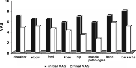
One hundred and nine patients in total, affected by different muscle-skeleton pathologies, underwent treatment. All patients were treated in ten sessions, one session per day. At the beginning and at the end of treatment, all patients were given a VAS (visu analogic scale), in order to value the short-term effect on the pain suffered. Figure1
shows the average results obtained by treated patients, with relevant VAS values, divided into different areas.
Most patients were affected by chronic pain. As a consequence even small initial and final VAS variations have to be read with enthusi-asm. The results point out the method efficacy in the orthopaedic area as a support in the rehabilitation intervention. As a result the thermal shock fits within the rehabilitation projects giving the patient all opportunities to reach the highest functional recovery.
References
1. Guy V, Lehmann JF, Stonebridge JB (1974) Therapeutic applications of electromagnetic power. Proceedings of IEEE 62(1)
2. Lehmann JF, De Lateur BJ. Cryotherapy (1982) In: Lehmann JF (ed) Therapeutic heat and cold, 3rd edn. Williams and Wilkins, Baltimore
3. Michlovitz SL, Nolan PN (2005) Modalities for therapeutic intervention. F.A. Davis Company, Philadelphia
Shoulder arthrodesis in brachial plexus injury:
our experience
M. Abate, N. Della Rosa, A. Russomando, A. Leti Acciaro, A. Landi
Department of Hand Surgery and Microsurgery, Policlinico di Modena (Modena, IT)
Objective We reviewed, trough outcomes’ studies, the results of shoulder arthrodesis in the sequelae of the brachial plexus injury. We compared data of literature with our own experience.
Material and methodsBetween 1994 and 2008 we performed eight (8) shoulder arthrodesis on eight (8) patients affected by sequelae of the brachial plexus injury. Aim of treatment was to reach shoulder stability in order to improve function of whole upper limb, to focus tendon transfers surgery just on the segment distal to the shoulder, to prevent potential pain linked to the phenomenon of shoulder sub-luxation. Contraindications to shoulder arthrodesis included paralysis of the trapetius, elevator scapulae, serratus anterior and rhomboid muscles. The position of the arthrodesis of the shoulder was at 30 degrees of abduction, 30 degrees of flexion and 30 degrees of internal rotation, keeping the body as reference point. The outcomes measures used in the evaluation were active ROM before and after the surgery. Global shoulder function was evaluated using the Frenchay test and the DASH test. The pain was evaluated by Mc Gill pain questionnaire. The complications were considered in our study.
ResultsThe mean follow-up was 4 years. The shoulder arthrodesis in the sequelae of the Brachial Plexus Injury allows improving function of shoulder and of whole upper limb trough the activation of
0 2 4 6 8 10
VAS
shoulder elbow foot knee hip muscle pathologies
hand backache
initial VAS final VAS
scapular-thoracic joint. We report measurement of pre and post-operative active ROM for each patient. The improvement of stability and function was the basic condition to perform palliative surgery for recovering elbow active flexion in some cases. The position of arthrodesis in 30degrees of abduction, flexion e internal rotation did not cause any problem to the patients when upper limb was in rest position because it did not overload scapular-thoracic joint. All patients were satisfied. No complications were recorded.
ConclusionsResults obtained from revision of our cases reflect lit-erature’s results. In patients with sequelae of the brachial plexus injury the shoulder arthrodesis improves upper limb function. It is the basic condition to perform tendon transfers on distal segments. Shoulder arthrodesis in 30degrees of abduction, flexion and internal rotation keeping body as reference point is the best position because it allows the development of potential muscles’ strength (trapetius, serratus anterior, elevator scapulae, rhomboids) and ensures to the patient the best upper limb position as regards scapular-thoracic joint.
Suggested readings
1. Richards RR et al (1985) Shoulder arthrodesis for treatment of brachial plexus palsy. Clin Orthop Rel Res 198:250–258 2. Ruhmann O et al (1999) Reconstructive operations for the
paralyzed shoulder in brachial plexus palsy: concept of treatment. Int J Care Inj 30:609–618
3. Ruhmann O et al (2005) Shoulder Arthrodesis: Indications, technique, results, and complications. J Soulder Elbow Surg 14:38–50
Total elbow replacement: our personal experience
M. Frattini, M. Corradi, F. Pogliacomi
Medical School of Orthopaedics and Traumatology and Unit of Orthopaedics, Traumatology and Functional Rehabilitation, Depart-ment of Surgical Sciences, University of Parma (Parma, IT)
In the second half of the 1900, total elbow replacement was developed. Implant’s choice depends on the underlying pathology, on the degree of damage of the capsuloligamentous structure and on surgeon’s prefer-ence. One of the most widely used models is the Coonrad-Morrey total elbow prosthesis, modified in 1981 by the Mayo Clinic.
Between January 2001 and January 2008, at the Unit of Orthopaedics of the University of Parma, 18 subjects were treated by Coonrad-Morrey total elbow replacement (10 cases of rheumatoid arthritis, 1 psoriatic arthritis, 1 gouty arthritis, 1 osteoarthritis, 2 pseudoarthrosis and 3 complex articular fractures). Patient mean age was 69 years. In all cases, posteromedial access was performed and the ulnar nerve was isolated. Immediately postoperatively the limb was kept immobilized in full extension and assisted passive motion begun 48 h after surgery. All subjects were clinically assessed by the Mayo Elbow Performance Score (MEPS), by the DASH (Disabilities of the Arm, Shoulder and Hand) questionnaire before and after surgery and by radiological investigation. Mean follow-up was 39.6 months (range 1–8 years). The final MEPS was: excellent in 14 cases (78%), good in 2 (11%), poor in 1 (5.5%), bad in 1 (5.5%). Fifteen patients (83%) reported full symptom resolution, whereas three patients (17%) reported moderate pain. Mean maximum flexion was 133.8 (range, 75–100), mean maximum extension 32.5(range, 20–60) and mean ROM 101.3. Mean pronation was 67.5(range, 50–80) and mean supination was 63.8(range, 40–80). Clinical examination revealed joint stability. Mean postoperative DASH was 26.6/100 (range, 1.6–92.2), versus a mean preoperative DASH of 60.7/100 (range, 35–100). Statistical analysis by the Wilcoxon test yielded a significant difference (p\0.02). Radiographic analysis showed that in 17 of the 18 cases,
bone implants positioned behind the flange were incorporated, and no prosthesis loosening or failing were observed.
Postoperatively, two patients exhibited signs of superficial infection which resolved by targeted antibiotic administration. One patient developed deep Staphylococcus infection. Radiographic investigation failed to show signs of mobilization and treatment consisted in tar-geted antibiotic administration associated with soft tissue surgical debridement. In one RA patient, prosthetic aseptic mobilization was observed 17 months postoperatively, requiring implant revision. The good clinical and functional outcomes reported in our report confirm the validity of total elbow replacement and of this prosthetic model which still ensures sufficient functional recovery of the upper limb to carry out essential daily living activities.
Coracoid pain test: a new clinical sign of shoulder
adhesive capsulitis
S. Carbone1, S. Gumina1, AR Vestri2, R. Postacchini3
1Department of Orthopaedics and Traumatology and
2Department of Experimental Medicine, University ‘‘La Sapienza’’
(Rome, IT);
3Department of Orthopaedics and Traumatology, University ‘‘Tor
Vergata’’ (Rome, IT)
Purpose Patients with adhesive capsulitis were clinically evaluated for establishing whether pain elicited by pressure on the coracoid area, which is just above the anatomical structures involved in the disease, may be considered a pathognomonic sign of this condition.
Material and methods The study group included 85 patients with primary adhesive capsulitis, 465 with rotator cuff tear, 48 with calci-fying tendonitis, 16 with glenohumeral arthritis, 66 with acromioclavicular arthropathy and 150 asymptomatic subjects. We aimed at evaluating whether digital pressure on the coracoid area evocated pain. Digital pressure was also carried out on the acromio-clavicular joint and the anterolateral subacromial area. The test was considered positive when pain on the coracoid region was more severe by three points or more (VAS scale) with respect to other areas.
Results The test was positive in 96.4% of patients with adhesive capsulitis. In rotator cuff tear, calcifying tendonitis, glenohumeral and acromioclavicular arthritis a positive test was found in 11.1, 14.5, 6.2 and 10.6% of patients, respectively. A positive result was obtained in 3/150 normal subjects (2%). If adhesive capsulitis was compared to the other four conditions, the test had a sensitivity of 0.96 and a specificity from 0.87 to 0.89. Respect to controls, the sensitivity and specificity was 0.99 and 0.98.
ConclusionsBecause this test is very specific and sensitive, it may be used to confirm or exclude diagnosis of adhesive capsulitis. The
coracoid pain test could be considered as a pathognomonic sign in physical examination of patients with stiff and painful shoulder.
The anatomic features of proximal radius and their
implication for osteosynthesis with plate
G. Giannicola, F.M. Sacchetti, E. Manauzzi, G. Bullitta, F. Postacchini, A. Vestri
Department of Orthopaedic Surgery, University ‘‘La Sapienza’’ (Rome, IT)
comminution. ORIF often gives unsatisfactory results because of the difficulty in restoring the head-neck off-set and the radial head incli-nation relative to its neck. In these cases radial head replacement may be indicated; however, there are no long-term studies on complications and survival of the implant. Recently precontoured plates for the proximal radius has been introduced but no trials have determined whether they are able to restore the normal anatomy of the radius. The latter is still partially unknown because no studies have analyzed the morphology of posterolateral aspect of radial head and neck (‘‘safe zone’’). Our study was aimed at: (1) determining the possible presence of anatomical variations of the safe-zone and (2) analyzing the ana-tomical congruence of precontoured plates to this zone.
Measurements, performed on 44 cadaver dry radii of adults, included: length of the radius, diameters and height of the radial head, and height and diameter of the neck of the radius. The radius of bending of the safe zone was also calculated.
The morphological evaluation of the ‘‘safe zone’’ of the radius revealed three different morphological types of this zone: A (flat) (25%), B (slightly concave) (63.6%) and C (markedly concave) (11.4%). Adherence of a precoundered plate (Acumed) to the bone surface of the safe zone was performed independently by three of us, and the gap between plate and bone was measured. Plate adaptability was good in Type B, scarce in Type C and absent in Type A. In conclusion, we identified three different morphologies of the safe zone, not previously described, and we found that the precountered plates now available can ensure a good restoration of anatomy only in one half of the human radii.
Treatment of traumatic hip dislocations associated
with acetabular fractures
F. Castelli, G. Pesenti, D. Capitani
C.U. Orthopaedics and Traumatology and Trauma Team, DEA Niguarda Ca’ Granda (Milan, IT)
BackgroundTraumatic dislocation of the hip is an extremely severe injury. Although previously considered an uncommon lesion, it is now seen more often as a result of MVA. In most cases this injury can result in a high incidence of complications. Early diagnosis, in poli-trauma patient too, and a early closed reduction constitute the gold standard of a proper treatment of this injury.
Material and methodsThree hundred and sixty-nine patients with surgical acetabular fractures were selected in a 70-month period, 209 (67.4%) were males and 101 (32.6%) females. Average age was 38.2 years (80.6%[20 years\60 years). In the present study 126 patients (34.14%) with hip dislocation associated with acetabular fracture were included. Seventy-one patients (56.34%) were pos-terior wall fxt, 2 (1.59%) pospos-terior column fxt, 3 (2.38%) pospos-terior wall?posterior column, 1 (0.79%) pure transverse fxt, 24 (19.05%) transverse?pw fxt. 101 patients (32.6%) had a posterior fracture-dislocation, 21 (6.7%) central and 4 (1.2%) anterior. Average follow-up was 62 months (range, 18–114). Every patient received an early closed reduction ([6 h from trauma) when directly admitted. Once the dislocation was reduced when surgical reconstruction was indicated it was preferably performed in the first 10 days after injury. Ten patients (3.2%) that reported a irreducible fracture-dislocation required an early open reduction and internal fixation.
ResultsIn 79 patients (62.69%) we obtained a stable and concentric closed reduction. In two cases of uncommon pattern of postero-superior wall fracture (1.59%) a secondary dislocation occurred, in 17 patients (13.49%) the hip remained unstable, 1 patient (0.79%) had an
associated femoral neck fracture, in 13 patients with posterior column plus posterior wall and transverse plus posterior wall fractures (10.31%), after closed reduction of the posterior dislocation, we obtained a central dislocation. Twenty-one patients (16.6%) received closed reduction[6 h. Six patients (28.5%), out of 21 patients that received a delayed reduction, developed AVN. One hundred and five patients (83.4.%) received closed reduction\6 h. Two patients (1.9%) out of 105 patients developed AVN. The average AVN rate in our study was 6.3% with eight cases of AVN out of 126 acetabular fracture-dislocations.
Conclusions In summary, this study suggest: (a) there are two dif-ferent mechanisms of intra-articular incarceration of bone fragments: (i) Primary incarceration with one or several fragments, perhaps pedunculated to the capsule that enter the acetabulum at the moment of injury and (ii) secondary incarceration with fragments maintaining a capsular flap pedicle that is drawn into the joint during the closed reduction; (b) early closed or open reduction\6 h constitute the gold standard of a proper treatment of this injury reducing the AVN rate of approximately 15 tim.
Damage control orthopaedics and early total care
F. Castelli, F. Sala, A. La Maida, O. Chiara, S. Cimbanassi, D. Capitani
C.U. Orthopaedics and Traumatology and Trauma Team, DEA Niguarda Ca’ Granda (Milan, IT)
Introduction Early fixation of long bone fractures in the multiple injured patient has been recognized for much of the past three decades as beneficial in minimizing secondary lung and remote organ failure. Although early fracture fixation is an expedient in patients with multiple injury ETC may be associated with post-traumatic systemic complications.
Material and methodsIn this study all patients from a consecutive series of 690 trauma patients with trauma team activation admitted between January 2004 and January 2006 to the Emergency Depart-ment of Niguarda Hospital in Milan were included when they fulfilled all of the following criteria: (1) directly admitted, (2) ISS[than 15 and (3) survival[than 24 h. Patients with fracture of long bones and/ or pelvis with a clear indication for operative fracture treatment and the necessity of immediate fracture fixation were treated according with DCO. All other patients fulfilling the inclusion criteria with minor fracture or not requiring immediate fixation formed the control group. ISS, RTS and Ps was calculated at the admission and evaluated later by the Trauma Leader. All injuries were classified with AO and Gustilo classification.
Conclusions The goals of damage control include stopping or lim-iting ongoing injury including both local soft-tissue injury and also remote organ injury secondary to the local release of inflammatory mediators to the systemic circulation further thought to prevent pul-monary complications by allowing patients to avoid the enforced supine position. This study was conducted retrospectively to evaluate the effectiveness of the Trauma Team organization and to evaluate the concept of DCO by immediate external fracture fixat