Available online at http://jddtonline.info REVIEW ARTICLE
A STUDY ON ANTIOXIDANT PROPERTIES OF DIFFERENT BIOACTIVE
COMPOUNDS
Ghosh Rajat*, Deb Panchali
Department of Pharmacy, Tripura University (A Central University), Suryamaninagar, Tripura-799022, India
*Corresponding Author’s E-mail: [email protected]
INTRODUCTION
An antioxidant is a chemical that reduces the rate of particular oxidation reactions in a specific context1. Vitamin C and Vitamin E were the first recognized antioxidants, but other substances that have powerful antioxidant properties have also been recognized such as:
Selenium, Carotenoids (Beta-carotene, Lutein,
Lycopene, Sulforaphane, Zeaxanthin, and Astaxanthin), Bioflavonoids (Anthocyanins, Proanthocyanidins, Quercetin, and Apigenin), Coenzymes, Soy Isoflavones (Genistein and Daidzein), and many less well-known compounds found in fruits and vegetables2.
Free radicals are atoms or groups of atoms with unpaired electrons and can be formed when oxygen interacts with certain molecules. Free radicals are highly reactive, that’s why they can start a chain reaction immediately once they are formed. They have the capability to damage cellular components such as DNA and the cell membranes when they react. The formation of free radicals can be happened during normal metabolic functions or introduced from the environment3. Free radicals are inherently unstable, since they contain extra energy. To reduce their energy load, free radicals react with certain chemicals in the body, and in the process, interfere with the cells’ ability to function normally. Free radicals are believed to play a major role in different health conditions, including the aging process, cancer, atherosclerosis, Parkinson's disease and Alzheimer's disease, ulcerative colitis, Crohn's disease, Diabetes mellitus, Rheumatoid Arthritis, AIDS, Renal diseases, Respiratory diseases and Eye diseases. To reduce the number of free radical-induced health problems, we need large amount of antioxidant nutrients along with less exposure to free radicals.
Oxygen is the major source of the potentially damaging free radicals, though it is essential for living. Free radicals are also there in the environment. Environmental sources which give rise to free radicals include exposure to ionizing radiation (from industry, sun exposure,
cosmic rays, and medical X-rays), ozone and nitrous oxide (primarily from automobile exhaust), heavy metals (such as mercury, cadmium, and lead), cigarette smoke (both active and passive), alcohol, unsaturated fat, and other chemicals and compounds from food, water, and air4.
Oxidation is a chemical reaction or process through which electrons are transferred between molecules by an oxidizing agent. Oxidation is a common and essential process in nature, but it can form free radicals inside human body which cause damage to healthy cells. This damage is known as oxidative stress5. Highly reactive oxygen species (ROS) and reactive nitrogen species (RNS) that possess an unpaired electron induce this stress. But cells are equipped to combat this oxidative attack with numerous cellular antioxidant defenses such as glutathione (GSH), superoxide dismutases, and catalase. During aging and various free radicals related disease states, these antioxidant defense systems can be changed leading to progressive oxidative damage and subsequent cell death and significant inactiveness. Changes in antioxidant level within specific cells and structures of the brain may be particularly useful in determining whether oxidative stress is benign or pathological6.
Molecules usually contain an even number, or paired electrons. Free radicals, however contain an uneven number of, or unpaired electrons. These electrons make free radicals unstable and highly reactive. Free radicals attack other molecules or healthy cell components in order to capture an electron, and thereby become stable. Subsequently, in the process, the original free radical creates a new free radical. A chain reaction of new free radicals is created due to this scavenging of electrons which eventually results in oxidative stress and damage to healthy cells.
ABSTRACT
Free radicals induce damage mainly to biomembranes and DNA due to peroxidation, which lead to tissue damage resulting in number of degenerative diseases. Antioxidants can neutralise the effect of these free radicals through various ways and may protect the body from harmful alterations. Antioxidants may be of different types viz. enzymatic, non-enzymatic and also found from plant sources. Nowadays, numerous synthetic antioxidant supplements are also found in the market. The antioxidative potential of samples can be assayed by different in vitro and in vivo methods. The present review describes a brief account of the free radical generation, their role in aging and other diseases, classification of antioxidants, their n atural and synthetic sources, possible mechanism and several assay methods of free radical scavenging activity of different bioactive compounds.
Rajat et al Journal of Drug Delivery & Therapeutics; 2014, 4(2), 105-115 106
As the brain is the main target organ of oxidative stress; hence the researchers are studying about the correlation between enhanced levels of free radicals and neurodegenerative conditions.
Although free radicals are considered as harmful, they do perform some beneficial functions in the immune system. Free radicals help in the functioning of white
blood cells as they attack bacterial invaders and other pathogens in the body. Free radicals are also thought to have a role in “redox signaling” process which assist to communicate between cells.
The main aim of antioxidants is to maintain the free radicals at optimum levels and not to completely destroy them as they do serve some useful functions also5.
Different types of free radicals/oxidants and their defense system
Free Radical Free Radical Scavenger
Hydroxy radical (OH.) (SOD), Mn-SOD, Cu-Zu-SOD
Peroxy radical (ROO.) Tocopherols, Ubiquinone
Oxidant Oxidant Scavenger
Superoxide anion (O2-) Superoxide dismutases
Hydrogen peroxide (H2O2) Catalase, glutathione peroxide (GPx)
Hydroperoxides (HOD) GPx, glutathione reductase
Transition metals (Fe2+, Cu+) Chelators
FREE RADICALS BEHIND AGING:
Free radicals accumulation is mostly responsible for human aging where the use of antioxidants can prevent the aging process by deterring the progression of free radicals. The most widely accepted free radical theory of aging finds that through normal metabolic processes, ultraviolet light, and environmental toxins, cells constantly produce free radicals which generate a chemical process known as oxidation. It causes cellular degeneration. This is considered a major contributor to the aging process.
Dr. Denham Harman identified the first free radical theory of aging, afterwards researchers have constantly been searching the authenticity of Dr. Harman’s theory. Some researchers opposed to the theory find that reactive oxygen species (ROS) have minimal influence on aging and suggest a certain amount of free radicals are required for cellular signaling7.
MECHANISM OF ACTION OF ANTIOXIDANTS
Oxidation can be initiated with the help of a number of chemical and physical phenomena, which occurs continuously in the presence of suitable substrates, until and unless a blocking defence mechanism occurs. The steps involved during oxidation via a free radical-mediated chain reaction are initiation, propagation, branching and termination. The process can be initiated with the help of external agents such as heat, light, ionizing radiation, by chemical initiation involving metal ions or metalloproteins etc.
Initiation:
LH + R∙ → L∙ + RH
Where LH denotes the substrate molecule, such as, a lipid; R∙ as the initiating oxidizing radical. The oxidation of the lipid generates a highly reactive allyl radical (L∙)
which can rapidly react with oxygen to form a lipid peroxyl radical (LOO∙).
Propagation:
L∙ + O2 → LOO∙
LOO ∙ + LH → L∙ + LOOH
The peroxyl radicals are the chain carriers of the reaction; they can further oxidize the lipid, producing lipid hydroperoxides (LOOH), which in turn break down to a wide range of compounds, including alcohols, aldehydes, alkyl formates, ketones and hydrocarbons, and radicals, including the alkoxyl radical (LO∙).
Branching:
LOOH → LO∙ + HO∙ 2 LOOH → LOO ∙ + LO∙ + H2O
Primary antioxidants (AH) when present in trace amounts, may either delay or inhibit the initiation step by reacting with a lipid radical or inhibit the propagation step by reacting with peroxyl or alkoxyl radicals. Secondary or preventative antioxidants are compounds that retard the rate of oxidation. This may be achieved in a number of ways, including removal of substrate or singlet oxygen quenching8.
LOO∙ + AH → LOOH + A
CLASSIFICATION OF ANTIOXIDANTS
ENZYMATIC ANTIOXIDANTS
Antioxidant enzymes like SOD, GPx and CAT are the first lines of defense against O2- and H2O2- mediated
injury.
peroxide, they are an important antioxidant defense in nearly all cells exposed to oxygen. These enzymes are found in all aerobic organisms and also present in mitochondria and cytosol. There are four families of SODs: Cu-SOD, Cu-Zn-SOD, Mn-SOD and Fe-SOD enzyme and has been detected in a large number of tissues and organisms, and is thought that it is present to protect the cell from damage caused by O2. – .
The SOD-catalysed dismutation of superoxide :
M(n+1)+ − SOD + O2− → Mn+ − SOD + O2
Mn+ − SOD + O2− + 2H+ → M(n+1)+ − SOD + H2O2
where M = Cu (n=1) ; Mn (n=2) ; Fe (n=2) ; Ni (n=2).
In this reaction the oxidation state of the metal cation oscillates between n and n+1.
Glutathione peroxidase (GPx) - Glutathione peroxidase (a selenium containing enzyme) involves in the reduction of H2O2 and lipid hydroperoxide (LO2H; generated
during lipid peroxidation) to water using reduced glutathione as substrate. It is found in both cytosol and mitochondria and is a well-known first line of defense against oxidative stress, which in turn requires glutathione as a cofactor. Its involvement is noticed during generation of nucleotide precursors of DNA via the reduction of ribonucleotides to deoxyribonucleotides. GPx catalyses the oxidation of reduced glutathione (GSH) to oxidized glutathione (GSSG) at the expense of H2O2, by its selenium dependency. Since, selenium is an
integral component of GPx, the measurement of this enzyme has been used as a functional index of selenium level. Low levels of selenium have been associated with a high risk of cardiovascular diseases and cancer in humans.
2GSH + H2O2 → GS–SG + 2H2O (glutathione peroxidase)
Catalase (CAT) - Catalase (CAT) is present in most cells and catalyses the decomposition of hydrogen
peroxide to water and oxygen. CAT is mainly localized in mitochondria and in subcellular respiratory organelles. The mechanism of action is as follows:
2H2O2 → 2H2O+ O2
In addition to the above enzymes, glutathione transferase, ceruloplasmin, hemoxygenase and possibly several other enzymes may participate in enzymatic control of oxygen radicals and their products9,10.
NON ENZYMATIC ANTIOXIDANTS
Antioxidative vitamins:- Antioxidative vitamins stimulates immune system, inhibit nitrosamine formation and alter metabolic activation of carcinogens. They inhibit DNA damage induced by ROMs and thus prevent genetic alterations. They protect the somatic cell from free radicals .
Vitamin A (Retinol, retinal) :- Vitamin A is a fat-soluble vitamin which includes the compounds -retinol and its esters, retinoldehyde and retinoic acid. It is essential for growth, maintenance of visual function, reproduction and differentiation of epithelial tissue. Beta-carotene protects human body from sun light damage. β-carotene is part of a family of chemicals called the carotenoids, which are found in many fruit and vegetables, as well as some animal products such as egg yolks. Biologically, β-carotene is most important as the precursor of vitamin A in the human diet. It has anti-oxidant properties along with preventing cancer and other diseases. It is an excellent scavenger of singlet oxygen (produced during photosensitivity). Both natural and synthetic analogues of vitamin A have been shown to be effective in various diseases. Carrots, squash, sweet potatoes, peaches, kale and apricots are rich sources of -carotene. Vitamin A is structurally related to carotene. Carotene is converted into vitamin A in the liver. Two molecules of vitamin are formed from on molecule of beta carotene shown in the figure below9,11.
CH3 C H3
CH3
C H3
C H3 CH3 CH3
CH3 CH3
CH3
C H3 CH3
CH3 CH3
H O CH3
dioxygenase enzyme break double bond
O2
Vitamin A Beta Carotene
2
Rajat et al Journal of Drug Delivery & Therapeutics; 2014, 4(2), 105-115 108
Vitamin E (Tocopherol) :- This is a lipid soluble vitamin, which occurs in plasma as a variety of tocopherols, major one is alpha-tocopherol. It scavenges peroxy radical intermediates in lipid peroxidation and is responsible for protecting PUFA (Poly unsaturated fatty acid) present in cell membrane and low-density lipoprotein against lipid peroxidation. In addition to this, it’s involvement in quenching of singlet oxygen and the reaction with peroxynitrite was noticed. Vitamin E works more efficiently in the lipid phase. Sources of vitamin E include wheat germ oil, nuts, seeds, whole grains, vegetable oil and fish liver oil. Chemical structure of Vitamin E is shown in the figure below9.
O C H3 C H3
CH3 O
H
CH3
CH3 CH3
CH3 CH3
Figure 2: Vitamin E12
Vitamin C (Ascorbic acid) :- Vitamin C is a water-soluble antioxidant in biological fluids and its presence is also necessary for normal metabolic functions of the body. It interacts directly with radicals like O2- and HO
-in plasma. It has been found that vitam-in C is superior to over-the-counter medicines in reducing the symptoms, duration and severity of colds. It helps in cardiovascular disease by protecting the linings of arteries from oxidative damage. It neutralizes reactive oxygen metabolites and reduces oxidative DNA damage and genetic mutations . The rich source of this vitamin is citrus fruits, green vegetables, raw cabbage and tomatoes. Chemical structure of Vitamin C is shown here9.
O
OH O
H
O O
H O
H H
Figure 3: Vitamin C12
Melatonin: Melatonin is a naturally occurring hormone found in animals and in some other living organisms, including algae. Unlike other antioxidants, melatonin does not undergo redox cycling. Melatonin, once oxidized, cannot be reduced to its former state because it forms several stable end-products upon reacting with free radicals. Therefore, it is referred to as a terminal antioxidant13.
Moreover numerous small molecules such as bilirubin, uric acid etc also act as antioxidants.
ANTIOXIDANT PLANTS
Plants are especially vulnerable to damage by active oxygen exposed to radiation, UV light. Hence, plants are equipped with numerous antioxidant defense systems that have resulted in certain numbers of very potent antioxidants. Beside plants, many of microbial and animal products as well as fermented products, seaweeds
and protein hydrolysates were found to be powerful antioxidants. Antioxidant activity of plants may be attributed to presence of phenols, flavonoids, xanthones, alkaloids, anthraquinones, phytosterols, steroids, amino acids, isothiocyanate indoles etc.
Several antioxidants of plant origin are experimentally proved and used as effective protective agents against oxidative stress. Some of them are given below:
Black pepper – Piperine, a major constituent of Piper nigrum, enhances the synthesis of glutathione9.
Resveratrol - It is a potent antioxidant with polyphenol compounds. It enhances nitric oxide activity and improve endothelial function. It is found in grapes and red wine7. Chemical structure of resveratrol is shown in Figure 4.
OH O H
OH
Figure 4: Resveratrol14
Lutein - Lutein is a xanthophyll in the carotenoid family (shown in figure 5). It is plentiful in green leafy vegetables such as spinach, kale and yellow carrots. Lutein was found to be present in the macula, a small area of retina responsible for central vision. It was noticed that lutein helps keep the eyes safe from oxidative stress and the high-energy photons of blue light. Lutein can protect vision from cataracts and age-related macular degeneration15.
C H3 CH3
O
H CH3
C H3 CH3
OH C
H3
CH3 CH3
CH3 CH3
Figure 5: Lutein16
Pine Bark Extract: Pine bark extract is a rich source of procyanidin oligomers, which increase antioxidant activity. It improves glutathione levels. The active components of pine bark extract act as anti-inflammatory agent.
Quercetin - Quercetin is a phytochemical with high antioxidant activity that has been shown to maintain blood pressure levels and thus benefit cardiovascular function. Quercetin can inhibit platelet aggregation, increase nitric oxide activity and improve endothelial function. It is found in apples and onions7.
Brand Name Formulation
Glutathione Complex Tablets
Glutathione Booster Capsules
Mega L- Glutathione Tablets
Reduced Glutathione 50mg Capsules
Gluton Inj 600 mg Injection
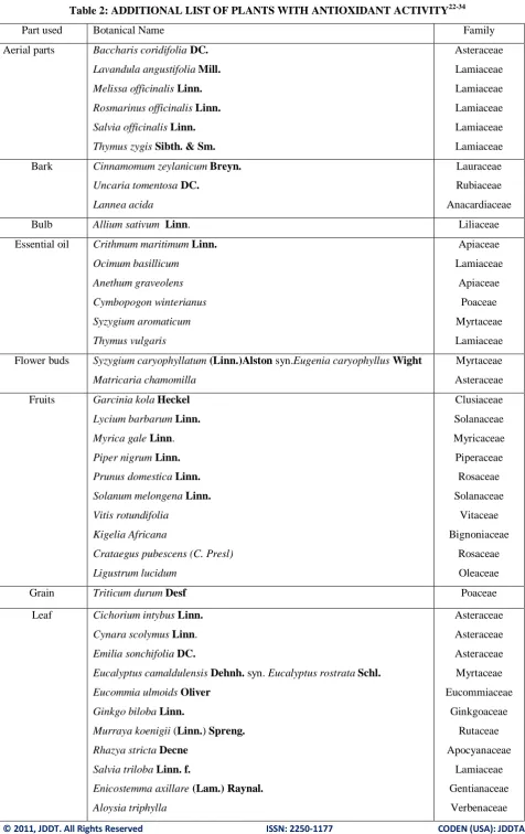
A brief description including part used, chemical constituents and other biological activities of the common antioxidant plants is given in Table 1 and an additional list of antioxidant plants is shown in Table 2.
TABLE 1: LIST OF ANTIOXIDANT PLANT SOURCES 19-21
S.N. Common Name Chemical Constituents Part
Used
Biological Activities
1 Akashabela Flavonoids, glycosides, lactones,
coumarins, dulcitol, bergenin
Stem Expectorant, anthelmintic, purgative, diuretic, in jaundice,antifertility drug.
2 Am/Mango Cyanogenetic glycosides,
mangiferin, quercetin, ellagic acid, gallic acid
Fruit Prevention of cancer (colon, breast, leukemia)
Leaf Antibacterial
Root Leucorrhoea, diarrhoea
3 Amla/Myrobalan Vitamin C (L-ascorbic acid,
polyphenols (ellagic acid, gallic acid, tannins)
Fruit Useful in burning sensations, piles, leprosy, inflammations, anaemia, urinary discharges, constipation
4 Ashwagandha Steroidal lactones, glycene,
withanine, withanolides
Leaf Immunomodulator
Root Analgesic
Seed Diuretic and hypnotic
5 Babchi Essential oil, fixed oil, resin,
bakuchiol
Seed Purgative, stomachic, stimulant,
anthelmintic, aphrodisiac, in scabies
6 Carrot Carotene, carotenoids, sugars,
glycosides, flavonoids,
quarternary bases
Root Used in bronchitis, piles, jaundice, tumours, leprosy, urinary complaints, chest troubles, aphrodisiac
7 Chirayita/Chiretta Xanthones, mangiferin, swertinin, chiratin, arginine
Whole plant
Febrifuge, laxative, anthelmintic, antimalarial, chronic fever
8 Karela/Bitter
Melon
Stearic acid, triterpene glycosides Fruit Fruit as stomachic, aphrodisiac, increases immunity
Leaf Antidiabetic Root Laxative, antipyretic Seed Stomachic, aphrodisiac
9 Makoi/Common
nightshade
Polyphenolic compounds,
flavonoids, steroids
Leaf Polyphenolic compounds, flavonoids, steroids
10 Mulethi/Liquorice Glycyrrhizin, flavonoids,
liquiritin, isoliquiritin
Root Diuretic, emmenagogue, in vomiting, asthma, peptic ulcer, bronchitis, for curing wounds
11 Safed
Chandan/Sandal
Volatile oil, santalol, α-santalol, β-santalol, β-sitosterol
Heart wood
Antipyretic, aphrodisiac, in heart diseases, bronchitis, small pox, used in perfumery
12 Saunf/Fennel Volatile oil, fenchone, anethole, limonene, estragole
Fruit oil
Stimulant, purgative, diuretic, useful in venereal diseases, vermicide
13 Tulsi/Sacred Basil Volatile oil, terpenoids, eugenol, thymol, estragole
Rajat et al Journal of Drug Delivery & Therapeutics; 2014, 4(2), 105-115 110
Table 2: ADDITIONAL LIST OF PLANTS WITH ANTIOXIDANT ACTIVITY22-34
Part used Botanical Name Family
Aerial parts Baccharis coridifoliaDC.
Lavandula angustifoliaMill.
Melissa officinalisLinn.
Rosmarinus officinalis Linn.
Salvia officinalis Linn.
Thymus zygis Sibth. & Sm.
Asteraceae
Lamiaceae
Lamiaceae
Lamiaceae
Lamiaceae
Lamiaceae
Bark Cinnamomum zeylanicumBreyn.
Uncaria tomentosa DC.
Lannea acida
Lauraceae
Rubiaceae
Anacardiaceae
Bulb Allium sativum Linn. Liliaceae
Essential oil Crithmum maritimumLinn.
Ocimum basillicum
Anethum graveolens
Cymbopogon winterianus
Syzygium aromaticum
Thymus vulgaris
Apiaceae
Lamiaceae
Apiaceae
Poaceae
Myrtaceae
Lamiaceae
Flower buds Syzygium caryophyllatum (Linn.)Alstonsyn.Eugenia caryophyllus Wight
Matricaria chamomilla
Myrtaceae
Asteraceae
Fruits Garcinia kolaHeckel
Lycium barbarumLinn.
Myrica galeLinn.
Piper nigrum Linn.
Prunus domestica Linn.
Solanum melongena Linn.
Vitis rotundifolia
Kigelia Africana
Crataegus pubescens (C. Presl)
Ligustrum lucidum
Clusiaceae
Solanaceae
Myricaceae
Piperaceae
Rosaceae
Solanaceae
Vitaceae
Bignoniaceae
Rosaceae
Oleaceae
Grain Triticum durumDesf Poaceae
Leaf Cichorium intybusLinn.
Cynara scolymusLinn.
Emilia sonchifoliaDC.
Eucalyptus camaldulensisDehnh. syn. Eucalyptus rostrataSchl.
Eucommia ulmoidsOliver
Ginkgo bilobaLinn.
Murraya koenigii (Linn.) Spreng.
Rhazya stricta Decne
Salvia triloba Linn. f.
Enicostemma axillare (Lam.) Raynal.
Aloysia triphylla
Asteraceae
Asteraceae
Asteraceae
Myrtaceae
Eucommiaceae
Ginkgoaceae
Rutaceae
Apocyanaceae
Lamiaceae
Gentianaceae
Ferula foetida
Camellia sinensis
Bacopa monnieri
Salvia hypoleuca
Apiaceae
Theaceae
Scrophulariaceae
Lamiaceae
Leaves & Fruit Ipomoea batatas (L.) Lam. Convolvulaceae
Leaves & Root Punica granatum Lythraceae
Peels & Seeds Citrus sinensis Rutaceae
Rhizome Zingiber officinale Rosc.
Curculigo orchioides Gaertn.
Zingiberaceae
Hypoxidaceae
Rhizome & Root Picrorrhiza kurroa Scrophulariaceae
Root Bryonia albaLinn.
Panax ginsengMey.
Tinospora cordifolia (Willd.) Miers. ex Hook. f.& Thoms.
Medicago sativa L.
Rhodiola rosea
Ferula hermonis
Polygonum multiflorum
Cucurbitaceae
Araliaceae
Menispermaceae
Fabaceae
Crassulaceae
Apiaceae
Polygonaceae
Seed Plantago asiatica Linn.
Cassia auriculata
Artemisia annua L.
Daucus carota
Momordica charantia
Plantaginaceae
Caesalpiniaceae
Asteraceae
Apiaceae
Cucurbitaceae
Shoot Asparagus racemosusWilld. Liliaceae
Tuber Solanum tuberosum Linn. Solanaceae
Whole plant Sida cordifolia
Dana racemosa
Yucca schidigera
Malvaceae
Asparagaceae
Asparagaceae
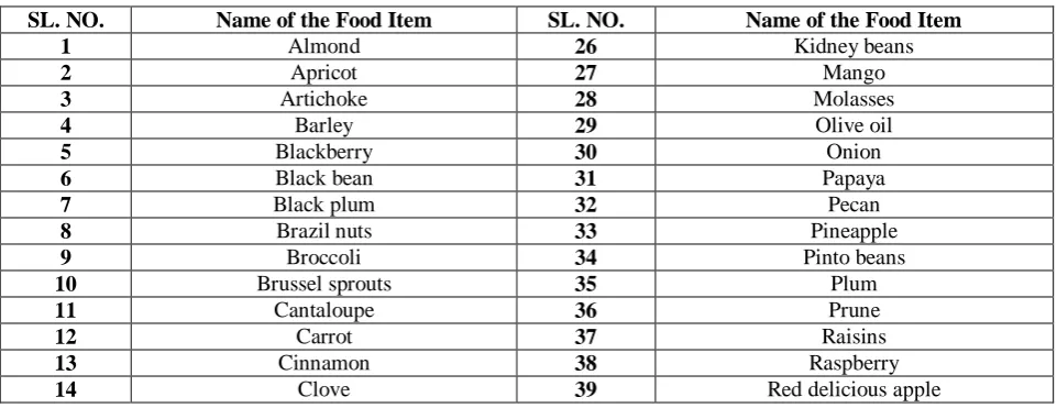
Some of the common antioxidant food sources are shown below:
Table 3: Common Antioxidant Food Sources35-39
SL. NO. Name of the Food Item SL. NO. Name of the Food Item
1 Almond 26 Kidney beans
2 Apricot 27 Mango
3 Artichoke 28 Molasses
4 Barley 29 Olive oil
5 Blackberry 30 Onion
6 Black bean 31 Papaya
7 Black plum 32 Pecan
8 Brazil nuts 33 Pineapple
9 Broccoli 34 Pinto beans
10 Brussel sprouts 35 Plum
11 Cantaloupe 36 Prune
12 Carrot 37 Raisins
13 Cinnamon 38 Raspberry
Rajat et al Journal of Drug Delivery & Therapeutics; 2014, 4(2), 105-115 112
15 Cocoa powder 40 Russet potato
16 Coffee 41 Salmon
17 Cottonseed oil 42 Small red beans
18 Cranberry 43 Sorghum
19 Cultivated Blueberry 44 Strawberry
20 Eggs 45 Sweet Cherry
21 Gala apple 46 Tomato
22 Ginger roots 47 Vanilla beans
23 Granny smith apple 48 Walnut
24 Green peppers 49 Watercress
25 Guava 50 Wild blueberry
ANTIOXIDANT ACTIVITY OF ESSENTIAL OILS
A study was done on 248 essential oils belonging to 18 botanical families for the presence of antioxidant potential. The antioxidant activities of the oils were tested by DPPH radical scavenging activity at three different concentrations of 5, 25 and 100 mg/ mL using % DPPH inhibition and then further evaluated by TLC. Thin layer chromatography regions responsible for the antioxidant activity were examined by GC/MS and the active components responsible for the activity were identified based on their mass spectra and GC retention times. Among them, 60 essential oils were found to be
active at a concentration of 100 mg/mL, 27 of them at 25 mg/mL & 17 of them were active at 5 mg/mL concentration. Essential oils belonging to the family Annonaceae, Apiaceae, Asteraceae, Burseraceae, Cupressaceae, Fabaceae, Geraniaceae, Lamiaceae, Lauraceae, Myrtaceae, Myristicaceae, Myoporceae, Piperaceae, Poaceae, Rosaceae, Rutaceae, Rubiaceae, Solanaceae and Moschidae were the most effective essential oils. Out of the 18 constituents identified, 7 of
them were aromatic hydrocarbons or phenolic
compounds and 14 of the identified compounds were oxygenated monoterpenoids40.
O CH3 C H3
C H3
C
H3 CH3
CH3
C
H3 CH3
CH3
OH
C
H3 CH3
CH3
OH
OH OH H
O OH
O
CH2
CH3
Camphor
Cymene Thymol Carvacrol Vanillin Eugenol
Figure 6: Chemical Structure of Active Antioxidant Compounds40
Another study was reported on the Oregano (Origanum vulgare L., ssp. hirtum) essential oils to reveal the antioxidative properties in which three different methods were employed to assess the activity namely β-carotene bleaching test, DPPH radical scavenging method and Thiobarbituric acid reactive species assay. 17 compounds were identified in hydrocarbons fraction with γ-terpinene (31.0%), p-cymene (22.1%), α-terpinene (10.4%) and transcaryophyllene (9.1%) as main components. There were 4 oxygen-containing compounds, out of which, thymol (47.3%) and carvacrol (46.4%) were the major ones. The phenolic fraction contained only 2 compounds, thymol (58.9%) and carvacrol (41.1%). It was seen that the BCB method can be helpful especially for the investigation of the antioxidant activity of essential oils. On the other hand, the DPPH method is faster than BCB method and it is useful in giving preliminary information of radical scavenging abilities of novel antioxidants. The method is sensitive and requires small sample amounts. The TBA method is preferable in order to obtain useful data in an environment similar to the real-life situation. Both methods, DPPH and TBA, similarly allow testing of both lipophilic and hydrophilic substances. In the
study, it was confirmed that the same antioxidant samples exhibit different antioxidative values depending on the concentration and the measured antioxidant parameter. This study confirmed that the oregano essential oil possess remarkable antioxidant properties. The antioxidant effect was due to the presence of thymol and carvacrol, but a possible synergistic effect among oxygen containing compounds could be suggested too41.
ANTIOXIDANT ASSAY METHODS
Antioxidant activity should not be concluded based on a single antioxidant test model. Generally various antioxidant assay methods are employed to assess the samples of interest. Researcher
has to critically verify methods of analysis before adopting the choice of method. Various in vitro and in vivo methods are described here:-
In-vitro methods
DPPH Radical Scavenging Activity - The molecule
1,1-diphenyl-2-picrylhydrazyl
molecule as a whole, so that the molecule does not dimerize. The delocalization of electron also gives rise to the deep violet color, characterized by an absorption band centered at about 517 nm. When a solution of DPPH is mixed with that of a substrate (AH) that can donate a hydrogen atom, then this gives rise to the reduced form and the colour changes from violet to colourless or pale yellow. Alam MN et al reported that the sample extract (0.2 mL) was mixed with methanol and 2 mL of DPPH solution (0.5 mM). After 30 min, the absorbance was measured at 517 nm. The percentage of the DPPH radical scavenging was calculated using the equation as given below:
% inhibition of DPPH radical = ([Abr – Aar] /Abr ) × 100
where Abr was the absorbance before reaction and Aar
was the absorbance after reaction has taken place.
Hydrogen Peroxide Scavenging (H2O2) Assay - H2O2
is rapidly decomposed into oxygen and water and this may produce hydroxyl radicals (OH.) that can initiate lipid peroxidation and cause DNA damage in the body. According to Alam MN et al, the ability of plant extracts to scavenge H2O2 can be estimated. In this method,
phosphate buffer was used as blank solution and the absorbance was checked at 230 nm. The percentage of hydrogen peroxide scavenging was calculated as follows:
% scavenged H2O2 = [(Ai - At)/Ai] × 100
where Ai was the absorbance of control and At was the
absorbance of test.
Nitric oxide scavenging activity – NO- is generated in biological tissues by specific nitric oxide
synthases, which metabolizes arginine to citrulline with the formation of NO- via a five electron oxidative reaction. Alam MN et al reported that, 2 mL of 10 mM sodium nitroprusside dissolved in 0.5 mL phosphate buffer saline (pH 7.4) is mixed with 0.5 mL of sample at various concentrations (0.2–0.8 mg/mL). After 150 min of incubation at 25oC, 0.5 mL of the incubated solution is withdrawn and mixed with 0.5 mL of Griess reagent [(1.0 mL sulfanilic acid reagent (0.33% in 20% glacial acetic acid at room temperature for 5 min with 1 mL of naphthylethylenediamine dichloride (0.1% w/v)]. After incubation for 30 min and its absorbance was measured at 546 nm. The amount of nitric oxide radical inhibition was calculated following this equation:
% inhibition of NO radical = [A0 – A1]/ A0 × 100
where A0 was the absorbance before reaction and A1 was
the absorbance after reaction with Griess reagent.
Trolox Equivalent Antioxidant Capacity (TEAC) method/ ABTS Radical Cation Decolorization Assay -
This method, uses a diode-array spectrophotometer to measurethe loss of color when an antioxidant is added to the blue–green chromophore ABTS. +(2,2-azino-bis(3-ethylbenzthiazoline-6-sulfonic acid)). The antioxidant reduces ABTS.+to ABTS and decolorize it. ABTS.+ is a stable radical not found in the human body. Trolox (6-hydroxy-2,5,7,8-tetramethylchroman-2 carboxylic acid), a water-soluble analog of vitamin E, was used as an antioxidant standard and absorbance is read (at 750 nm) after 5 min. TEAC values can be calculated from the
Trolox standard curve and expressed as Trolox equivalents (in mM).
Total radical-trapping antioxidant parameter (TRAP) method - This method is based on the protection provided by antioxidants on the fluorescence decay of R-phycoerythrin (R-PE) during a controlled peroxidation reaction. The antioxidative potential is evaluated by measuring the decay in decoloration. 120 μL of diluted sample is added to 2.4 mL of phosphate buffer (pH 7.4), 375 μL of bidistilled water, 30 μL of diluted R-PE and 75 μL of ABAP; the reaction kinetics at 38 oC is recorded for 45 min by a luminescence spectrometer. TRAP values are calculated from the length of the lag-phase due to the samplecompared with standard.
Ferric reducing-antioxidant power (FRAP) assay - It is based on the reduction of the complex of ferric iron and 2,3,5-triphenyl-1,3,4-triaza-2-azoniacyclopenta-1,4-diene chloride (TPTZ) to the ferrous form at low pH. According to Alam MN et al, 3 mL of prepared FRAP reagent is mixed with 100 μL of diluted sample; the absorbance at 593 nm is recorded after a 30 min incubation at 37 oC. FRAP values can be obtained by comparing the absorptionchange in the test mixture with those obtained from increasing concentrations of Fe3+ and expressed as mM of Fe2+ equivalents per kg (solid food) or per L (beverages) of sample.
Superoxide radical scavenging activity (SOD) -
Although superoxide anion is a weak oxidant, it ultimately produces powerful and dangerous hydroxyl radicals as well as singlet oxygen, both of which contribute to oxidative stress. The superoxide anion radicals are generated in 3.0 mL of Tris–HCl buffer (16 mM, pH 8.0), containing0.5 mL of nitroblue tetrazolium (NBT) (0.3 mM), 0.5 mLNADH (0.936 mM) solution, 1.0 mL extract and 0.5 mL, Tris–HCl buffer (16 mM, pH 8.0). The reaction is initiated by adding 0.5 mL phenazine methosulfate (PMS) solution(0.12 mM) to the mixture, incubated at 25 oC for 5 min and then the absorbance is measured at 560 nm against a blank sample.
Thiobarbituric acid (TBA) method – Thefinal sample concentration of 0.02% w/v was used in this method. 2 mL of 20% trichloroacetic acid and 2 mL of 0.67% of thiobarbituric acid were added to 1 mL of sample solution.The mixture was placed in a boiling water bath for 10 min and then centrifuged after cooling at 3000 rpm for 20 min. The absorbance activity of the supernatant was measured at 552 nm and recorded after it has reached its maximum.
Hydroxyl radical scavenging activity - Hydroxyl radical is one of the potent reactive oxygen species inthe biological system that reacts with polyunsaturated fatty acid moieties of cell membrane phospholipids and causes damageto cell. Thereaction mixture (1.0 mL) consist of 100 μL of 2-deoxy- Dribose (28 mM in 20 mM KH2PO4-KOH buffer, pH 7.4), 500 μL of the extract, 200 μL EDTA (1.04 mM) and 200 μM FeCl3 (1:1 v/v), 100 μL of H2O2 (1.0 mM) and 100 μL ascorbicacid (1.0
Rajat et al Journal of Drug Delivery & Therapeutics; 2014, 4(2), 105-115 114
min. After cooling, absorbance is measured at 532 nm, against ablank sample42.
Hypochlorite scavenging assay – Thereaction mixture contained taurine (150 mM), sodium
hypochlorite (600 mM), reference compounds (variable concentrations) and phosphate saline buffer (pH 7.4) in a final volume of 1 mL. The solution was mixed thoroughly and potassium iodide (2 M) was added. A yellow coloration was developed and the absorbance was read at 350 nm43.
Other methods include Oxygen radical absorbance capacity (ORAC) method, Hydroxyl radical averting capacity (HORAC) method, Peroxynitrite radical scavenging activity, Reducing power method (RP), Phosphomolybdenum method, Ferric thiocyanate (FTC) method, DMPD (N,N-dimethyl-p-phenylene diamine dihydrochloride) method, β-carotene linoleic acid method /conjugated diene assay, Copper-phenanthroline (Cu-phen) assay, Deoxyribose assay, Xanthine oxidase method, Cupric ion reducing antioxidant capacity (CUPRAC) method, Metal chelating activity, LOX/RNO method, Galvinoxyl radical-scavenging assay44, Oxidative hemolysis inhibition assay (OxHLIA)[44], Total oxyradical scavenging activity(TOSC)45.
In-vivo methods
Glutathione-S-transferase (GSt) – These enzymes catalyze the reaction of such compounds with the -SH group of glutathione, thereby neutralizing their electrophilic sites and rendering the products more water-soluble. Thereaction mixture (1 mL) consisted of 0.1 N potassiumphosphate (pH 6.5), 1 nM/L GSt, 1 M/L l-chloro-2, 4-dinitrobenzene as substrate and a suitable amount of cytosol (6 mg protein/mL). The reaction mixture is incubated at 37 oC for 5 min and the reaction is initiated by the additionof the substrate. The increase
in absorbance at 340 nm was measured
spectrophotometrically.
Catalase (CAT) - Catalase activity can be determined in erythrocyte lysate. 50 μL of the lysate is added to a
cuvette containing 2 mL of phosphate buffer (pH 7.0) and 1 mL of 30 mM H2O2. Catalase activity is measured
at 240 nm for 1 min using spectrophotometer. The molar extinction coefficient of H2O2, 43.6M cm_1 was used to
determinethe catalase activity.
γ-Glutamyl transpeptidase activity (GGT) assay - The serum sample is addedto a substrate solution containing glycylglycine, MgCl2 and g-Glutamyl-p-nitroanilide in
0.05 M tris (free base), pH 8.2. Themixture is incubated at 37 oC for 1 min and the absorbanceread at 405 nm at 1 m interval for 5 m.
Superoxide dismutase (SOD) method - It is estimated in the erythrocyte lysate prepared from the 5% RBC suspension. To 50 μL of the lysate, 75 mMof Tris–HCl buffer (pH 8.2), 30 mM EDTA and 2 mM ofpyrogallol are added. An increase in absorbance is recorded at 420 nm for 3 min by spectrophotometer42.
Others include Ferric reducing ability of plasma, LDL
assay, Reduced glutathione (GSH) estimation,
Glutathione peroxidase (GSHPx) estimation, Glutathione reductase (GR) assay, Lipid peroxidation (LPO) assay, Cellular antioxidant activity (CAA) assay46.
CONCLUSION
REFERENCES
1. http://www.sciencedaily.com/articles/a/antioxidant.htm 2. http://www.chiro.org/nutrition/Antioxidants.shtml 3. http://www.rice.edu/~jenky/sports/antiox.html 4. http://www.truestarhealth.com/Notes/2802005.html 5.
http://voices.yahoo.com/what-free-radicals-why-they-harmful-7891348.html?cat=5
6. Freeman LR, Keller JN, Oxidative Stress And Cerebral Endothelial Cells : Regulation Of The Blood-brain-barrier And Antioxidant Based Interventions, Biochimica et Biophysica Acta, 2012, 1822, 822-829.
7. http://www.cenegenicspost.com/archives/Cenegenics%20Car diovascular%20ANTIOX%20NOV.pdf
8. Pisoschi AM, Negulescu GP (2011) Methods For Total Antioxidant Activity determination: A Review, Biochem & Anal Biochem, 1(1), 1-10.
9. http://farmacists.blogspot.in/2009/05/role-of-antioxidants-in-biological.html
10. http://en.wikipedia.org/wiki/Reactive_oxygen_species 11. http://chemwiki.ucdavis.edu/Biological_Chemistry/Vitamins,
_Cofactors_and_Coenzymes/Vitamin_A
12. Jin H, Kanthasamy A, Ghosh A, Anantharam V, Kalyanaraman B, Kanthasamy AG, Mitochondria-targeted Antioxidants For Treatment Of Parkinson’s Disease : Preclinical And Clinical Outcomes, Biochimica et Biophysica Acta, 2013, xxx, xxx-xxx.
13. Lobo V, Patil A, Phatak A, Chandra N, Free Radicals, Antioxidants And Functional Foods : Impact On Human Health, Pharmacogn Rev, 2010, 4(8), 118-126.
14. Olazabal CN, Condori J, Olazabal LN, Medina-Boliver F, Differential Induction Of Antioxidants Stilbenoids In Hairy Roots Of Vitis rotundifolia Treated With Methyl jasmonate And Hydrogen Peroxide, Plant Physiology and Biochemistry, 2014, 74, 50-69.
15. http://en.wikipedia.org/wiki/Lutein
16. Laguerre M, Lecomte J, Villeneuve P, Evaluation Of The Ability Of Antioxidants To Counteract Lipid Oxidation : Existing Methods, New Trends And Challenges, Progress In Lipid Research, 2007, 46, 244-282.
17. http://www.seacoast.com/topic.php?health=glutathione+brand +name+india
18. http://www.igenericdrugs.com/?s=Glutathione
19. Gupta VK, Sharma SK, Plants As Natural Antioxidants, Natural Product Radiance, 2006, 5(4), 326-334.
20. https://www.academia.edu/939417/Medicinal_Properties_of_ Mango_Mangifera_indica_
21. http://medicinalplantinindia.blogspot.in/2011/06/withania-somnifera-ashwagandha-seeds.html
22. Javdan N, Estakhr J, In vitro Antioxidant Studies Of Various Extracts Of Salvia hypoleuca, Research Journal of Pharmacology, 2011, 5(6), 86-89.
23. Singh RP, Murthy KNC, Jayaprakasha GK, Studies On The Antioxidant Activity Of Pomegranate (Punica granatum) Peel And Seed Extracts Using In Vitro Models, Journal of Agricultural and Food Chemistry, 02/2002, 50(1), 81-6. 24. Padda MS, Phenolic Composition And Antioxidant Activity
Of Sweet Potatoes [Ipomoea batatas (L.) Lam], A Dissertation Submitted To The Graduate Faculty Of The Louisiana State University and Agricultural And Mechanical College In Partial Fulfillment Of The Requirements For The Degree Of Doctor Of Philosophy In The Department Of Horticulture, May 2006, 1-98.
25. Deore SL, Khadabadi SS, Bhagure L, Ghorpade DS, In vitro
Antimicrobial And Antioxidant Studies On Enicostemma axillare (Lam.) Raynal. Leaves, Natural Product Radiance, 2008, 7(5), 409-412.
26. Doshi GM, Shahare MD, Aggarwal GV, Pillai PG, Desai SK, Evaluation Of In-Vitro Antioxidant Methods Of Cassia auriculata, Dey Pharmacia Lettere, 2011, 3(3), 297-305. 27. Ali HFM, El-Beltagi HS, Nasr NF, Evaluation Of Antioxidant
And Antimicrobial Activity Of Aloysia triphylla, Electronic Journal of Environmental, Agricultural and Food chemistry, 2011, 10(8), 2689-2699.
28. Rana MG, Katbamna RV, Padhya AA, Dudhrejiya AD, Jivani NP, Sheth NR, In Vitro Antioxidant And Free Radical Scavenging Studies Of Alcoholic Extract Of Medicago Sativa
L., ROM. J. BIOL. – PLANT BIOL, 2010, 55(1), 15-22. 29. Pawa RS, Jain A, Sharma P, Chaurasiya PK, Singour PK, In
Vitro Studies On Sida cordifolia Linn. For Anthelmintic And Antioxidant Properties, Chinese Medicine, 2011, 2, 47-52. 30. Rong-zhen Z, Dao-wei Z, Oxidative Stress And Role Of
Natural Plant Derived Antioxidants In Animal Reproduction, Journal of Integrative Agriculture, 2013, 12(10), 1826-1838 31. He K, Xuegang L, Xiaoli Y, Yuen L, Li X, Chen X et al, A
Mitochondria-based Method For The Determination Of Antioxidant Activities Using 2’, 7’ – dichlorofluorescin diacetate Oxidation, Food Research International, 2012, 48, 454-461.
32. Laus MN, Tozzi D, Soccio M, Fratianni A, Panfili G, Pastore D, Dissection Of Antioxidant Activity Of Durum Wheat (Triticum durum Desf.) Grains As Evaluated By The New LOX/RNO Method, Journal of Cereal Science, (2012), 56, 214-222.
33. Bentayeb K, Vera P, Rubio C, Nerin C, The Additive Properties Of Oxygen Radical Absorbance Capacity (ORAC) Assay : The Case Of Essential Oils, Food Chemistry, (2014), 148, 204-208.
34. Qureshi MI, Abdin MZ, Ahmed J, Iqbal M, Effect Of Long-term Salinity On Cellular Antioxidants, Compatible Solute And Fatty Acid Profile Of Sweet Annie (Artemisia annua L.), Phytochemistry, 2013, 95, 215-223.
35. http://www.menshealth.com/nutrition/surprising-antioxidants 36.
http://www.webmd.com/food-recipes/20-common-foods-most-antioxidants
37. http://my.clevelandclinic.org/heart/prevention/nutrition/food-choices/anti-oxidants.aspx
38. http://www.buzzle.com/articles/antioxidant-rich-foods-list-of-antioxidants-in-food.html
39. http://www.naturalnews.com/036992_antioxidants_best_sour ces_foods.html
40. Saleh MA, Clark S, Woodard B, Deolu-Sobogun SA, Antioxidant And Free Radical Scavenging Activities Of Essential Oils, Ethnicity & Disease, 2010, 20, 78-82. 41. Kulisic T, Radonic A, Katalinic V, Milos M, Use Of Different
Methods For Testing Antioxidative Activity Of Oregano Essential Oil, Food Chemistry, 2004, 85, 633-640.
42. Alam MN, Bristi NJ, Rafiquzzaman M, Review On In Vivo
and In Vitro Methods Evaluation Of Antioxidant Activity, Saudi Pharmaceutical Journal, 2013, 21, 143-152.
43. Soobrattee MA, Neergheen VS, Luximon-Ramma A, Aruoma OI, Bahorun T, Phenolics As Potential Antioxidant Therapeutic Agents : Mechanism And Actions, Mutation Research, 2005, 579, 200-213.
44. Tai A, Sawano T, Yazama F, Ito H, Evaluation Of Antioxidant Activity Of Vanillin By Using Multiple Antioxidant Assays, Biochimica et Biophysica Acta, 2011, 1810, 170-177.
45. http://foodscience.cornell.edu/research/labs/liu/upload/08-11-IFT-Food-Tech-lab-antioxidant-activity-assay.pdf