ISSN (e): 2250-3021, ISSN (p): 2278-8719
Vol. 09, Issue 5 (May. 2019), ||S (XI) || PP 76-80
International Conference on Innovations in Engineering, Technology, Science & Management – 76 | Page
Segmentation of Liver Organ using Marker Watershed
Transform Algorithm for CT Scan Images
Nisha Wankhade
1, Shraddha Sangewar
1, Dr. Prema Daigavane
21
(Department of Information Technology, YCCE /RTM Nagpur University, India)
2(Department of t, GHRCE /RTM Nagpur University, India)
Abstract:
The Liver is a largest gland in the body. Distinctdiseases affected on the liver. Liver diseases is one of the most serious health problem worldwide. For detecting the liver diseases the Segmentation Technique is essential. Segmentation is used for the classification of liver diseases. The liver diseases are focal or diffused is easily understood by the physician using segmentation. We use CT scan image for segmentation but the noise is present in the image. Therefore preprocessing is applied on the image for the removal of noise. In this paper, Watershed Transform segmentation Algorithm is used because it produce complete division of images in separate region even if contrast is poor. Therefore this method could be achieved 92.1% accuracy.Keywords :
Liver Diseases; Preprossesing; Segmenation; Watershed Transform AlgorithmI.
Introduction
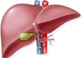
The introduction of the paper should explain the nature of the problem, previous work, purpose, and the contribution of the paper. The contents of each section may be provided to understand easily about the paper. (10) In the Human body Liver is a vital internal organ. It is a metabolic organ in the body and perform different functions ie produce a protein, cholesterol and bile acid. Livers main job is to filter blood coming from the digestive tract, before passing it to the rest of the body. In metabolic function of the body, the circulatory system is used to circulate blood by the heart and carried out to the liver. Every minute 1-1.5 litres of blood is transported to the liver through portal veins. The hepatic artery brings oxygen-rich blood to the liver similarly the portal vein transports nutrient.[1-4]The portal veins are used to passed to the blood through the gastrointestinal tract and absorbed large amounts of nutrients. The primary factor of liver diseases are excessive consumption of drugs, poison and drinking too much alcohol. The liver affects different diseases like cirrhosis, hepatitis virus A,B,C , cancer etc. This diseases are affect on any age and no age limit. The main symptoms of this diseases are weakness, excessive weight loss or gain, pain in the upper right abdomen where the liver is present. Fig. 1 show the healthy liver that perform a normal functions[5].
Fig. 1. Structure of Healthy liver
less time consuming. This paper is formed as follows Section II presents preprocessing the, Section III explains the segmentation. Section IV explains the result.
II.
Preprocessing
Image Preprocessing is an crucial and challenging factor in medical image processing. It is use to process the image so that segmentation and feature extraction algorithm work correctly. There are different ways for pre process the image like image re-sampling, manual correction, gray scale contrast enhancement and noise removal. Most of the abdominal CT images are noisy and the edges of objects are not cleared enough in the image. The preprocessing is use to enhance the manipulation of datasets and also improve the visual appearance of images. Generally medical images like MRI, CT etc. always contain a large amount of noise caused by equipment, operator performance, and the environment, which can lead to serious inaccuracies[7-10]. So that filtering method is useful to improving the visual appearance and for removal of noise it gives more accurate values. Before applying filter, image sharpened is use to enhance quality of edges without adding new details and improve eye visualization[11-12].
A. Weiner Filter
The noise is removed using Weiner filter. Weiner filter preserves edges as well as high frequency areas. It takes 3 inputs: i).Image to be filtered, ii).Point Spread Function (PSF) with which noise is distributed and iii). Noise-to-Signal Ratio (NSR). This is basically a restoring method, which performs an optimal trade off between inverse filtering and noise smoothing [8]. Weiner filter is the MSE-optimal stationary linear filter i.e. it minimizes the mean square error (MSE) which is given as the difference between some desired response and the actual filter output. This filter is not suitable for non-stationary inputs. It perform image filtering adaptively by computing local image variance. It performs a little smoothing for large local variance. Weiner filter requires more time than linear filtering and works best provided that the noise is constant power additive noise such as Gaussian noise. The restoration process using this filter incorporates both the degradation function and statistical characteristic of noise.
S
η(u,v)=
N(u,v)
2= power spectrumof the noise
S
f(u,v)=
F(u,v)
2= power spectrumof the
undegraded
image
III.
Segmentation
International Conference on Innovations in Engineering, Technology, Science & Management – 78 | Page shape, and texture, surface visualization, compression image segmentation and image registration is used. Correct segmented results are very useful for the analysis, predication and diagnoses.
A. Watershed Transform
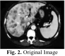
The Watershed is a powerful region based image segmentation algorithm. Watershed was introduced by Beucher and Lantuejoul, Since its introduction it has been widely studied and achieved encouraging results in image segmentation . Its basic idea is to consider image as a topography topology of geodesy Fig. 2, The pixel gray value of each point of image stands for the every local minimum value, altitude of that point, and its impact region are known as the collection basin and the borders of the basin form the watershed[11]. Intuitively, the watershed of a function is composed of the various locations from which a water droplet could flow towards different minima. The immersion process is simulated from the heights of local minima. The water level rises in each basin and when two basins meet, a watershed is created between them.
Fig. 2. Watershed Segmentation simplified in 2 dimension
IV.
Result
The procedure took for watershed segmentation is described details as follows[9]: Step 1: Convert image to grayscale and enhance the image contrast.
Fig. 2. Original Image Fig. 3. Histogram of image
Step 2: Use the Gradient Magnitude as the Segmentation Function.
Fig. 4. Gradient Magnitude Fig. 5. Watershed Transform
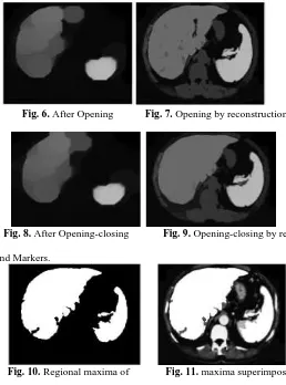
Fig. 6. After Opening Fig. 7. Opening by reconstruction
Fig. 8. After Opening-closing Fig. 9. Opening-closing by reconstruction
Step 4: Compute Background Markers.
Fig. 10. Regional maxima of Fig. 11. maxima superimpose
Opening –closing reconstruction on original image
Step 5: Modify the Segmentation Function.
Fig. 12. Modified regional Fig. 13. Thresholded opening- closing maxima. by superimposed on original image.
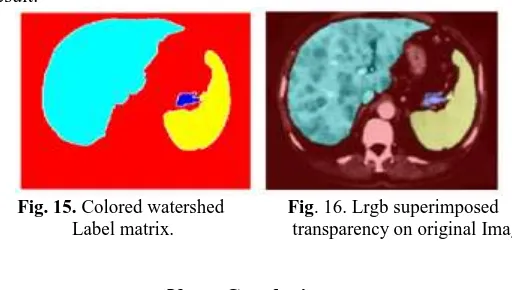
International Conference on Innovations in Engineering, Technology, Science & Management – 80 | Page Step 7: Visualize the Result.
Fig. 15. Colored watershed Fig. 16. Lrgb superimposed Label matrix. transparency on original Image
V.
Conclusion
In this paper, Image Segmentation method is important step in any image processing method can be proceeded. The CT scan image has noise. Preprocessing is a step to clear the image noise before applying the watershed segmentation and for this Weiner filter is used which gives good result as compared to other filters. This segmentation gives region of interest, minimum probability to identify the background and the affected region in CT scan images. It also used to separate the touching objects in the images. This method avoid the over segmentation and identification of main edge of the images. The proposed method gives 92.1% accurate result as comparing with other used techniques.
References
[1] Suhuai Luo, Jesse S. Jin, Stephan K. Chalup, Guoyu Qian, “A Liver Segmentation Algorithm Based on Wavelets and Machine Learning” The University of Newcastle, Australia 978-0-7695-3645-3/09 $25.00 © 2009 IEEE DOI 10.1109/CINC.2009.225. [2] Nasrul Humaimi Mahmood, Noraishikin Zulkarnain and Nor Saradatul Akmar Zulkifli “Ultrasound Liver Image
Enhancement Using Watershed Segmentation Method” International Journal of Engineering Research and Applications (IJERA) ISSN: 2248-9622 Vol. 2, Issue 3, May-Jun 2012
[3] Niket Amoda1, Ramesh K Kulkarni2 “Image Segmentation and Detection using Watershed Transform and Region Based Image Retrieval” International Journal of Emerging Trends & Technologyin Computer Science (IJETTCS) ISSN 2278-6856 Volume 2, Issue 2,March – April 2013
[4] Gunasundari S , Janakiraman S, “A Study of Textural Analysis Methods for the Diagnosis of Liver Diseases from
Abdominal Computed Tomography”, International Journal of Computer Applications (0975 – 8887)
Volume 74– No.11, July 2013.
[5] Yen-Wei Chen, IEEE Member, Masaki Kaibori, Tsukasa Shindo, Kousuke Miyawaki, Tsukasa Shindo, Amir H. Foruzan, Tomoko Tateyama, Xian- Hua Han, Kosuke Matsui, Takumi Tsuda, A-Hon Kwon, “Computer-Aided Liver Surgical Planning System Using CT Volumes”, 35th Annual International Conference of the IEEE EMBSOsaka, Japan, 3 - 7 July, 2013.
[6] Akanksha Sharma, Parminder Kaur, “Optimized Liver Tumor Detection and Segmentation Using Neural Network”, International Journal of Recent Technology and Engineering (IJRTE) ISSN: 2277-3878, Volume-2, Issue-5, November 2013.
[7] Smriti Sahu, Maheedhar Dubey, Mohammad Imroze Khan, Jitendra Kumar “Comparative Evaluation of Filters for Liver Ultrasound Image Enhancement”, International Journal of Emerging Trends and Technology in Computer Science (IJETICS) ISSN 2278-6856 Volume 2, Issue 1, January – February 2013.
[8] Belgherbi. Aicha, Bessaid. Abdelhafid “ Morphological Segmentation of the Spleen From Abdominal CT Images” I.J. Image, Graphics and Signal Processing,2014,456-62DOI: 10.5815/ijigsp.2012.04.08
[9] Suhuai Luo1, Xuechen Li1, Jiaming Li2 “Review on the Methods of Automatic Liver Segmentation from Abdominal Images”, Journal of Computer and Communications, 2014, 2, 1-7 Published Online January 2014 The University of Newcastle, Australia; 2The CSIRO Computational Informatics, Australia.
[10] Kanitkar, Sayali Satish, N. D. Thombare, and S. S. Lokhande. "Detection of lung cancer using marker-controlled watershed transform." Pervasive Computing (ICPC), 2015 InternationalConference on. IEEE, 2015.
[11] Anisha P R, Kishor Kumar Reddy C, Narasimha Prasad L V., “A Pragmatic approach for Detecting Liver Cancer using Image Processing and Data Mining Techniques”, Space (Spacecraft prelaunch automatic checkout equipment) -2015,Dept of ECE,K L University.