University of Pennsylvania
ScholarlyCommons
Publicly Accessible Penn Dissertations
1-1-2012
The Function of Hedgehog and Wnt Signaling
Pathways in Otic Development
Alexander Brown
University of Pennsylvania, [email protected]
Follow this and additional works at:http://repository.upenn.edu/edissertations
Part of theDevelopmental Biology Commons, and theGenetics Commons
This paper is posted at ScholarlyCommons.http://repository.upenn.edu/edissertations/495
For more information, please [email protected].
Recommended Citation
Brown, Alexander, "The Function of Hedgehog and Wnt Signaling Pathways in Otic Development" (2012).Publicly Accessible Penn Dissertations. 495.
The Function of Hedgehog and Wnt Signaling Pathways in Otic
Development
Abstract
The inner ear is a complex sensory organ essential for hearing and balance. During embryonic development, the inner ear depends on signaling information originating from the embryonic hindbrain to establish dorsoventral and anteroposterior identity. The Hedgehog (Hh) and Wnt signaling pathways are active in the hindbrain and implicated in otic development, but their exact mechanisms of action remained unclear. We investigated the function of Hh in ear development using a mouse model where we conditionally inactivated Hh signaling in the otic vesicle, a transient embryonic structure that gives rise to the inner ear, while leaving nearby Hh dependent tissues unaffected. We found Hh signaling within the otic vesicle functions to establish ventral otic identity and drive the proliferation of cochlear-vestibular ganglion (cvg) neuroblasts that will innervate the ear. We identified presumptive Hh target genes in the developing inner ear using microarrays. Several of these presumptive Hh targets are known to function in ear development or hearing. We also identified many novel targets that have not been characterized in the ear. Many of these novel presumptive Hh target genes are expressed in the ventral otic vesicle, a region that will give rise to the cochlear duct. To interrogate the function of Wnt signaling in ear development, we used a Wnt responsive inducible Cre recombinase (TopCreERT2) to genetically label cells at different stages of ear development. We found cells that make up dorsal, vestibular, structures and cvg neurons are Wnt responsive for prolonged periods of ear development. In the cochlear duct, we found both sensory and support cells originate from a Wnt responsive population. Surprisingly, we found the Wnt responsive population of cochlear progenitors was also labeled using a cre recombinase expressed from the Gbx2 locus. TopCreERT2 and Gbx2 expression overlap in the dorsomedial wall of the otic vesicle, suggesting this region is a likely source for auditory cells.
Degree Type
Dissertation
Degree Name
Doctor of Philosophy (PhD)
Graduate Group
Cell & Molecular Biology
First Advisor
Douglas Epstein
Keywords
Ear, Hedgehog, neurogensis, Wnt
Subject Categories
THE FUNCTION OF HEDGEHOG AND WNT SIGNALING PATHWAYS IN OTIC DEVELOPMENT
Alexander S. Brown
A DISSERTATION
in
Cell and Molecular Biology
Presented to the Faculties of the University of Pennsylvania
in
Partial Fulfillment of the Requirements for the
Degree of Doctor of Philosophy
2012
Supervisor of Dissertation:
___________________________
Douglas J. Epstein, PhD, Associate Professor, Genetics
Graduate Group Chairperson:
___________________________
Daniel S. Kessler, PhD, Associate Professor, Cell & Developmental Biology
Dissertation Committee:
Jonathan Raper, PhD, Professor, Neuroscience
Edward Morrisey, PhD, Professor, Cell & Developmental Biology Nadia Dahmane, PhD, Assistant Professor, Neurosurgery
THE FUNCTION OF HEDGEHOG AND WNT SIGNALING PATHWAYS IN OTIC DEVELOPMENT
COPYRIGHT 2012
Alexander Stanley Brown
This work is licensed under the Creative Commons Attribution-NonCommercial-ShareAlike 3.0 License
To view a copy of this license, visit
Dedication
I would like to dedicate this thesis to my parents, who have always supported
me, even when they didn’t understand what I was doing or why it seemed so
Acknowledgements
This this would not exist without the help of many people. I am deeply indebted to
my wife Christine, who fed me when I was too distracted to eat, who patiently
listened to my terrible ideas for experiments, and who tolerated the nights I
worked so late I didn’t come home. I’m also grateful to my advisors, Mike
Klymkowsky who convinced me to apply to graduate school, and Doug Epstein
who shepherded me through grad school and oversaw the work that makes up
ABSTRACT
THE FUNCTION OF HEDGEHOG AND WNT SIGNALING PATHWAYS IN OTIC
DEVELOPMENT
Alexander S. Brown
Douglas J. Epstein
The inner ear is a complex sensory organ essential for hearing and balance.
During embryonic development, the inner ear depends on signaling information
originating from the embryonic hindbrain to establish dorsoventral and
anteroposterior identity. The Hedgehog (Hh) and Wnt signaling pathways are
active in the hindbrain and implicated in otic development, but their exact
mechanisms of action remained unclear. We investigated the function of Hh in
ear development using a mouse model where we conditionally inactivated Hh
signaling in the otic vesicle, a transient embryonic structure that gives rise to the
inner ear, while leaving nearby Hh dependent tissues unaffected. We found Hh
signaling within the otic vesicle functions to establish ventral otic identity and
drive the proliferation of cochlear-vestibular ganglion (cvg) neuroblasts that will
innervate the ear. We identified presumptive Hh target genes in the developing
inner ear using microarrays. Several of these presumptive Hh targets are known
to function in ear development or hearing. We also identified many novel targets
that have not been characterized in the ear. Many of these novel presumptive Hh
target genes are expressed in the ventral otic vesicle, a region that will give rise
development, we used a Wnt responsive inducible Cre recombinase
(TopCreERT2) to genetically label cells at different stages of ear development.
We found cells that make up dorsal, vestibular, structures and cvg neurons are
Wnt responsive for prolonged periods of ear development. In the cochlear duct,
we found both sensory and support cells originate from a Wnt responsive
population. Surprisingly, we found the Wnt responsive population of cochlear
progenitors was also labeled using a cre recombinase expressed from the Gbx2
locus. TopCreERT2 and Gbx2 expression overlap in the dorsomedial wall of the
Attributions
The work described in chapter two was previously published in:
Brown, AS., Epstein, DJ. (2011). Otic ablation of smoothened reveals direct and indirect requirements for Hedgehog signaling in inner ear development.
Development 138, 3967-76.
Table of Contents
.
Dedication
!
iii
Acknowledgements
!
iv
.
Abstract
!
v
Attributions
!
vii
List of Tables
!
xi
List of Figures
!
xii
.
Chapter 1: Introduction
!
1
Function of the inner ear!1
Mechanosensory cells!1
The vestibular system!2
The auditory system!4
The Hedgehog signaling pathway!6
The Wnt Signaling Pathway!11
Development of the inner ear!13
Morphogenesis!13
Neurogenesis!17
. The prosensory domain!18
Chapter 2: Requirements for Hedgehog signaling in otic
development
!
19
Introduction!19
Materials and Methods!23
Results!25
Cochlear, but not vestibular, morphogenesis is dependent on direct Hh signaling within the otic epithelium!28
Direct Hh signaling within the otic epithelium establishes ventral otic identity!29
Ngn1 expression is not directly dependent on Hh signaling!32
Opposing roles for Wnt and Fgf signaling pathways in cvg neurogenesis !34
Shh is a mitogen for cvg progenitors!36
Auditory and vestibular neurons are specified in Smoecko embryos!38
Discussion!40
.
Chapter 3: Hedgehog target genes in otic development
!
47
Introduction!47
Materials and Methods!48
Results!51
Identification of genes with reduced expression in Smoecko mutants!51
Identification of putative Shh dependent transcription factors!52
Categorizing genes by co-expression!55
Ventral expression of down regulated genes!57
Conclusions!59
Chapter 4: A Fate Map of Wnt responsive cells
!
61
Introduction!61Materials and Methods!62
Results!64
Wnt responsive cells contribute to the cochlear duct!64
TopCre activity is present in multiple cochlear cell types!66
. Contribution of Gbx2 fate mapped cells to the cochlear duct!70
A population of TopCre+, Gbx2+, Ngn1- cells are cochlear sensory progenitors!70
Conclusions!72
List of Tables
...
List of Figures
...
Figure 1.1: Mechanosensory hair cells! 1
... Figure 1.2: Sensory patches of the inner ear! 3
...
Figure 1.3: The organ of Corti! 4
... Figure 1.4: The Hedgehog signaling pathway! 7
...
Figure 1.5: The Wnt signaling pathway! 11
...
Figure 1.6: Inner ear development! 13
Figure 2.1: Foxg1:Cre activity!...26 Figure 2.2: Inactivation of Hedgehog signaling in Smoecko otic vesicles!...27
... Figure 2.3: Cochlear morphogenesis depends on direct Hh signaling! 28 Figure 2.4: Loss of ventral otic markers in Smoecko embryos!...30
... Figure 2.5: Neurogenic patterning is indirectly regulated by Shh! 32 Figure 2.6: Extrinsic signals regulate Tbx1 expression!...35
... Figure 2.7: CVG formation depends on direct and indirect Hh signaling! 36
... Figure 2.8: A mitogenic role for Hh on cvg progenitors! 37 Figure 2.8: Specification of both spiral and vestibular neurons in Smoecko ...! 39
... Figure 2.10: A model of direct and indirect roles of Shh! 42 Figure 3.1: Loss of ventral tissue in Smoecko otic vesicles!...51
Figure 3.2 GO terms enriched in Smoecko!...52
... Figure 3.3 Relative expression of transcription modulators across tissues! 54
... Figure 3.4 Transcription effectors grouped by coexpression! 56 Figure 3.5 Genes with reduced expression in Smoecko are ventrally expressed!..58
... Figure 4.1: Temporal dependence of TopCre activity! 65 Figure 4.2: TopCre+ cells contribute to all cell types in the organ of Corti!...66
... Figure 4.3: Cell movement in the anterior otic vesicle! 68 Figure 4.4: Gbx2Cre/+ cells contribute to the cochlear duct!...69
Chapter 1: Introduction
Function of the inner ear
The ability to detect gravity, and its counterpart acceleration, is present
throughout the animal kingdom. To this end, different detection schemes have
been employed throughout evolution ranging from simple structures like
Johnston’s organ in insects1, 2 to the complex, multipart vertebrate inner ear.
Mechanosensory cells
! The basic information gathering unit of the inner ear is the
mechanosensory hair cell (Figure 1.1). These highly specialized cells are
polarized with actin based protrusions lining the apical surface. These actin
based microvilli, termed stereocilia, are the site of mechanosensation3, 4. The
distribution of stereocilia on the apical surface of the cell is not random. Instead,
they form in a cluster or chevron, as a result the majority of sterocilia have a
common orientation5. The common orientation of steociliary bundles makes hair
Figure 1.1: Mechanosensory hair cells
In the mouse, mechanosensory hair cells are located within the inner ear (A). Displacement is detected
cells most sensitive to particular vectors of displacement6, 7. The tips of stereocilia
are physically linked together, and this linkage is essential for hearing8. The tip
links do not force stereocilia to move as a group, a property due to their physical
structure independent of their tip links9. Instead, displacing the stereocilliary
bundle pulls on the tip links causing ion channels of uncertain identity to open.
The newly opened channels allow calcium and potassium ions to enter the cell10.
This influx of cations depolarizes the cell leading to a local increase of Ca2+ at
the base of the hair cell adjacent to the synapse. The local increase of calcium
causes the release of glutamate containing vesicles into the synapse11, 12,
completing the transduction of mechanical energy to neural impulse. The
stereociliary bundle is an exquisitely sensitive motion detector, displacing it as
little as 600 pm leads to detectible changes in membrane voltage potential13.
! Hair cells located in the inner ear synapse with neurons in the VIIIth
cranial nerve, which carries information to the auditory or vestibular nucleus in
the central nervous system. Although an individual sensory hair cell is capable of
transducing motion into neural impulses, the functions of hearing and balance
depend on a variety of additional cell types and the physical structure of the ear
itself.
The vestibular system
! In mammals, the inner ear contains six groups of sensory hair cells, five
that detect acceleration and one that detects sound (Figure 1.2). The structures
vestibulum. Vestibular
structures can be further
divided into the three cristae,
each housed in an ampulla
located at the base of a
semicircular canal, that
detect angular acceleration
(Fig. 1.2 blue shading), and
the utricule and saccule that
detect linear acceleration
and gravity (Fig. 1.2 green
shading). The hair cells of
the utriclular and saccular maculae are covered with otoconia, a mixture of
protein and CaCO3 crystals14 whose mass imparts inertia. The inertia of the
otoconia makes the utricle and saccule sensitive to gravity and linear
acceleration15. Alternatively, the cells in each crista are covered by a cupula, a
protein matrix that helps distribute the force imparted by circulating endolymph
within the semicircular canal. Angular acceleration, for example a turn of the
head, displaces endolymph within the canals which stimulates ampullar hair
cells7. The inner ear is the organ that perceives balance, and to do so each
vestibular structure must function correctly. Blocking only the formation of the
lateral semicircular canal severely disrupts balance in mice16, and improper
stimulation of the lateral ampulla in humans results in benign paroxysmal Figure 1.2: Sensory patches of the mammalian inner ear
The vestibulum contains structures to detect gravity (green), and angular acceleration (blue). The auditory apparatus (magenta) detects sound. Anterior semicircular canal (asc), lateral
positional vertigo (bppv). Fortunately, bppv is readily treated by a specific series
of head movements designed to reposition rouge otoconia particles that may
have drifted into the lateral canal17. The use of semicircular canals in the inner
ear is evolutionarily ancient with examples spread across at least 500 million
years of evolution ranging from lamprey to human18, 19.
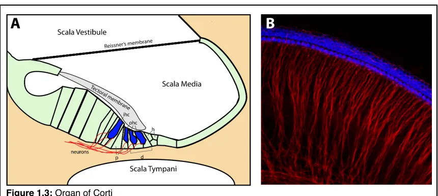
The auditory system
In mammals the auditory organ, the cochlear duct, contains a stripe of
mechanosensory hair cells along its length that respond to different frequencies
of sound (Fig 1.2 magenta shading). The structure of the cochlear duct, and
within it the organ of Corti that houses the hair cells (Figure 1.3), plays an
essential role in hearing. Amphibians and birds hear using an analogous
structure, the basal papilla, which also houses a collection of sensory cells but in
a different arrangement than the cochlea.
Figure 1.3: Organ of Corti
! The incredible sensitivity of the mammalian cochlea is due to three
characteristics: the ability of hair cells to detect tiny displacements, the physical
structure of the cochlea to dissect complex sounds into pure tones, and the
movement of outer hair cells to physically amplify sounds.
! The cochlear duct houses three fluid filled channels, the scala vestibule,
scala media, scala tympani (Fig. 1.3A). Vibrations that makeup sound are
transduced by the middle ear to generate waves of pressure in the endolymph of
the scala vestibule and scala media, which displace the basilar membrane
housing the organ of Corti. Each frequency of sound creates a different pressure
wave along the length of the cochlear duct. These different pressure waves,
maximally displace a unique region of the basilar membrane20, allowing a limited
section of cochlear to respond a unique frequency of sound. Frequency
selectivity creates a tonotopic map, where basal regions of the cochlear duct
respond to high frequency sound, while more apical regions respond to
increasingly lower frequencies. This tonotopic map is reflected in the innervation
pattern of the cochlear nucleus in the brainstem. Each frequency of sound
detected by different hair cells leads to a spatially distinct innervation pattern21.
! Although changing the mechanical properties of the basilar and tectoral
membranes alters resonant properties of the ear, the physical structure of the
cochlea is not the sole cause of hair cell stimulation. An additional active
amplification step22 increases sensitivity 100 fold using force generated by the
outer hair cells. For this amplification step, changes in transmembrane voltages
frequency that matches a sound stimulus23, 24. This increases the amplitude of
stimulation on the inner hair cell. These differences in hair cell properties, where
inner hair cells detect vibration and outer hair cells amplify vibration, are reflected
in their innervation patterns. Inner hair cells are synapsed by multiple afferent
spiral ganglion neurons, while multiple outer hair cells can be innervated by a
single efferent neuron (Fig. 1.3B).
! Of all the sensory systems, the inner ear has the finest temporal resolution
where hair cells respond on the order of microseconds25, and exquisite sensitivity
with the ability to detect acceleration as small as 10-6g26. Despite all the
complexities of the inner ear, its embryonic origin and development is controlled
by a limited number of cell signaling pathways. These pathways are often used
repeatedly during development to create remarkably different cells and tissues
depending on the time and context of signal activity. A variety of genetic studies
and embryo extirpation experiments support roles for the Hedgehog and Wnt
signaling pathways in establishing dorsoventral polarity in the ear27, 28, which in
turn, guides the formation of the vestibulum and cochlear duct29.
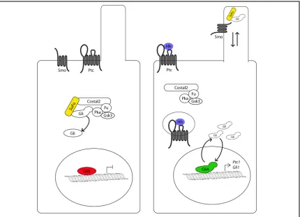
The Hedgehog signaling pathway
Since its discovery in the fruit fly Drosophila Melanogaster30, the Hedgehog
signaling pathway has been found to function in the patterning, proliferation and
frequently results in birth defects and a variety of cancers, making the pathway
medically important. In the context of the developing ear, the ligand Sonic
Hedgehog (Shh)29, 27, the transducing protein Smoothened (Smo)31, and the
transcriptional effectors Gli2 and Gli332 have been studied experimentally.
! Although core components of the Hedgehog pathway are conserved from
fly to human, important differences have evolved among species. In all cases,
the first step of a cell’s response to hedgehog signaling begins at the cell
membrane when the a ligand, Hedgehog in flies, or any of the three ligand Sonic Figure 1.4: The Hedgehog signaling pathway
In the cell on the left, the Hh pathway is inactive because it has not received any ligand. Gli is
phophorylated and proteolyitically cleaved into a transcriptional repressor. The cell on the right is exposed to ligand, allowing Ptc to clear from the cilia and be internalized. This allows Smo to move onto the cilia, and recruit Sufu and Gli to the cilia tip. Full length Gli protein accumulates, and a labile fraction is able to enter the nucleus and serve as a transcriptional activator. Two direct targets of Hh signaling are Ptc1 and
Hedgehog (Shh), Indian Hedgehog (Ihh), Desert Hedgehog (Dhh) proteins in
mammals33-35, binds a complex of receptor proteins. This receptor complex must
contain the twelve pass transmembrane protein Patched (Ptc), which
antagonizes Hh signal transduction in the absence of ligand36. In the absence of
Ptc, unrestrained Hh signaling occurs which can lead to lethal embryonic defects.
Less severe cases of Ptc disruption result in Gorlin’s syndrome which is
characterized by increased frequencies of basal cell carcinoma37, 38 and
medulloblastoma39. Ptc was originally characterized as the primary Hh receptor40,
41. However, an increasing number of co-receptors have been found to be
necessary for signal transduction, including Ihog and Boi42 in fly and their
mammalian homologs Cdo and Boc43, as well as vertebrate specific co-receptors
Gas144, 45 and LRP246.
! Structurally Ptc is similar to cholesterol transport proteins like NPC-147,
and bacterial RND permeases48. Despite the apparent similarity to membrane
transport proteins the exact mechanism of Ptc activity is unclear. However, Ptc
has been observed to function in a catalytic manner49 that may involve lipid or
sterol intermediates50. Further support for the idea that Ptc functions to modulate
the levels of small molecules comes from the structure of antagonists to the Ptc
target Smoothened (Smo). The first discovered Smo antagonist, cyclopamine51, a
steroidal alkaloid isolated from veratum californicum was identified for causing
cyclopia in offspring of pregnant livestock that consumed the plant5253. Additional
small molecule Smo antagonists have been discovered54, but their divergent
! Hh binding to the Ptc complex relieves antagonism of the eight pass
transmembrane protein Smoothened (Smo). Smo activity is essential for Hh
signal transduction55. At this point, the hedgehog pathway begins to diverge
between flies and vertebrates. In flies, active Smo antagonizes an intracellular
complex containing the kinesin Costal256, 57, the kinases PKA58 and Fused (Fu)59,
and the novel protein Suppressor of Fused (Sufu)60. When this protein complex
functions in the absence of Hh, it sequesters and phosphorylates the zinc finger
transcription factor Cubitus Interruptus (Ci)61, 62. Ci contains an N-terminal
transcriptional repression domain and a C-terminal activation domain, allowing it
to function as a transcriptional activator or repressor in a signal dependent
manner63, 64. The phosphorylation of Ci marks the C-terminus for degradation65
creating a truncated repressor isoform consisting of the N-terminal repressor
domain and DNA binding zinc fingers.
! In vertebrates, the primary cilium is an essential site of hedgehog
signaling. The cilium is a microtubule based organelle that protrudes from the cell
surface and functions as a signaling center and as a sensor for the local
environment. The requirement for cilia in Hedgehog signal transduction was
initially discovered in mouse embryos mutant for members of the IFT family of
ciliary transport proteins66, 67. These mutants had poorly formed or missing cilia
and phenotypically resembled embryos mutant for Gli transcription factors, the
three vertebrate homologs of Ci68. These observations lead to a model where: In
response to ligand, Ptc is displaced from the cilium69. Clearance of Ptc is
through an unknown mechanism71 leading to an accumulation of effector proteins
such as Sufu and full length Gli proteins, at the cilia tip72. Sufu directly interacts
with Gli proteins to promote the formation of truncated Gli repressor isoforms and
assists in forming labile Gli activator isoforms73. Divergently from flies, Sufu
serves as a prominent vertebrate inhibitor of hedgehog signaling74. However,
Sufu successfully antagonizes signaling in the absence of cilia75, calling into
question whether the accumulation of effector proteins at the cilia tip plays a
functional role in signal transduction. Other aspects of hedgehog signal
transduction appear well conserved. The Costal2 homolog Kif776-78 functions in a
complex with PKA, CK1 and GSK3 to mark Gli for degradation65, or to establish
labile full length transcriptional activator.
! The endpoint of the hedgehog signaling pathway is the differential
expression of target genes in response to relative levels of activator and
Gli-repressor proteins. Using differential activator/Gli-repressor activity generates many
possible responses to ligand and allows Hedgehog to function as a morphogen,
specifying different cell fates in a time and concentration dependent manner79.
Shh emanating from the floor plate and notochord specifies different classes of
neurons along the dorsoventral axis of the neural tube. In this case, different
concentrations of Shh are reflected in different amounts of activator or
Gli-repressor activity80-82. A similar logic is seen in the developing limb bud, where
Shh originating from the zone of polarizing activity (ZPA)35 establishes a gradient
of Gli3 repressor activity to specify individual digits83. The hedgehog receptor and
signaling, establishing a negative feedback loop84. This regulatory loop prevents
runaway signaling, and may function to help a cell interpret different levels
hedgehog ligand85.
The Wnt Signaling Pathway
The Wnt signaling pathway has many more ligands and receptors than the
hedgehog pathway, as well as multiple extracellular signaling inhibitors86-88. This
plethora of ligand, receptor, and inhibitor combinations feed into a pathway that
can have multiple readouts including changes in transcription, cytoskeletal Figure 1.5: The Wnt signaling pathway
In the canonical Wnt/β-catenin pathway the cadherin subunit β-catenin is phosphorylated and degraded
by a cytoplasmic destruction complex consisting of the scaffold protein Axin2, APC and Ck1, Gsk3β
kinases, while TCF/LEF family members bind DNA in complex with Groucho transcriptional repressors. In the presence of Wnt ligand, Dvl recruits Axin to the cell membrane disrupting the β-catenin destruction
complex. β-catenin accumulates and enters the nucleus where it displaces Groucho and activates
remodeling, and activation of heterotrimeric G proteins to modulate intracellular
calcium levels 89-92.
! Wnt ligands are lipid modified proteins93 that interact with one or more of
the 10 Frizzled (Frz) receptors94 and the obligate co-receptor LRP5/695-97.
Mammals have 19 Wnt ligands, which are expressed in partially overlapping
patterns, and there seems to be some variability as to how a given ligand or
receptor activates the Wnt pathway98. Once ligand is bound, Frz recruits
disheveled (Dlv)99, which in turn recruits the tumor suppressor adenomatous
polyposis coli (APC)100and the scaffold protein Axin2101. This recruitment of APC
and Axin to the cell membrane disrupts the catenin destruction complex. The
β-catenin destruction complex consisting of APC, Axin102, Glucose Synthase
Kinase 3β (Gsk3β)103, Casein kinase I (CKI)104-106 phosphorylates the
cytoskeletal protein β-catenin, leading to its ubiquitylation and degradation107.
The disruption of the destruction complex in the presence of Wnt signal leads to
an accumulation of β-catenin, allowing it to enter the nucleus and interact with
TCF/LEF transcription factors108-110. The recruitment of β-catenin to TCF/LEF
displaces Groucho corepressors111-113, activating transcription. This
transcriptional activation can be detected by increased expression of Axin114 or
the use of reporter constructs driven by synthetic promoters consisting of
multimerized TCF/LEF consensus binding sites115, 116.
! In addition to the canonical β-catenin mediated pathway, some Wnt
ligands function through the planar cell polarity (PCP) pathway during ear
signaling with the β-catenin pathway including Frz119 and Dlv120-122, but can signal
through a Rho123, Rac124, Jun-kinase125, cascade to control the cytoskeleton.
Ultimately, a cell displays polarized distribution of PCP effector proteins, where
Frz,Dlv are enriched in a domain distinct from the effector proteins Prk, and
Vangl126-128. This molecular polarity is then reflected in the shape and
organization of the cell giving rise to tissue wide properties.
! In vertebrates, the most striking examples of tissue wide organization due
to PCP signaling are convergent extension movements necessary for neural tube
closure, cochlear duct outgrowth, and the the orientation of hair cells within the
cochlear duct.
Development of the inner ear
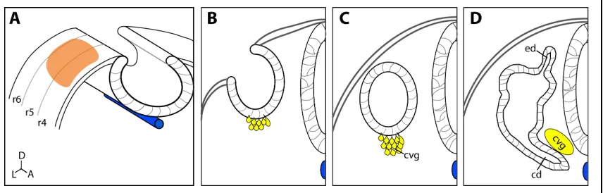
Morphogenesis
A variety of fate mapping experiments in chick and mouse reveal that an
overwhelming majority of the cells that make up the inner ear come from a
FIgure 1.6 Early otic Development
Transverse sections through the developing hindbrain show ear development starts as the otic placode (A, orange). As development progresses the placode invaginates to form the otic cup (B) and neuroblasts begin to delaminate (yellow). The otic cup closes to form the otic vesicle (C), and delaminating
common embryonic origin, the ectoderm adjacent to the developing hindbrain.
Series of heterotopic grafting experiments, where different regions of ectoderm
are replaced within age matched embryos, revealed that amphibians and fish
have large regions of ectoderm competent to contribute to the ear. This
competency is largely due to the activity of several Wnt and Fgf signals, whose
identity varies between organisms. In zebrafish Fgf8 and Fgf3 are necessary for
otic induction129, a role filled in the mouse by Fgf3 and Fgf10130, 131, and Fgf19
and Wnt8c in chick132. In mouse, the region of otic competency is gradually
restricted and a portion of the ectoderm thickens creating the otic placode by 10
somites of age. The choice between otic placode and cranial ectoderm seems to
be governed by Wnt/β-catenin signaling, as ectopic activation of the pathway
leads to expanded placodes at the expense of ectoderm and inhibited Wnt
signaling results in microvesicles that arrest early in development133.
! As otic development progresses roughly to the 15 somite stage, the
placode beings to invaginate forming the otic cup. Fate mapping experiments in
chick show that cells are already organized into presumptive dorsal, ventral,
anterior and posterior regions at this time point134. This presumptive
regionalization is reinforced by extirpation experiments in salamander and chick,
where rotating the otic placode resulted in defects in structures along the
anteroposterior axis, but not along the dorsoventral axis. Rotating the otic
vesicle a few hours later resulted in defects along the dorsoventral axis135, 136.
These results suggest that the anteroposterior axis becomes fixed in
reprogrammed from an initial pattern to that of its host suggests the developing
ear receives positional information from nearby tissues. Although the invaginating
otic placode displays regionalized gene expression and restricted cell fates, there
remains a possibility for additional cell movement. Cells have been observed
migrating into the otic vesicle137, 138, while fish forgo otic cup formation all
together, instead relying on cavitation to hollow a mass of cells into the otic
vesicle139.
! The factors responsible for axis specification and the development of otic
structures may vary between species. In mouse and chick, Wnt signals
emanating from the dorsal hindbrain28 and Hedgehog emanating from the ventral
neural tube and notochord29, 27 establish the dorsoventral axis in the otic vesicle,
while a wave of retinoic acid signaling imparts anteroposterior polarity140.
Conversely, in frogs and fish, hedgehog activity largely establishes posterior
identity141-143, and promotes ventromedial identity144 while Fgf signaling
establishes anterior identity145. This is use of different signaling pathways to
create an evolutionarily conserved organ is somewhat puzzling. A role for Hh
signaling in auditory development is common among each of these examples,
yet significant differences remain. One commonality is that Hh is required for the
cochlear duct in mammals, and the fish auditory organ, the posterior macula.
Additionally, in both mice and fish Hh promotes the proliferation of cvg
progenitors. Yet Shh antagonizes hair cell formation in mice146, while Hh
promotes late forming saccular hair cells in fish through the regulation of atoha1
of convergent evolution. These molecular differences likely help explain the
dramatic morphological differences in auditory structures between tetrapods and
teleost fish, but more study is required for a full understanding of the molecular
mechanisms that pattern the otic vesicle.
! The ventral outgrowth that gives rise to the cochlear duct is governed by
at least two overlapping signaling pathways. Hedgehog signaling within the otic
vesicle establishes ventral otic identity establishing gene expression patterns to
support cochlear duct outgrowth31, 27. As the cochlear duct develops, Gli-activator
activity is required for full elongation32. Cochlear duct outgrowth also depends on
convergent extension governed by Wnt5a118, Wnt7117, and multiple PCP effector
proteins147-149. Although Wnt and Hh both coordinate cochlear duct outgrowth,
there are distinct differences in their mutant phenotypes. Altering Hh activity
truncates the cochlear duct and induces ectopic patches of sensory hair cells, but
does not cause hair cell orientation defects within the organ of Corti146.
Perturbing PCP signaling also truncates the cochlear duct, but differs from Hh by
randomizing hair cell orientation within the organ of Corti.
! The dorsal outgrowths that give rise to the semicircular canals will
undergo an even more dramatic series of morphogenetic changes. The initial
domain of the canals is defined by the expression of the homeobox transcription
factor Dlx5150. The cells that makeup a region within the growing out pouches,
termed the fusion plate, will ultimately die or be resorbed into the canal proper.
The action of the fusion plate depends on the expression of Netrin1 (Ntn1)151, but
Nonetheless, Ntn1 expression is necessary for the wave of apoptosis152 and
possibly cell movement that create the canals. Surprisingly, canal formation does
not seem to be affected by mutations in PCP components, indicating that the
dramatic morphogenesis that occurs in the developing canals functions
independently of PCP signaling.
Neurogenesis
! Like the majority of inner ear cells, the neurons that innervate the ear trace
their origin to the otic placode. The presumptive neurogenic domain that contains
the cells that create in the anteroventral region of the otic placode and vesicle.
This region is initially defined by the expression of the neurogenic master
regulator Neurogenin 1 (Ngn1)153. Cells within this neurogenic domain proliferate,
then express NeuroD154 and begin to delaminate from the otic vesicle. After
delaminating, neuroblasts begin to express Islet-1 (Isl1)155 and cease to
proliferate. The newly delaminated neuroblasts aggregate to form the
cochlear-vestibular ganglion (cvg), which will ultimately split, giving rise to the spiral
ganglion innervating the cochlea and Scarpa’s ganglion innervating the
vestibulum. The exact process that selects a neuroblast for auditory of vestibular
fate is poorly understood. Fate mapping studies showed auditory neurons are
generated slightly later in development than vestibular neurons156, and birth
dating studies reached similar conclusions157.
! During development an overabundance of inner ear neurons are
generated. These excess neurons compete for survival factors expressed by
maturation. These include brain derived neurotrophic factor (bdnf) and
neurotrophin 3(Nt3). Mutants lacking both factors have an almost complete loss
of inner ear neurons158. The amount of pruning following this overproduction of
neuroblasts is remarkable. In the cvg up to half of all neurons born will fail to
receive sufficient trophic support die during development159, 160.
The prosensory domain
! The medial wall of the developing cochlear duct contains a region fated to
give rise to auditory hair cells, termed the prosensory domain. This region
consists of an equivalence group defined by the expression Sox2161 and Notch
signaling components. The presumptive hair cells express high levels of notch
ligands Dll1162, Jagged2, Jagged1163, which signal through Notch1 in adjacent
support cells164. Differential notch activity in the prosensory domain limits the
number of sensory hair cells through a classic lateral inhibition mechanism. As
presumptive hair cells begin to differentiate, they begin to express Atoh1, a factor
necessary and sufficient for hair cell fate165, 166, while cells with high levels of
Notch activity become support cells, and maintain high levels of the cell cycle
inhibitor P27kip1167, 168. Several factors in addition to Sox2 and Notch function
within the prosensory domain. Shh has been shown to antagonize Notch activity
in the prosensory domain and limit the number of sensory hair cells146, and Wnt/
β-catenin signaling is sufficient to transform auditory hair cells to a vestibular fate
in chick169. Regardless of the identity of input signals, the entire prosensory
domain undergoes terminal mitoses by e15.5157, and further growth growth is
Chapter 2: Requirements for Hedgehog signaling
in otic development
Introduction
The mammalian inner ear is a sensory organ with dual roles in sound and
motion detection. The partitioning of these functions within the inner ear to
auditory and vestibular components occurs early in embryonic development,
allowing each of these senses to operate independently170. The auditory portion
of the inner ear, the cochlea, derives from the ventral outgrowth of the otic
vesicle, which progressively extends and coils as it matures. Mechanosensory
hair cells lining the cochlear duct from base to apex respond to sound waves in a
tonotopic manner, and transmit information along auditory (spiral) neurons to
sound processing centers in the brain21, 171. Vestibular structures, on the other
hand, mostly derive from dorsal out-pockets of the otic vesicle and through
incompletely understood mechanisms are sculpted into the three semicircular
canals, utricle and saccule170, 172. Sensory patches associated with each of these
structures detect angular movements of the head (semicircular canals) and linear
acceleration along the horizontal (utricle) and vertical (saccule) planes. Vestibular
neurons innervating each of these sensory patches transmit sensory information
to visual, vestibular and proprioceptive centers to coordinate balance173.
The hindbrain is a critical source of signals necessary for dorsoventral
vestibular components29, 174-176, 28, 177. Members of the Wnt and Hedgehog (Hh)
families play prominent roles in establishing dorsoventral identity within the otic
epithelium. Wnt1 and Wnt3a secreted from the dorsal hindbrain regulate the
expression of dorsal otic determinants, such as the homeodomain transcription
factors Dlx5 and Dlx6 28, 178. Consequently, vestibular morphogenesis is
completely impaired in Wnt1-/-;Wnt3a-/- mutants28. Sonic hedgehog (Shh),
secreted from the floor plate of the hindbrain and notochord, opposes the
dorsalizing effects of Wnts by repressing Dlx5, and activating ventral otic genes,
including the transcriptional regulators Otx2 and Pax227, 28. The failure to regulate
the ventral otic program in Shh-/- embryos results in cochlear agenesis179-181, 27, 182. Interestingly, Shh-/- embryos also display profound deficits in vestibular
development including, malformations of the semicircular canals, utricle, saccule
and endolymphatic duct. Each of these morphological defects can be traced back
to alterations in otic vesicle patterning genes27. For example, the misexpression
of Otx1 and Gbx2 in the Shh mutant otocyst likely explains the absence of the
lateral semicircular canal and endolymphatic duct, respectively183, 184.
Shh also functions in inner ear neurogenesis. The cochlear and vestibular
neurons that make up the VIIIth cranial nerve originate from progenitors in the
anteroventral region of the otic vesicle that express Ngn1, a neural determinant
required for their specification153. The establishment of the neurogenic domain is
one of the earliest signs of asymmetry along the anteroposterior axis of the otic
vesicle. The T-box containing transcription factor Tbx1, is expressed in a
to the anterior portion of the otocyst185. Shh-/- embryos show a significant
reduction in Ngn1 expression, suggesting a possible involvement in the
regulation of anteroposterior identity within the otic vesicle, although the
underlying mechanism has not been elucidated27.
What remains uncertain from these previous studies is the extent to which
the inner ear phenotype in Shh-/- embryos can be attributed to a direct loss of
Shh signaling within the otic epithelium versus an indirect consequence that the
absence of Shh has on tissues surrounding the inner ear. The hindbrain and
periotic mesenchyme are sources of other signals essential for inner ear
development that are also disrupted in Shh-/- embryos29, 186, 176, 187, 27, 28, 188, 189.
Thus, their misregulation could also explain the inner ear defects observed in
Shh-/- mutants.
The best evidence in support of Shh acting directly on the otic epithelium
comes from the observation that Gli1, a transcriptional target of the Shh pathway,
is expressed in a graded manner along the dorsoventral axis of the otocyst, with
higher levels detected ventrally, closer to the source of Shh, and lower levels
tapering off dorsally32. While suggestive, this result does not resolve the
functional significance of this signaling gradient. The analysis of single and
compound mutants in Gli2 and Gli3, the transcriptional mediators of Shh
signaling, support a model whereby reciprocal gradients of Gli activator and Gli
repressor function are required to shape inner ear morphology along the entire
dorsoventral axis in response to Shh32. Of particular interest was the finding that
removing a wild type allele of Gli3 (Shh-/-;Gli3+/-). This result suggests that Shh
promotes vestibular morphogenesis by reducing Gli3 repressor function32.
However, it does not address the tissue specificity of this action. Recovery from
the vestibular defects in Shh-/-;Gli3+/- embryos could equally be explained by the
reduction of Gli3 repression in the inner ear as it could the neural tube, which
also shows improvements in patterning and morphology compared to Shh
-/-embryos32, 81.
In order to distinguish between the primary requirements for Shh in inner
ear development from its secondary roles in surrounding tissues, we generated
conditional mutants in which Smoothened (Smo), an essential Hh signal
transduction component, was selectively inactivated in the otic epithelium
(Smoecko). Our results demonstrate that Shh acts directly on the otic epithelium to
regulate ventral target genes that are necessary for the outgrowth of the cochlear
duct and saccule. On the other hand, the development of dorsal otic derivatives
is indirectly dependent on Shh, as these vestibular structures were absent or
malformed in Shh-/- mutants but maintained in the ears of Smoecko embryos. The
role of Hh signaling in cochlear-vestibular ganglion (cvg) formation is more
complex, as it is dependent on both direct and indirect signaling mechanisms.
Our data suggest that the loss of cvg neurons in Shh-/- animals is partly due to an
increase in Wnt responsiveness in the otic vesicle (indirect signaling), resulting in
the ectopic expression of Tbx1 in the neurogenic domain and subsequent
repression of Ngn1 transcription. An unanticipated role for Shh as a mitogen for
signaling). These data contribute to a better understanding of the intrinsic and
extrinsic signaling properties of Shh during inner ear development.
Materials and Methods
Animals
Foxg1Cre/+ and Smoloxp/loxp mouse lines were described elsewhere190, 191. Smoloxp/ loxpmice were maintained on a mixed Swiss-Webster, C57BL6/J background. Shh +/- 192 and RosaGfp/Gfp193 mice were obtained from Jackson Labs (Bar Harbor,
ME). Tbx1+/- mice were provided by J. Epstein194. Topgal mice were provided by
E. Fuchs115.
Immunohistochemistry
For immunohistochemistry, embryos were fixed in 4% paraformaldehyde for 1
hour, cryoprotected in 30% sucrose overnight, mounted in OCT embedding
media (Sakura Finetek Torrence, CA) and snap frozen. Embryos were sectioned
at 14 μm and stained with DAPI and the following antibodies: Mouse anti-Islet 1
(DSHB) 1:100, Rabbit anti-Phospho Histone H3 (Cell Signaling Technology,
Danvers, MA) 1:1000, Rabbit anti-cleaved caspase 3 (Cell Signaling Technology)
1:1000, Rabbit anti-MyosinVIIa (Proteus Biosciences Ramona, CA) 1:300,
Mouse anti-Neurofilament (DSHB) 1:200, Chicken anti-GFP (Aves Labs, Tigard,
OR) 1:1000, Mouse anti-Gata3 (Santa Cruz Biotechnology, Santa Cruz, CA)
1:50. Primary antibodies were detected with one of the following secondary
antibodies: Donkey anti-mouse IGG conjugated to Cy3 (Jackson
Donkey anti-Rabbit IGG conjugated to Cy3 or Alexa488; or Goat anti-Chicken
IGG conjugated to Alexa488.
In situ hybridization
For section in situ hybridization, embryos were processed in the same manner as
for immunohistochemistry. Sections were rehydrated in PBS containing 0.1%
Tween-20, and hybridization was performed as in 195. For antibody detection after
in situ hybridization the following modifications were made: Proteinase K
treatment was omitted. After completion of the BM purple reaction, slides were
washed three times in PBS-Tween, fixed for 10 minutes with 4%
paraformaldehyde then washed three times in PBS-Tween. Slides were then
incubated in primary antibody and the immunohistochemistry protocol was
followed. Whole mount in situ hybridization was carried out as in 196 using
digoxigenin –UTP labeled riboprobes.
Embryo culture
Embryo roller culture was performed as described in 197. Briefly, E9.5 embryos
were collected in ice-cold L-15 media without damaging the yolk sac. Embryos
were grown under 95% O2: 5% CO2 at 37°C in 100% rat serum (Gemini
Bio-Products, West Sacramento CA) supplemented with 0.175 mg/ml glucose, 2 mM
glutamine, 1x Penn-Strep. Embryos were re-gassed every 12 hours. LiCl
treatment: embryos carrying a TopGal transgene were cultured with increasing
amounts of LiCl to determine an optimal concentration (50 mM LiCl) that
inhibitor: 3mg/ml EMD341603 dissolved in DMSO was added to culture media to
a final concentration of 25µM.
Inner ear paint fill
Inner ear paint fills were performed essentially as described in 172, with the
exception that White-out Plus (Bic Corp. Milford CT) was used to fill the inner
ears instead of latex paint.
Cell counts
The total number of cells in the cvg was determined by counting Isl1+, cRet- cells
in each sequential section through the entire otic vesicle. Bright field images of
section in situ hybridizations were inverted, assigned a color and merged with
DAPI and antibody channels in Image J. Cells were hand counted using the cell
counter plug-in in Image J.
Area measurements
To determine the percent of otic vesicle expressing Tbx1, the area of positive
staining in lateral whole-mount views was traced in ImageJ and measured, and
then divided by the total area of the otic vesicle.
Results
Inactivation of Hedgehog signaling in the otic epithelium
To determine the specific requirements of Hedgehog (Hh) signaling in the
inner ear, we generated embryos in which a floxed allele of Smo (Smoloxp), an
essential mediator of Hh signaling, was selectively inactivated in the otic
particularly advantageous for our
studies because it is active in all otic
progenitors well in advance of when
Shh signaling is known to be required
in the otic vesicle190, 27. Moreover, cre
showed negligible expression in tissues
surrounding the otic vesicle including
the neural tube and periotic
mesenchyme (Fig. 2.1). In all
experiments described below, at least
three to five Foxg1cre/+; Smo
loxp/-embryos (herein referred to as Smoecko
for ear conditional knockout of Smo)
were compared to an equal number of control littermates (Foxg1cre/+; Smoloxp/+
and Smoloxp/-). No differences were seen in ear morphology or vesicle patterning
between Foxg1cre/+ and Foxg1+/+ genotypes.
We first assessed the effect of deleting Smo in the inner ear by examining
the expression of Gli1 and Ptc1, two transcriptional targets of Hh signaling. In
control embryos, Gli1 expression initiated weakly at E9.5 in the ventral most
region of the otic vesicle (Fig. 2.2A, n=4). At this stage, Ptc1 was not yet detected
in the otic epithelium despite its strong expression in other Shh responsive cell
types, including the ventral neural tube and periotic mesenchyme (Fig. 2.2C,
n=3). By E10.5, Shh signaling intensified resulting in robust Gli1 and Ptc1 Figure 2.1: Foxg1Cre activity
A transverse section through an E10.5 Foxg1Cre/ +;RosaGfp/+embryo stained for GFP (green), Isl1
staining in ventral regions of the otic vesicle and along the medial wall in a
ventral (high) to dorsal (low) gradient (Fig. 2.2E,G,n=3 and 3, respectively) and
32. Smoecko embryos consistently failed to express Gli1 and Ptc1 in the otic
epithelium at both stages analyzed, yet robust expression of these markers was
observed in the neural tube and periotic mesenchyme (Fig.
2.2B,D,F,H,n=4,3,3,3). Therefore, the disruption to Hh signaling was both
specific and complete in the inner ears of Smoecko embryos.
Figure 2.2:Inactivation of Hedgehog signaling in Smoecko otic vesicles.
In situ hybridization for Gli1 (A,B,E,F) and Ptc1 (C,D,G,H) on transverse sections through the otic vesicle of control and Smoecko embryos. Arrowheads indicate staining within the otic epithelium. At E9.5, Gli1 was
detected in the otic epithelium of control (A), but not Smoecko (B) embryos. Ptc1 was not detected in the
otocyst of either control (C), or Smoecko (D) embryos at this stage. By E10.5, both Ptc1 and Gli1 were
detected in the otic epithelium of control (E,G), but not Smoecko (F,H) embryos. Ptc1 and Gli1 were also
detected in the neural tube and periotic mesenchyme of control and Smoecko embryos. Abbreviations: D,
Cochlear, but not vestibular, morphogenesis is dependent on direct Hh
signaling within the otic epithelium
Shh-/- embryos show profound vestibular and auditory defects including
cochlear agenesis, missing or malformed semicircular canals, as well as absence
of the utricle, saccule and endolymphatic duct (Fig. 2.3A,C). If these defects are
wholly attributed to the loss of Shh signaling in the otic epithelium, then they
should be recapitulated in Smoecko embryos. On the other hand, if some, or all, of
these phenotypes result from secondary consequences of perturbing Shh
signaling in tissues surrounding the inner ear, then they should be milder in
Smoecko embryos.
We visualized the gross anatomy of Smoecko and control inner ears by
paint-fill at E15.5 (Fig. 2.3A,B). At this stage, the morphology of the inner ear has
reached near full maturity in wild type embryos. The vestibulum, comprising the Figure 2.3: Cochlear, but not vestibular, morphogenesis is directly dependent on Hh signaling.
Medial view of inner ear paint fills at E15.5. (A) Control inner ears reveal the morphology of the anterior, posterior and lateral semicircular canals (asc,psc,lsc), endolymphatic duct (ed), common crus (cc) utricle (u) saccule (s), and cochlear duct (cd). (B) Smoecko inner ears lacked a cochlear duct and saccule, but all
other structures were present. (C) Shh-/- inner ears possessed an anterior semicircular canal, but all other
three semicircular canals, utricle, saccule, endolymphatic duct and common crus
were readily discerned, and the cochlear duct had elongated and coiled 1.5 turns
(Fig. 2.3A). Ventral ear structures, namely the cochlear duct and saccule, were
entirely absent in Smoecko embryos (n=14 ears), a phenotype similar to that
observed in Shh-/- mutants (Fig. 2.3B,C). Remarkably, all dorsal otic derivatives,
including the semicircular canals, utricle and endolymphatic duct, were present in
Smoecko embryos (Fig. 2.3B). The appearance of dorsal vestibular structures in
Smoecko embryos contrasts with the pronounced vestibular dysmorphology
observed in Shh-/- mutants and suggests that dorsal otic derivatives are not
directly dependent on Shh for their development. Conversely, the consistent loss
of ventral inner ear structures in Smoecko and Shh-/- embryos suggests that Shh
signaling, acting directly on the otic epithelium, is required for cochlear duct
outgrowth and saccule formation.
Direct Hh signaling within the otic epithelium establishes ventral otic
identity
At E10.5, the otic vesicle displays regionalized patterns of gene
expression that mark competency domains for subsequent development into
distinct adult structures170, 198. Several of these otic patterning genes are
misexpressed in Shh-/- embryos27. In order to distinguish the genes that are
dependent on Hh signaling within the otic epithelium from those that are
misregulated due to the secondary effects of disrupting Shh in neighboring
Pax2, Otx2 and Gata3 are three transcription factors expressed in partially
overlapping domains in the ventral otocyst, which are necessary for cochlear
duct development179, 180, 199, 200, 181, 182. Pax2 is broadly expressed throughout the
otic placode before becoming restricted to the ventromedial wall of the otic
vesicle (Fig. 2.4A and data not shown). Otx2 is also expressed in the ventral
region of the otocyst (Fig. 2.4B). The pattern of Gata3 expression at early stages
of otic development is dynamic, but is then localized to the elongating cochlear
duct and spiral ganglion (Fig. 2.4C) and 199. Each of these genes was previously
Figure 2.4: Loss of ventral otic markers in Smoecko embryos.
In situ hybridization on transverse sections through the otic vesicle of control (A,B,C,D,E,F), Smoecko
(G,H,I,J,K,L) and Shh-/-(M,N,O,P,Q,R) embryos at E10.5. Arrowheads point to ventral otic expression of
(A)Pax2, (B)Otx2, and (C)Gata3 in control embryos, which was absent in Smoecko (G,H,I) and Shh -/-(M,N,O) embryos. Otx1 expression in the lateral wall of the otocyst of control embryos (D) was not altered in Smoecko mutants (J), but shifted ventrally in Shh-/-(P). The expression of dorsal otic markers Gbx2 (E,K) and Dlx5 (F,L) was similar between control and Smoecko embryos. The ventral extent of Dlx5
shown to be downregulated in Shh-/- embryos (Fig. 2.4 M-O) and 27. A
comparable reduction in the expression of Pax2, Otx2 and Gata3 was observed
in Smoecko embryos (Fig. 2.4G-I, n=3), suggesting that Shh signaling within the
otic vesicle is required for ventral otic identity and subsequent cochlear duct
morphogenesis.
Otx1 is expressed in the lateral wall of the otic vesicle at E10.5 and is
required for lateral semicircular canal formation (Fig. 2.4D)183, 201, 202. A
significant ventral shift in the expression of Otx1 was observed in the otic vesicle
of Shh-/- embryos (Fig. 2.4P), which likely explains the absence of the lateral
semicircular canal in these mutants27. In Smoecko embryos, Otx1 was properly
localized to the lateral wall of the otocyst, indicating that Hh signaling within the
otic epithelium is not required for lateral otic identity (Fig. 2.4J, n=3). This result
also suggests that the lateral semicircular canal defect in Shh-/- embryos is an
indirect consequence of perturbing Shh signaling in tissues adjacent to the inner
ear.
The additional vestibular dysmorphologies observed in the ears of Shh
-/-mutants can also be explained by patterning changes in the otic vesicle. For
instance, the expression of Gbx2, a homeodomain containing transcription factor
required for endolymphatic duct formation184, is not maintained in the
dorsomedial otocyst of Shh-/- mutants (Fig. 2.4Q) and 27. Moreover, the dorsal
otic expression of Dlx5, a homeodomain containing transcription factor required
for semicircular canal development203, 150, 204 is expanded ventrally in Shh-/-
suggested that Shh was necessary for the expression of certain dorsal otic genes
(Gbx2), while antagonizing the expression of others (Dlx5, Topgal). However, the
regulation of these dorsal otic genes by Shh appears to be indirect as neither is
misexpressed in Smoecko embryos (n=3) (Fig. 2.4E,F, K,L). These data also
indicate that the antagonistic interaction between Shh and Wnt signaling
pathways responsible for setting up the dorsoventral axis of the otocyst does not
stem from a cell intrinsic mechanism within the otic epithelium, but must reside
from interplay between these pathways outside of the ear.
Ngn1 expression is not directly dependent on Hh signaling
The neurons that make up the VIIIth cranial nerve and innervate the
sensory patches within the inner ear originate from a common progenitor pool in
the anteroventral region of the otic vesicle. The bHLH transcription factor, Ngn1,
Figure 2.5: Neurogenic patterning is indirectly regulated by Shh.
Lateral surface views of embryos stained by whole mount in situ hybridization for Ngn1 (A-D), and Tbx1
(E-H) at E9.5 arranged by genotype. In control embryos, Ngn1 (A) marks the neurogenic domain,
whereas, Tbx1 (E) shows a complementary pattern of expression. In Shh-/- embryos, Ngn1 expression
was reduced (B), while Tbx1 was expanded into the anterior otocyst (F). Shh-/-;Tbx1+/-embryos, showed
restored expression of Ngn1 (C), despite the partial expansion of Tbx1 into the presumptive neurogenic domain (G). Smoecko embryos revealed a similar pattern of expression for Ngn1 (D), and Tbx1 (H)
is expressed in these neuroblast progenitors (Fig. 2.5A), and is required for their
specification153. The spatial restriction of Ngn1 to the anteroventral otic domain is
mediated, in part, by the repressive action of Tbx1, a T-box containing
transcription factor expressed in a complementary pattern to Ngn1 (Fig. 2.5E)
and 185. In Tbx1-/- embryos, Ngn1 is ectopically expressed resulting in the
posterior expansion of neuroblast progenitors185.
Previous studies demonstrated that Ngn1 expression is greatly reduced in
Shh-/- embryos, causing a significant reduction to the size of the
cochlear-vestibular ganglia (cvg) (Fig. 2.5B, n=3) and 27. However, the mechanism
underlying the regulation of Ngn1 transcription by Shh was unclear. Novel insight
to this problem came from our observation that Tbx1 expression had expanded
into the neurogenic domain of Shh-/- mutant otic vesicles (Fig. 2.5E-G, n=5). This
raised the possibility that the failure to repress Tbx1 is an effect of the loss of
Shh, and not Ngn1 function. Alternatively, the downregulation in Ngn1 may have
prompted the expansion of Tbx1. Since Tbx1 was not expanded in the otic
vesicle of Ngn1-/- mutants, the latter prospect was ruled out (data not shown). To
address the former possibility, we generated embryos lacking a wild type allele of
Tbx1 on a Shh-/- mutant background. We reasoned that if Tbx1 was responsible
for the repression of Ngn1 in Shh-/- embryos, then reducing its dosage should
restore Ngn1 transcription. Notably, the pattern of Ngn1 expression in
Shh-/-;Tbx1+/- embryos was greatly enhanced compared to Shh-/- mutants and
closely resembled that of controls (Fig. 2.5C, n=3). Thus, Shh indirectly regulates
We next determined whether the repression of Tbx1 from the neurogenic
domain was a direct or indirect action of Shh on the otic epithelium. Both Tbx1
and Ngn1 were properly localized to their respective otic territories in Smoecko
embryos (Fig. 2.5D,H, n=3 and 6, respectively), arguing that their misregulation
in Shh-/- mutants was a secondary consequence of disrupting Shh signaling in
tissues extrinsic to the inner ear.
Opposing roles for Wnt and Fgf signaling pathways in cvg neurogenesis
If Shh is not acting directly on the otic epithelium to regulate the
anteroposterior positioning of the neurogenic lineage, then what is the
responsible signal(s)? Select members of the Wnt and Fgf families appeared to
be excellent candidates based on prior studies. For instance, Wnts secreted
from the dorsal hindbrain were shown to partially suppress the neurogenic
lineage28. Whereas, Fgfs were shown to both repress and activate neuronal
determinants in the otic epithelium205-207. In Fgf3-/-; Fgf10-/- mouse embryos,
neuroblast progenitors were ectopically expressed in the posterior otocyst207.
Conversely, the pharmacological inhibition of Fgf signaling in the chick otic
vesicle caused a dramatic reduction in the expression of Ngn1 and NeuroD and a
corresponding loss of cvg neurons206. These seemingly contradictory results
may be attributed to species-specific differences in Fgf signaling activity and/or
temporal differences in Fgf ligand utilization.
To investigate whether modulation of Wnt or Fgf signaling pathways could
embryos in the presence or absence of the canonical Wnt signaling agonist, LiCl
208, or Fgf signaling antagonist, EMD 341608 209, and assayed the expression of
Ngn1 and Tbx1. Wild type embryos harvested at E9.5 and cultured in control
media for 18 hours showed proper anterior and posterior expression of Ngn1 and
Tbx1, respectively (Fig. 2.6A,D, n=8/9 and 17/20, respectively). However, when
embryos were cultured in either LiCl (n=5/5) or EMD 341608 (n=6/8), they
showed a consistent and profound downregulation of Ngn1 in the anterior otocyst
(Fig. 2.6B,C). These results confirm that Wnt signaling antagonizes, while Fgf
signaling is necessary for, Ngn1 expression in the mouse otocyst. Interestingly,
the anterior otic expansion of Tbx1 was only observed in embryos cultured in LiCl
(n=6/8), and not EMD 341608 (n=0/15) (Fig. 2.6E-G, P<0.001, unpaired t-test).
Thus, heightened Wnt signaling better recapitulated the a/p polarity defects
observed in Shh-/- embryos than did Fgf inhibition. The upregulation of canonical
Figure 2.6: Extrinsic signals regulate Tbx1 expression.
Lateral surface views of embryos stained by whole mount in situ hybridization for Ngn1 (A-C), and Tbx1
(D-F), after being cultured for 18 hours in the presence of control media (A,D), 50 mM LiCl (B,E), or 25
μM of the FgfR inhibitor EMD 341603 (C,F). Control embryos showed a normal pattern of Ngn1 (A) and