R E S E A R C H A R T I C L E
Open Access
Minimal detectable difference of the finger
and wrist range of motion: comparison of
goniometry and 3D motion analysis
Lisa Reissner
1*, Gabriella Fischer
2, Renate List
2,3, William R. Taylor
2, Pietro Giovanoli
1and Maurizio Calcagni
1Abstract
Background:The measurement of finger and wrist range of motion (ROM) is of great importance to clinicians
when assessing functional outcomes of therapeutic interventions and surgical procedures. The purpose of the study was to assess the repeatability of ROM measurements of the hand joints with manual goniometer and 3D motion capture system and to calculate the minimal detectable difference for both methods.
Methods:Active finger and wrist joints ROM of 20 healthy volunteers were assessed using a manual goniometer
and 3D motion capture system. Minimal detectable difference (MDD) and standard error of measurement (SEM) were calculated for both measurement systems and compared within the same task. Maximal ROM of all joints was registered twice on two different days to evaluate the test-retest repeatability. The intraclass correlation coefficients (ICC) was calculated and examined to determine if reliability≥0.70 existed.
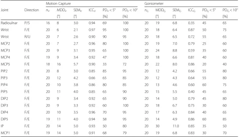
Results:MDD for the 3D motion capture was between 5 and 12° except for the metacarpophalangeal joint (MCP)
1, interphalangeal joint (IP), and MCP5. SEM values lay between 2 and 4° for all joints except for the MCP5, IP, and MCP1. For the goniometric measurements, MDD and SEM were between 12–30° and 4–11°, respectively. The reliability criterion (ICC > 0.7) was achieved for the ROM measurement with the 3D motion capture system for 94% of the joints and in only 65% of the joints with the manual goniometer.
Conclusions:Joint ROM assessed with 3D motion analysis showed higher test-retest agreement demonstrating
overall better repeatability for this method. Because of the smaller measurement error, the 3D motion capture system has a smaller MDD. Only individual test-rest differences bigger than the MDD can be considered as real changes, and therefore, in an experimental situation, the use of a more precise measurement method can greatly reduce the number of subjects needed for a statistical significance. Goniometer measurements of some joints should be carefully interpreted, due to a low repeatability and reliability.
Trial registration:This study is approved by the Ethical Committee Zurich (Kek-ZH-Nr: 2015-0395).
Keywords:Manual goniometer, 3D motion capture, Range of motion, Motion analysis, Minimal detectable
difference
© The Author(s). 2019Open AccessThis article is distributed under the terms of the Creative Commons Attribution 4.0 International License (http://creativecommons.org/licenses/by/4.0/), which permits unrestricted use, distribution, and reproduction in any medium, provided you give appropriate credit to the original author(s) and the source, provide a link to the Creative Commons license, and indicate if changes were made. The Creative Commons Public Domain Dedication waiver (http://creativecommons.org/publicdomain/zero/1.0/) applies to the data made available in this article, unless otherwise stated.
* Correspondence:[email protected]
1Division of Plastic Surgery and Hand Surgery, University Hospital Zurich,
Introduction
The measurement of finger and wrist postures is one of the important parameters for the clinicians when assessing the outcomes of therapeutic interventions and compare them. Joint angular measurements are also essential for hand therapists to record the pro-gress of rehabilitation. It is therefore important for clinicians and researchers to have complete and rele-vant information on the accuracy, repeatability, and reliability of these measurements. While the manual goniometer is commonly used in clinical practice as a tool to measure joint angles, 3D motion capture sys-tems are increasingly applied in research to measure hand motion [1–5]. Moreover, 3D motion capture systems allow the dynamic evaluation of all hand joints simultaneously [6–8]. They determine the pos-ition of skin markers highly accurate. The main ad-vantages of the manual goniometry are that it is cheap, fast, and does not require data post-processing or joint angle calculations, and the main drawback is that it relies on rater’s performance for the quality of measurements. For evaluative instruments that are used to measure changes in the same subject over time, the ability to detect minimal clinically important differences is essential. Hence, it is fundamental to know the size of the measurement error is required not only for the selection of the appropriate measure-ment tool, but also for the interpretation of the data and the comparison between different studies.
Trained therapists generally have adequate intrarater reliability for the measurement of wrist and finger pos-tures; however, some joints are easier to assess [9–12]. When standard goniometry is used, variability between 2 and 7° occurs in joint angle measurements of the hand [13–15]. The validation of goniometer measurement was done in splinted positions, which means that the force applied on the joint by the examiner was neutralized. This is not the case in real life where joints are examined looking for the actual angles and not a predefined one [16, 17]. Moreover, there are few studies comparing manual goniometer measurement and 3D motion cap-ture, but none of them taking into account all the joints of the hand [16].
Sample size calculation (power) is a standard require-ment for high-quality studies. Minimal detectable differ-ence (MDD) and standard error of measurement (SEM) are among the most important parameters for its calcu-lation. If we can reduce them, this will result in a smaller sample size.
Therefore, the aim of this study was to assess the re-peatability of ROM of the hand joints with manual goni-ometer and 3D motion capture system and to calculate the minimal detectable difference and the standard error of measurement for both methods.
Material and methods
Participants and protocol
Finger and wrist joints motion of 20 healthy right-hand dominant volunteers (ten men, ten women) with mean age 28 years (SD 4.7) were assessed with 3D motion capture system and mea-sured with manual goniometer. In order to assess the test-retest repeatability for both methods, each par-ticipant was tested on two different days. The same hand surgeon performed all goniometric measure-ments, and the same examiner placed the skin markers in both sessions. The local ethics committee approved the study, and all participants provided written informed consent for their data to be used for this analysis (Kek-ZH-Nr: 2015-0395).
Manual goniometric measurements
The protocol of the goniometric measurement followed the recommendations of the American Society for Hand Therapists for the wrist joint and the finger joints [17–19]. Each volunteer placed their elbow on the table with the forearm in neutral position. Dorsal placement of a plastic goniometer (Zimmer®) on the wrist was applied. For the measure-ment of the distal interphalangeal joint (DIP), raters instructed patients to maximal extend the metacarpo-phalangeal joint (MCP) while maximal flexion of the proximal interphalangeal (PIP) joint and DIP joint, making a hook fist. During pronation and supination of the forearm, the volunteer was sitting with the shoulder in 0° of flexion, extension, abduction, and rotation so that the upper arm was close to the side of the body. The elbow was in 90° of flexion, and the goniometer was placed just proximal to the radial and ulnar styloid process while performing maximal pro-nation and supipro-nation.
3D motion capture system and setup
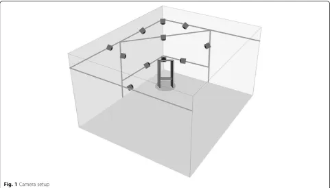
An optoelectronic motion capture system consisting of 11 fixated infrared cameras (VICON® MX3+ and VICON® MX3 motion capture system, Oxford Metrics Ltd., UK) and the corresponding software VICON®-Nexus (version 2.3) were used for data collection. The capture volume was approximately 50 × 50 × 50 cm3, and the cameras were positioned such that the markers were always visible by at least two cameras, avoiding hiding of markers (Fig. 1). The cameras have a reso-lution of 659 × 493 pixel, and recordings were carried out with a frequency of 100 Hz.
had a diameter of 9 mm and 5 mm, respectively. For the hand and fingers, hemispherical markers with a diameter of 3 mm were chosen. We attached the markers with a skin-compatible adhesive tape.
Motion tasks
First, a natural reference position with the hand lying on a flat surface and a 40° wedge between the second finger and the thumb was recorded. After-wards, each volunteer performed a set of basic mo-tion tasks:
– Pronation-supination (P/S) of the forearm – Flexion-extension (F/E) and radial-ulnar deviation
(R/U) and of the wrist
– Combined F/E in the MCP, PIP, and DIP joints of the long fingers (make a fist)
– F/E of the PIP and DIP joints of the long fingers (without movement in the MCP joints)
– F/E of the thumb
The tasks aimed to detect joint ROM in a single move-ment plane. Furthermore, they were used to calculate func-tional joint axes and centers. Each trial started and ended with the hand in a neutral position and consisted of three cycles (e.g., flexion-extension-flexion-extension-flexion-ex-tension). Five valid repetitions of each task were recorded per session.
Data processing and data analysis
For the kinematic description of the hand, 18 seg-ments were defined and considered as rigid bodies. At least three markers per segment are needed to allow kinematic analysis in all three movement planes. The kinematic model of the fingers was based on the assumption that only motion around the flexion axis is possible for the PIP and DIP joints. Therefore, kinematics of the long fingers could be assessed with a reduced marker concept using only two longitudinally aligned markers per segment. The F/E angles were calculated by means of the vectors between the markers of each finger segment similar to Metcalf et al. but with a marker proximal or distal of the joint defining the segment vector instead of markers on the joint [20, 21].
standards defined by the International Society of Biomechanics [23, 24].
After checking recorded data for errors, the data were summarized using descriptive statistics. All analyzed pa-rameters and their abbreviations are defined in Table1. For all abbreviations of these parameters, a subscripted character G refers to the goniometer measurement and subscripted character V refers to the analysis by means of the motion capture system.
For the dynamic trials recorded by the motion cap-ture system, the minimum and maximum joint angle (e.g., maximum flexion and extension position) was determined for each trial and averaged within the five trials of the same session to obtain the ROM. The individual test-retest difference (DIFF) of the ROM was determined for each subject. To quantify the test-retest repeatability of the ROM, SEM and intra-class correlation coefficient (ICC) were calcu-lated within the same task and method for all joints.
It is recommended that ICC values need to be greater than or equal to 0.70 to be considered acceptable as a clinically meaningful measurement tool [25]. Ac-cording to de Vet et al., the SEM represents measure-ment error and equals the square root of the variance of differences [26]. Only changes of the ROM that ex-ceed the variability induced by the method can be regarded as real changes [27]. Therefore, based on the SEM, the minimal detectable difference (MDD) of both methods was calculated. In addition, the per-centage of subjects with absolute test-retest differ-ences below 5° and 10° (PD < 5°/PD < 10°) were determined for both methods, respectively.
Statistical analysis
Two-tailed ttests (p≤0.05) were performed to compare motion analysis and goniometry regarding the mean test-retest difference (meanDV vs. meanDG) as well as
the average ROM (MeanromV vs. MeanromG). For a
healthy population, we assumed ROM to be constant over time. Therefore, DIFFG and DIFFV are considered
as an estimation of measurement error, and a limit of 5° was set based upon the accuracy of the manual goniom-eter shown to be around 5° in literature [28]. The null hypotheses were that:
(H01) Within the same method, meanD equals zero,
(H02) DIFF lies within 5°, and (H03) the ROM is
equiva-lent when measured with the goniometer or the motion capture system.
H01 was rejected, if the absolute value of the ratio of
meanD and standard deviation of difference (SDD) ex-ceeds the 5% level of agreement (|T| > 1.96; p≤0.05). H02 was rejected, if the estimated precision represented
by the SEM was > 5°.
Results
For the 3D motion capture system, no valid joint angle could be calculated for four subjects at the radioulnar joint and for one subject at the MCP5 joint due to issues with visibility of markers or a lost marker at the elbow. Furthermore, one subject had a misplaced marker on the thumb, affecting MCP1 cal-culations, and one subject had a shifted marker affect-ing MCP4 and MCP5 angle calculations. Therefore, these values had to be excluded from further data analysis after visual inspection of the recorded data. The available data are reported in the second column of Table 2.
There was a wide range of maximum ROM among the healthy subjects (SDromV 9°, SDromG 10°) (Tables 3
and 4). The two-tailed t test revealed significant (p< 0.05) differences of the Meanrom derived from the
MeanromG and MeanromV differ by almost 57°, with
the lower values measured with the 3D motion cap-ture system.
The results of the test-retest parameters are pre-sented in Table 2. No statistically significant
difference (H01) of the ROM (p level≤0.05) was
found between the first and second measurement for both methods in all joints.
For the 3D motion capture method, MDDV lay
be-tween 5 and 12° except for the MCP1, IP, and MCP5.
Table 2Test-retest results of goniometer and 3D motion analysis measurement
Motion Capture Goniometer
Joint Direction nV MDDV SEMV ICCV PDV< 5° PDV< 10° nG MDDG SEMG ICCG PDG< 5° PDG< 10°
[°] [°] [%] [%] [°] [°] [%] [%]
Radioulnar P/S 16 8 3.0 0.94 69 100 20 19 6.8 0.35 45 65
Wrist F/E 20 6 2.1 0.97 95 100 20 18 6.4 0.87 50 75
Wrist R/U 20 7 2.6 0.90 90 95 20 18 6.5 0.72 55 65
MCP2 F/E 20 7 2.7 0.96 80 100 20 19 7.0 0.79 25 60
MCP3 F/E 20 9 3.1 0.95 65 100 20 24 8.8 0.59 35 60
MCP4 F/E 19 9 3.4 0.92 47 100 20 18 6.6 0.81 40 60
MCP5 F/E 18 16 5.7 0.90 33 72 20 22 8.0 0.86 20 40
PIP2 F/E 20 8 3.0 0.85 85 95 20 12 4.2 0.66 55 80
PIP3 F/E 20 12 4.2 0.66 65 85 20 12 4.3 0.64 55 80
PIP4 F/E 20 10 3.8 0.86 80 85 20 13 4.6 0.60 60 75
PIP5 F/E 20 11 4.0 0.85 65 90 20 15 5.5 0.40 45 65
DIP2 F/E 20 9 3.4 0.92 65 90 20 14 5.0 0.79 45 80
DIP3 F/E 20 9 3.3 0.92 60 100 20 18 6.7 0.75 30 60
DIP4 F/E 20 10 3.5 0.96 70 95 20 17 6.3 0.84 40 65
DIP5 F/E 19 11 4.0 0.94 58 95 20 14 4.9 0.86 60 85
IP F/E 20 14 5.0 0.93 50 80 20 30 11.0 0.85 35 50
MCP1 F/E 19 14 5.0 0.91 68 79 20 19 6.8 0.83 30 70
Table 1Kinematic parameters
Parameter Abbreviation Description/formula
Measurement parameter-determined for each subject in measurement session 1 + 2 (test-retest)
Range of motion ROMG1/G2,ROMV1/V2 Maximum range of motion measured within a method and session
Test-retest difference DIFFG/DIFFV Individual test-retest difference of ROM: DIFFG= ROMG1−ROMG2/
DIFFV= ROMV1−ROMV2
Test-retest and repeatability parameter—between test and retest within the same method over all subjects
Mean difference meanDG/meanDV Mean DIFFG/DIFFV
Standard deviation of the difference SDDG/SDDV Standard deviation of DIFFG/DIFFV
Standard error of measurement SEMG/SEMV Estimated measurement precision according to de Vet24:
SEM = SDD/√2
Minimal detectable difference MDDG/MDDV MDD = 1.96 × SDD
% within 5° or 10° PDG< 5° or < 10°/PDV< 5° or < 10° Percentage of subjects with an absolute |DIFFG|/|DIFFV| smaller than
5° or 10°
Intraclass correlation coefficient ICCG/ICCV Intraclass correlation coefficient (3,1) of the test-retest measurement
of the ROM
Method comparison—between measurement methods
Mean ROM MeanromG/MeanromV Mean ROM of test and retest of each subjects averaged over all
subjects
Table 3Range of motion (mean and SD) for goniometer and 3D motion capture measurement
Measurement day 1 Measurement day 2
Motion Capture Goniometer Motion Capture Goniometer Pvalue
Joint Direction MeanromV1 SDromV1 MeanromG1 SDromG1 Pvalue MeanromV2 SDromV2 MeanromG2 SDromG2
[°] [°] [°] [°] [°] [°] [°] [°]
Radioulnar P/S 116 10 174 8 < 0.0001 114 10 170 8 < 0.0001
Wrist F/E 150 9 141 14 0.570 150 10 139 12 0.499
Wrist R/U 51 7 56 10 0.663 51 6 58 10 0.573
MCP2 F/E 109 10 114 12 0.787 109 9 116 13 0.663
MCP3 F/E 114 10 114 11 0.991 113 10 115 13 0.902
MCP4 F/E 113 11 115 10 0.883 110 10 119 14 0.644
MCP5 F/E 115 16 120 14 0.795 112 13 123 19 0.624
PIP2 F/E 118 6 109 6 0.275 118 6 113 7 0.617
PIP3 F/E 119 6 109 6 0.234 118 6 112 7 0.487
PIP4 F/E 122 7 109 6 0.186 121 8 112 7 0.411
PIP5 F/E 105 7 101 5 0.705 106 9 103 8 0.775
DIP2 F/E 80 10 84 9 0.751 83 10 84 9 0.899
DIP3 F/E 93 9 95 12 0.927 95 9 94 9 0.921
DIP4 F/E 84 13 87 12 0.882 85 13 89 12 0.833
DIP5 F/E 89 13 89 10 0.996 88 12 90 11 0.906
IP F/E 93 14 103 23 0.720 92 13 104 21 0.643
MCP1 F/E 72 15 71 14 0.947 70 12 72 11 0.964
Significant difference between motion capture and goniometer measurement
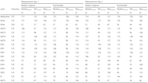
Table 4Range of motion (median and range) for goniometer and 3D motion analysis measurement
Measurement day 1 Measurement day 2
Motion Capture Goniometer Motion Capture Goniometer
Joint Direction MedianromV1 MinromV1 MaxromV1 MedianromG1 MinromG1 MaxromG1 MedianromV2 MinromV2 MaxromV2 MedianromG2 MinromG2 MaxromG2
[°] [°] [°] [°] [°] [°] [°] [°] [°] [°] [°] [°]
Radioulnar P/S 117 101 136 172 160 190 114 99 137 170 156 182
Wrist F/E 151 129 166 141 102 168 150 127 165 139 105 160
Wrist R/U 52 37 62 57 42 82 52 40 62 58 44 84
MCP2 F/E 108 91 132 112 90 138 108 94 127 116 96 136
MCP3 F/E 112 98 137 115 98 134 112 99 133 115 96 138
MCP4 F/E 112 100 138 115 96 134 107 93 130 119 100 148
MCP5 F/E 116 90 151 121 98 151 110 93 137 123 94 162
PIP2 F/E 118 110 132 108 98 118 119 109 128 113 102 132
PIP3 F/E 120 109 129 108 98 118 118 108 131 112 102 126
PIP4 F/E 123 110 135 108 100 120 122 105 133 112 100 122
PIP5 F/E 105 90 120 102 84 110 106 83 119 103 90 114
DIP2 F/E 79 65 98 84 66 104 84 66 100 84 62 96
DIP3 F/E 92 77 107 94 70 114 98 81 109 94 76 108
DIP4 F/E 87 58 103 89 68 114 85 61 105 89 64 104
DIP5 F/E 93 61 107 91 74 108 88 70 108 90 70 110
IP F/E 93 67 114 101 66 152 92 58 111 104 70 160
For the goniometric measurements, MDDGwas between
12 and 30°.
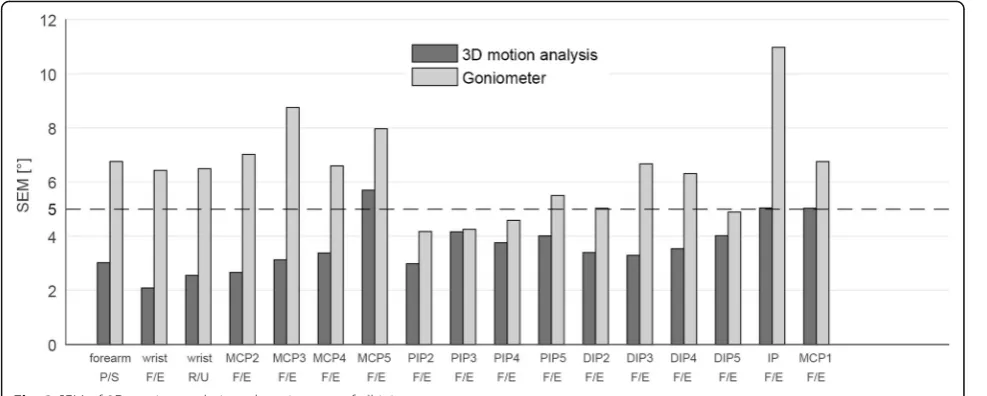
The observed precision of the measurement repre-sented by the SEM is displayed in Fig. 3 and Table 2. SEMV lied below the limit of 5° for all joints except
for the MCP5 (SEMV 5.7°), IP (SEMV 5.0°), and
MCP1 (SEMV 5.0°). For the goniometric
measure-ments, SEMG exceeds the limit of 5° (SEMG 5.0–
11.0°) in all joints, except for PIP2-4 and DIP5 (SEMG
4.2–4.9°).
ICC ranged from 0.35 to 0.87 (ICCG) and 0.66 to 0.97
(ICCV) for the goniometric and 3D motion capture
mea-surements, respectively (Table 2). Six out of 17 joints (radioulnar, MCP3, PIP2-5) did not achieve the reliability criterion with the manual goniometer. In comparison, the ICCVvalue for the 3D motion capture system was higher
for all degrees of freedom, and the ROM measurements with the motion capture system met the reliability criter-ion for all joints except for the PIP3.
Overall, 43% and 67% of the goniometric measure-ments had a test-retest difference below 5° (PDG< 5°)
and 10° (PDG< 10°), respectively (Table 2). The
corre-sponding percentages for the 3D motion capture sys-tem were 67% (PDV< 5°) and 92% (PDV< 10°),
respectively. The DIP5 was the only joint for which slightly more volunteers had an angular difference of less than 5° for the goniometric measurements compared to the 3D motion capture system. For all other joints, the 3D motion capture system had a higher percentage of individuals with small (< 5°) inter-session differences.
Discussion
In this study, we assessed the repeatability of ROM measurements of the hand joints with 3D motion
capture system, compared them with manual goniom-etry, and calculated the MDD for both methods. We measured all joints of the fingers and the wrist.
We observed method differences from −57 to + 11°, where negative values indicate higher ROM when ex-amined with the goniometer (MeanromG> MeanromV).
Since the true value of the joint angle is unknown, the comparison between the two methods serves as the first step in the validation of the new motion analysis protocol. Skin movement relative to the bone is the biggest source of error in motion ana-lysis with skin markers [29, 30]. Longitudinal rota-tions are more affected from skin movement artifacts, therefore leading to an under-/overesti-mation of the joint angle in motion analysis [30, 31]. In agreement, we found the highest method dis-agreement (57°) and the only significant difference for the pronation-supination movement. Difficulties to measure the radioulnar joint with a goniometer (ICC 0.35G, SEMG 6.8°) might have further
contrib-uted to the large difference between the methods. Armstrong et al. suggest that the lack of precision in goniometric measurement could be technique related, as the current method of measuring true forearm rotation involves placing a flat goniometer along the curved surface of the flexion/extension crease of the wrist [32]. The observed significant difference between the methods for measuring forearm rotation indicates that adjust-ments to the methodology are necessary. Schmidt et al. propose a procedure to reduce the influence of skin movement artifacts by looking at the hand rotation during pronation-supination instead of the rotation of the forearm itself [33]. Implementation of such simple corrections might further improve the presented meas-urement method.
A comparison with a gold standard, such as an im-aging technique, would be a possibility to estimate the accuracy of the measurements. However, the val-idity of the data is beyond the scope of this article. Still, our results reveal that the measurement system used to obtain the ROM has to be considered for clinical data interpretation. Therefore, a specific norm database for every method is highly recommended.
n a clinical setting, methods are often used to evaluate the effects of interventions or monitor changes over time within the same subject. There-fore, a focus on agreement parameters is recom-mended by de Vet et al. [26]. The SEM and MDD express measurement error in the same unit as the original value, which facilitates clinical interpretation. In contrast to ICC, SEM and MDD are not influ-enced by variability among the sample [34]. Hence, their values can be transferred to various groups of patients.
Averaged over all analyzed joints, the MDDV was
10°, compared to 18° MDDG. Therefore,
measure-ments by means of a motion capture system allow us to recognize smaller changes in joint mobility than with goniometer. This means that we have to be very careful in the clinical setting to interpret a change in the ROM as a true change, or just as a measurement error. For the wrist joint flexion/exten-sion and radial/ulnar deviation, MDDG was 18°.
Macedo and Magee examined the passive ROM of the wrist with a universal goniometer in 12 healthy adults. They found a MDDG for the wrist flexion of
11° and for the wrist extension of 8°, which is lower than our MDDG, but higher than our MDDV [35].
The MDDG for the finger joint lied between 12°
and 24°. Ellis and Bruton examined the finger joints with a goniometer, but in a splinted position, so they had MDD (reported as 95% confidence interval of difference) of between 4° and 5° [1].
Overall, the calculated precision of the ROM mea-surements was SEMV 3.6° and SEMG 6.4° for the 3D
motion capture system and goniometer, respectively. The mean values of all repeatability parameters indi-cate higher test-retest agreement for the 3D motion capture method. For the wrist joint, the SEMG with
the goniometer was 6.4° (F/E) and 6.5° (R/U). LaS-tayo and Wheeler assessed the passive ROM of the wrist with a universal goniometer in 120 patients with wrist conditions. They reported SEM between 5.6° and 8.1°, like Macedo and Magee with SEM be-tween 2.9° and 7.4°, compare to Horger who calcu-lated a SEM between 2.6° and 4.5° [12, 35, 36]. Our results consider the ROM of the movement, so both measurement points (e.g., maximum flexion and
extension position) are affected from independent error associated with the placement of the goniom-eter, whereas the other studies showed the results of each direction separately.
For the finger joints, SEMG was between 4.2° and
8.8°. Stam et al. evaluated 20 healthy subjects with a goniometer while holding cylinders with different diameter and had a SEM between 4° and 6°, similar to our results [37].
In comparison with previous repeatability goniom-etry studies, the intrarater reliability for the active ROM of the middle finger found in our study lies within the range of the intra- and interrater reliabil-ity (ICC 0.43 to 0.99) determined by Lewis et al. [2]. Solgaard et al. assessed intraobserver SDD for the goniometry of the wrist of 5.2–8° [38]. Compared to these findings, our results for wrist goniometry are slightly higher. In contrast, the 3D motion capture of the wrist ROM had better repeatability than the goniometry results in both studies.
Compared to previous measurements by 3D mo-tion capture, we found excellent test-retest reliability on the wrist (ICC 0.90–0.97). The corresponding values in Levanon et al. were only good (ICC 0.77– 0.83) [39]. In contrast, the root-mean-square error in our study was 5.3°, whereas Sancho-Bru et al. found smaller errors in repeatability (3.4°) [8]. In that study, a different marker set was used and the re-peatability was assessed for grasping different objects while we analyzed the maximum ROM. It is possible that variability of the ROM movement is bigger than in specific grasping tasks, but to quantify the source of error, a validation would be needed.
Limitations and achievements
In this study, we do not simply compare two different measurement tools but rather two different measurement procedures. Therefore, the comparison includes methodo-logical differences in addition to the measurement system itself, which might have influenced the reported repeatability.
study, but rather as an advantage of the motion ana-lysis method.
The main difference between the two protocols was the amount of measurements of each joint angle. When using the motion capture system, the maximum ROM of the dynamic trials could be aver-aged within the session, which might have compen-sated for outliers. In contrast, with the manual goniometer, each parameter was measured only once per session, as otherwise the rater could recall the values. This difference has likely contributed to the better results for the 3D motion capture system. Still, the study implements both methods such as they are usually applied in a clinical setting. It quan-tifies the test-retest repeatability of a realistic appli-cation, where usually a single surgeon or therapist measures the ROM to monitor change during treat-ment. We are aware that we cannot make a statement about the accuracy of both methods, the 3D motion capture system and the goniometry. A comparison with a radiological examination would be necessary for this. As a first step, we concentrated on the re-peatability of both methods and on their comparison, since repeated tests to assess changes are very com-mon in the clinical setting and the research [26].
We missed few measurements because of marker loss. This could happen while they are attached only with a double-sided adhesive tape, and can get lost or displaced, which is a disadvantage in motion ana-lysis. The advantage of the 3D motion capture sys-tem is the dynamic evaluation of the wrist and all finger joints simultaneously. Therefore, it can be ap-plied for the assessment of the ROM as well as dy-namic functional tasks, such as activities of daily living. The main advantages of the manual goniom-etry are that it is much easier to implement in the clinical setting. Our study shows that in applications where the goniometer is not precise enough, motion analysis is a possible alternative due to its lower MDD. The choice of the method has to be in ac-cordance with the research question and the ex-pected or clinically relevant change in joint ROM.
Conclusion
In conclusion, the MDD of the 3D motion capture system is smaller than of the goniometer measure-ment. This is particularly important in an experi-mental setup where a higher degree of precision is requested. In the clinical research, better MDD per-mits relevant reduction of the sample size.
Abbreviations
DIFF:Test-retest difference; DIP: Distal interphalangeal joint; ICC: Intraclass correlation coefficient; IP: Interphalangeal joint; MCP: Metacarpophalangeal
joint; MDD: Minimal detectable difference; PIP: Proximal interphalangeal joint; ROM: Range of motion; SEM: Standard error of measurement
Acknowledgements
We would like to thank Vera Beckmann and the Hand Therapy Department of the University Hospital of Zurich for their support in this study.
Funding
This research received no specific grant from any funding agency in public, commercial, or not-for-profit sectors.
Availability of data and materials
The datasets generated and analyzed during the current study are available in the password-protected System of the University Hospital Zurich repository. The datasets used and/or analyzed during the current study are available from the corresponding author on reasonable request.
Authors’contributions
LR, GF, and RL captured, analyzed, and interpreted the data and wrote the manuscript. MC and PG supervised and created parts of the manuscript. WRT wrote part of the manuscript and corrected it. MC was the principal investigator of the study and acted as a supervisor of LR. All authors read and approved the final manuscript.
Ethics approval and consent to participate
The study is approved by the Ethical Committee Zurich (Cantonal Ethical Committee number: Kek-ZH-Nr: 2015-0395).
Contact: Ethical Committee Zurich, Stampfenbachstrasse 121, 8090 Zurich E-mail:[email protected]
Tel: +41 43 2597970
Lisa Reissner, Gabriella Fischer, Renate List, William R. Taylor, Pietro Giovanoli, and Maurizio Calcagni approved the human protocol for this investigation that all investigations were conducted in conformity with ethical principles of research according to the declaration of Helsinki and that informed consent for participation in the study was obtained.
Consent for publication
Not applicable.
Competing interests
The authors declare that they have no competing interests.
Publisher’s Note
Springer Nature remains neutral with regard to jurisdictional claims in published maps and institutional affiliations.
Author details
1
Division of Plastic Surgery and Hand Surgery, University Hospital Zurich, University of Zurich, Raemistrasse 100, 8091 Zurich, Switzerland.2Institute for
Biomechanics, ETH Zurich, Zurich, Switzerland.3Human Performance Lab,
Schulthess Clinic, Zurich, Switzerland.
Received: 5 January 2019 Accepted: 30 April 2019
References
1. Ellis B, Bruton A. A study to compare the reliability of composite finger flexion with goniometry for measurement of range of motion in the hand. Clin Rehabil. 2002;16:562–70.
2. Lewis E, Fors L, Tharion WJ. Interrater and intrarater reliability of finger goniometric measurements. Am J Occup Ther. 2010;64:555–61. 3. Chiu HY, Su FC, Wang ST, Hsu HY. The motion analysis system and
goniometry of the finger joints. J Hand Surg Br. 1998;23:788–91. 4. Kuo LC, Su FC, Chiu HY, Yu CY. Feasibility of using a video-based motion
analysis system for measuring thumb kinematics. J of Biomech. 2002;35: 1499–506.
5. Rash GS, Belliappa PP, Wachowiak MP, Somia NN, Gupta A. A demonstration of validity of 3-D video motion analysis method for measuring finger flexion and extension. J of Biomech. 1999;32:1337–41.
definition of healthy motion patterns. Clin Biomech (Bristol, Avon). 2016;31: 47–58.
7. Degeorges R, Parasie J, Mitton D, Imbert N, Goubier JN, Lavaste F. Three-dimensional rotations of human three-joint fingers: an optoelectronic measurement. Preliminary results. Surg Radiol Anat. 2005;27:43–50. 8. Sancho-Bru JL, Jarque-Bou NJ, Vergara M, Pérez-Gonzalez A. Validity of a
simple videogrammetric method to measure the movement of all hand segments for clinical purposes. Proc Inst Meech Eng H. 2014;228:182–9. 9. Ellis B, Bruton A, Goddard JR. Joint angle measurement: a comparative
study of the reliability of goniometry and wire tracing for the hand. Clin Rehabil. 1997;11:314–20.
10. Groth HG, Ehretsman RL. Goniometry of the proximal and distal interphalangeal joints, Part I: a survey of instrumentation and placement preferences. J Hand Ther. 2001;14:18–22.
11. Hamilton GF, Lachenbruch PA. Reliability of goniometers in assessing finger joint angle. Phys Ther. 1969;49:465–9.
12. LaStayo PC, Wheeler DL. Reliability of passive wrist flexion and extension goniometric measurements: a multicenter study. Phys Ther. 1994;74:162–76. 13. Boone DC, Azen SP, Lin CM, Spence C, Baron C, Lee L. Reliability of
goniometric measurements. Phys Ther. 1978;58:1355–60.
14. Mayerson NH, Milano RA. Goniometric measurement reliability in physical medicine. Arch Phys Med Rehabil. 1984;65:92–4.
15. Wise S, Gardner W, Sableman E, et al. Evaluation of a fiber optic glove for semi-automated goniometric measurements. J Rehabil Res Dev. 1990;27: 411–24.
16. Cook JR, Baker NA, Cham R, Hale E, Edfern MS. Measurements of wrist and finger postures: a comparison of goniometric and motion capture techniques. J Appl Biomech. 2007;23:70–8.
17. Norkin C, White J. Measurement of joint motion: a guide to goniometry. Fifth ed. Philadelpihia: F.A. Davis; 2016.
18. Casanova J. ASHT clinical assessment recommendations. 3rd ed. Chicago: American Society of Hand Therapists; 2013.
19. Engstrand C, Krevers B, Kvist J. Interrater reliability in finger joint goniometer measurement in Dupuytren’s disease. Am J Occup Ther. 2012;66:98–103. 20. Metcalf CD, Notley SV, Chappell PH, Burridge JH, Yule VT. Validation and
application of a computational model for wrist and hand movements using surface markers. IEEE Trans Biomed Eng. 2008;55:1199–210.
21. Metcalf CD, Notley SV. Modified kinematic technique for measuring pathological hyperextension and hypermobility of the interphalangeal joints. IEEE Trans Biomed Eng. 2011;58:1224–31.
22. List R, Gülay T, Stoop M, Lorenzetti S. Kinematics of the trunk and the lower extremities during restricted and unrestricted squats. J strength Cond Res. 2013;27:1529–38.
23. Grood ES, Suntay WJ. A joint coordinate system for the clinical description of three-dimensional motions: application to the knee. J Biomech Eng. 1983;105:136–44.
24. Wu G, van der Helm FC, Veeger HE, Makhsous M, Van Roy P, Anglin C, Nagels J, Karduna AR, McQuade K, Wang X, Werner FW, Buchholz B. International Society of Biomechanics. ISB recommendation on definitions of joint coordinate systems of various joints for the reporting of human joint motion–Part II: shoulder, elbow, wrist and hand. J Biomech. 2005;38: 981–92.
25. Streiner DL, Gr N. Health measurement scales: a practical guide to their development and use. New York: Oxford University Press; 2003.
26. De Vet HC, Terwee CB, Knol DL, Bouter LM. When to use agreement versus reliability measures. J Clin Epidemiol. 2006;59:1033–9.
27. Weir JP. Quantifying test-retest reliability using the intraclass correlation coefficient and the SEM. J Strength Cond Res. 2005;19:231–40. 28. Clarkson H. Musculoskeletal assessment (2nd ed). Philadelphia, PA:
Lippincott, Williams&Wilkins.
29. Cappozzo A, Catani F, Leardini A, et al. Position and orientation in space of bones during movement: experimental artefacts. Clin Biomech. 1996;11:90–100.
30. Leardini A, Chiari L, Della Croce U, et al. Human movement analysis using stereophotogrammetry Part 3. Soft tissue artifact assessment and compensation. Gait and Posture. 2005;21:212–25.
31. Cappozzo AF, Catani F, Croce UD, Leardini A. Position and orientation in space of bones during movement: anatomical frame definition and determination. Clin Biomech. 1995;10:171–8.
32. Amstrong AD, MacDermid JC, Chinchalkar S, Stevens RS, King GJ. Reliability of range-of-motion measurement in the elbow and forearm. J Shoulder Elbow Surg. 1998;7:573–80.
33. Schmitd R, Disselhorst-Klug C, Silny J, Rau G. A marker-based measurement procedure for unconstrained wrist and elbow motions. J Biomech. 1999;32: 615–21.
34. Stratford PW, Goldsmith CH. Use of the standard error as a reliability index of interest: an applied example using elbow flexor strength data. Phys Ther. 1997;77:745–50.
35. Macedo LG, Magee DJ. Effects of age on passive range of motion of selected peripheral joints in healthy adult females. Physiother Theory Pract. 2009;25:145.
36. Horger MM. The reliability of goniometric measurements of active and passive wrist motions. Am J Occup Ther. 1990;44:342.
37. Stam HJ, Ardon MS, Den ouden AH, Schreuders TAR, Roebroeck ME. The compangle: a new goniometer for joint angle measurements of the hand. Eur Medicophys. 2006;42:37.
38. Solgaard S, Carlsen A, Kramhoft M, Petersen VS. Reproducibility of goniometry of the wrist. Scand J Rehabil Med.1986;18:5–7.